-
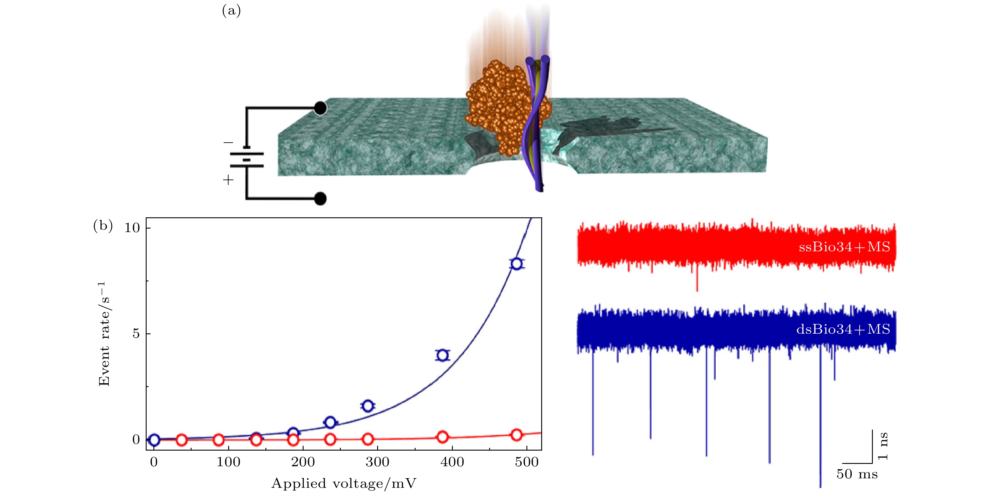
Nanopore sensors have become important tools for analyzing biomarkers, including but not limited to nucleic acids, proteins, and other biomolecules that play important roles in life. Though the nanopores themselves have no selectivity towards target molecules, higher sensitivity of nanopore sensing to the target biomarkers could be achieved with the help of the specificity enhancement technology. In this work, the basic principles of nanopore sensing are first introduced, then methods of modifying nanopore surface as well as the development and application of those selectivity enhancement technologies of nanopore sensing in recent years are reviewed. These enhancement technologies primarily fall into two categories: surface functionalization and molecular probes. Surface functionalization is further categorized based on the types of functional molecules used, while molecular probes are classified according to carrier forms. Finally, in this paper several challenges that nanopore sensing continues to encounter are discussed and some suggestions are made for its future development.
-
Keywords:
- nanopore /
- specificity /
- molecular probe /
- functionalization
[1] Yamazaki H, Hu R, Henley R Y, Halman J, Afonin K A, Yu D, Zhao Q, Wanunu M 2017 Nano Lett. 17 7067
 Google Scholar
[2] Kim J D, Lee Y G 2014 Biomed. Opt. Express 5 2471
 Google Scholar
[3] Verschueren D, Shi X, Dekker C 2019 Small Methods 3 1800465
 Google Scholar
[4] Fologea D, Gershow M, Ledden B, McNabb D S, Golovchenko J A, Li J 2005 Nano Lett. 5 1905
 Google Scholar
[5] Xue L, Yamazaki H, Ren R, Wanunu M, Ivanov A P, Edel J B 2020 Nat. Rev. Mater. 5 931
 Google Scholar
[6] Elaguech M A, Bahri M, Djebbi K, Zhou D, Shi B, Liang L, Komarova N, Kuznetsov A, Tlili C, Wang D 2022 Food Chem. 389 133051
 Google Scholar
[7] Beamish E, Tabard-Cossa V, Godin M 2019 ACS Sens. 4 2458
 Google Scholar
[8] Wang L, Han Y J, Zhou S, Guan X Y 2014 Biosens. Bioelectron. 62 158
 Google Scholar
[9] Oh S, Lee M K, Chi S W 2019 ACS Sens. 4 2849
 Google Scholar
[10] He L, Tessier D R, Briggs K, Tsangaris M, Charron M, McConnell E M, Lomovtsev D, Tabard-Cossa V 2021 Nat. Commun. 12 5348
 Google Scholar
[11] Chen X H, Zhou S, Wang Y J, Zheng L, Guan S, Wang D Q, Wang L, Guan X Y 2023 TrAC Trends Anal. Chem. 162 117060
 Google Scholar
[12] Ying Y L, Zhang J, Gao R, Long Y T 2013 Angew. Chem. Int. Ed. 52 13154
 Google Scholar
[13] Haque F, Li J H, Wu H C, Liang X J, Guo P X 2013 Nano Today 8 56
 Google Scholar
[14] Li S J, Xia N, Yuan B Q, Du W M, Sun Z F, Zhou B B 2015 Electrochim. Acta 159 234
 Google Scholar
[15] Ali M, Neumann R, Ensinger W 2010 ACS Nano 4 7267
 Google Scholar
[16] Ding D F, Gao P C, Ma Q, Wang D G, Xia F 2019 Small 15 1804878
 Google Scholar
[17] Wang H Y, Gu Z, Cao C, Wang J, Long Y T 2013 Anal. Chem. 85 8254
 Google Scholar
[18] Piguet F, Ouldali H, Pastoriza-Gallego M, Manivet P, Pelta J, Oukhaled A 2018 Nat. Commun. 9 966
 Google Scholar
[19] Liu Y, Zhang S Y, Wang Y Q, Wang L Y, Cao Z Y, Sun W, Fan P P, Zhang P K, Chen H Y, Huang S 2022 J. Am. Chem. Soc. 144 13717
 Google Scholar
[20] Hou G L, Zhang H C, Xie G H, Xiao K, Wen L P, Li S H, Tian Y, Jiang L 2014 J. Mater. Chem. A 2 19131
 Google Scholar
[21] Guo L P, Liu Y C, Zeng H O, Zhang S P, Song R Y, Yang J, Han X, Wang Y, Wang L D 2024 Adv. Mater. 36 2307242
 Google Scholar
[22] Ying Y L, Hu Z L, Zhang S L, Qing Y J, Fragasso A, Maglia G, Meller A, Bayley H, Dekker C, Long Y T 2022 Nat. Nanotechnol. 17 1136
 Google Scholar
[23] Mayer S F, Cao C, Dal Peraro M 2022 iScience 25 104145
 Google Scholar
[24] Lee K, Park K B, Kim H J, Yu J S, Chae H, Kim H M, Kim K B 2018 Adv. Mater. 30 1704680
 Google Scholar
[25] Wanunu M, Meller A 2007 Nano Lett. 7 1580
 Google Scholar
[26] Anderson B N, Muthukumar M, Meller A 2013 ACS Nano 7 1408
 Google Scholar
[27] Soni N, Chandra Verma N, Talor N, Meller A 2023 Nano Lett. 23 4609
 Google Scholar
[28] Tang Z P, Lu B, Zhao Q, Wang J J, Luo K F, Yu D P 2014 Small 10 4332
 Google Scholar
[29] Schneider G F, Xu Q, Hage S, Luik S, Spoor J N H, Malladi S, Zandbergen H, Dekker C 2013 Nat. Commun. 4 2619
 Google Scholar
[30] Yusko E C, Prangkio P, Sept D, Rollings R C, Li J, Mayer M 2012 ACS Nano 6 5909
 Google Scholar
[31] Li Q, Ying Y L, Liu S C, Lin Y, Long Y T 2019 ACS Sens. 4 1185
 Google Scholar
[32] Feng S L, Chen C T, Wang W, Que L 2018 Biosens. Bioelectron. 105 36
 Google Scholar
[33] Wilson D S, Szostak J W 1999 Annu. Rev. Biochem. 68 611
 Google Scholar
[34] Zhou J, Rossi J 2016 Nat. Rev. Drug Discovery 16 181
[35] Negrier C, Shima M, Hoffman M 2019 Blood Rev. 38 100582
 Google Scholar
[36] Jaberi N, Soleimani A, Pashirzad M, Abdeahad H, Mohammadi F, Khoshakhlagh M, Khazaei M, Ferns G A, Avan A, Hassanian S M 2018 J. Cell. Biochem. 120 4757
[37] Bock L C, Griffin L C, Latham J A, Vermaas E H, Toole J J 1992 Nature 355 564
 Google Scholar
[38] Rotem D, Jayasinghe L, Salichou M, Bayley H 2012 J. Am. Chem. Soc. 134 2781
 Google Scholar
[39] Reynaud L, Bouchet-Spinelli A, Janot J M, Buhot A, Balme S, Raillon C 2021 Anal. Chem. 93 7889
 Google Scholar
[40] Cao M Y, Zhang L J, Tang H R, Qiu X, Li Y X 2022 Anal. Chem. 94 17405
 Google Scholar
[41] Chou J, Shahi P, Werb Z 2014 Cell Cycle 12 3262
[42] Qiu X, Dong J Y, Dai Q S, Huang M M, Li Y X 2023 Biosens. Bioelectron. 240 115594
 Google Scholar
[43] Peinetti A S, Lake R J, Cong W, Cooper L, Wu Y, Ma Y, Pawel G T, Toimil-Molares M E, Trautmann C, Rong L, Mariñas B, Azzaroni O, Lu Y 2021 Sci. Adv. 7 eabh2848
 Google Scholar
[44] Wu D, Wu T T, Liu Q, Yang Z C 2020 Int. J. Infect. Dis. 94 44
 Google Scholar
[45] Wu F, Zhao S, Yu B, Chen Y M, Wang W, Song Z G, Hu Y, Tao Z W, Tian J H, Pei Y Y, Yuan M L, Zhang Y L, Dai F H, Liu Y, Wang Q M, Zheng J J, Xu L, Holmes E C, Zhang Y Z 2020 Nature 579 265
 Google Scholar
[46] Kim D, Lee J Y, Yang J S, Kim J W, Kim V N, Chang H 2020 Cell 181 914
 Google Scholar
[47] Ma W H, Xie W Y, Tian R, Zeng X Q, Liang L Y, Hou C J, Huo D Q, Wang D Q 2023 Sens. Actuators B 377 133075
 Google Scholar
[48] Albrecht C, Kuznetsov A S, Appert-Collin A, Dhaideh Z, Callewaert M, Bershatsky Y V, Urban A S, Bocharov E V, Bagnard D, Baud S, Blaise S, Romier-Crouzet B, Efremov R G, Dauchez M, Duca L, Gueroult M, Maurice P, Bennasroune A 2020 Front. Cell Dev. Biol. 8 611121
[49] Kwak D K, Kim J S, Lee M K, Ryu K S, Chi S W 2020 Anal. Chem. 92 14303
 Google Scholar
[50] Zhang X, Galenkamp N S, van der Heide N J, Moreno J, Maglia G, Kjems J 2023 ACS Nano 17 9167
 Google Scholar
[51] Thakur A K, Movileanu L 2018 Nat. Biotechnol. 37 96
[52] Ali M, Yameen B, Neumann R, Ensinger W, Knoll W, Azzaroni O 2008 J. Am. Chem. Soc. 130 16351
 Google Scholar
[53] Liu Y, Xuan W M, Cui Y 2010 Adv. Mater. 22 4112
 Google Scholar
[54] Fortuna A, Alves G, Falcão A 2013 Biomed. Chromatogr. 28 27
[55] Wang J, Prajapati J D, Gao F, Ying Y L, Kleinekathöfer U, Winterhalter M, Long Y T 2022 J. Am. Chem. Soc. 144 15072
 Google Scholar
[56] Ramirez J, He F, Lebrilla C B 1998 J. Am. Chem. Soc. 120 7387
 Google Scholar
[57] Kim B Y, Yang J, Gong M, Flachsbart B R, Shannon M A, Bohn P W, Sweedler J V 2009 Anal. Chem. 81 2715
 Google Scholar
[58] Han C P, Hou X, Zhang H C, Guo W, Li H B, Jiang L 2011 J. Am. Chem. Soc. 133 7644
 Google Scholar
[59] Xie G H, Tian W, Wen L P, Xiao K, Zhang Z, Liu Q, Hou G L, Li P, Tian Y, Jiang L 2015 Chem. Commun. 51 3135
 Google Scholar
[60] Jia W D, Hu C Z, Wang Y Q, Gu Y M, Qian G, Du X Y, Wang L, Liu Y, Cao J, Zhang S Y, Yan S, Zhang P K, Ma J, Chen H Y, Huang S 2021 Nat. Commun. 12 5811
 Google Scholar
[61] Jia W D, Hu C Z, Wang Y Q, Liu Y, Wang L Y, Zhang S Y, Zhu Q, Gu Y M, Zhang P K, Ma J, Chen H Y, Huang S 2022 ACS Nano 16 6615
 Google Scholar
[62] Wang Y Q, Fan P P, Zhang S Y, Wang L Y, Li X Y, Jia W D, Liu Y, Wang K F, Du X Y, Zhang P K, Huang S 2022 ACS Nano 16 21356
 Google Scholar
[63] Talaga D S, Li J 2009 J. Am. Chem. Soc. 131 9287
 Google Scholar
[64] Firnkes M, Pedone D, Knezevic J, Döblinger M, Rant U 2010 Nano Lett. 10 2162
 Google Scholar
[65] Sze J Y Y, Ivanov A P, Cass A E G, Edel J B 2017 Nat. Commun. 8 1552
 Google Scholar
[66] Wanunu M, Dadosh T, Ray V, Jin J, McReynolds L, Drndić M 2010 Nat. Nanotechnol. 5 807
 Google Scholar
[67] Carlsen A T, Zahid O K, Ruzicka J A, Taylor E W, Hall A R 2014 Nano Lett. 14 5488
 Google Scholar
[68] Zahid O K, Wang F, Ruzicka J A, Taylor E W, Hall A R 2016 Nano Lett. 16 2033
 Google Scholar
[69] Ilktac A, Kalkan S, Caliskan S 2020 Int. J. Clin. Pract. 75 e13935
[70] Mosquera-Sulbaran J A, Pedreañez A, Carrero Y, Callejas D 2021 Rev. Med. Virol. 31 e2221
 Google Scholar
[71] Tatsuoka T, Okuyama T, Takeshita E, Oi H, Noro T, Mitsui T, Yoshitomi H, Oya M 2020 Surgery Today 51 397
[72] Wu J, Liang L Y, Zhang M K, Zhu R, Wang Z, Yin Y J, Yin B H, Weng T, Fang S X, Xie W Y, Wang L, Wang D Q 2022 ACS Appl. Mater. Interfaces 14 12077
 Google Scholar
[73] Zou Z, Yang H, Yan Q, Qi P, Qing Z H, Zheng J, Xu X, Zhang L H, Feng F, Yang R H 2019 Chem. Commun. 55 6433
 Google Scholar
[74] Saha K, Agasti S S, Kim C, Li X, Rotello V M 2012 Chem. Rev. 112 2739
 Google Scholar
[75] Wang H, Yang R H, Yang L, Tan W H 2009 ACS Nano 3 2451
 Google Scholar
[76] Billinge E R, Broom M, Platt M 2013 Anal. Chem. 86 1030
[77] Blundell E L C J, Vogel R, Platt M 2016 Langmuir 32 1082
 Google Scholar
[78] Hernández-Neuta I, Pereiro I, Ahlford A, Ferraro D, Zhang Q, Viovy J L, Descroix S, Nilsson M 2018 Biosens. Bioelectron. 102 531
 Google Scholar
[79] Kühnemund M, Nilsson M 2015 Biosens. Bioelectron. 67 11
 Google Scholar
[80] Rosen C B, Rodriguez-Larrea D, Bayley H 2014 Nat. Biotechnol. 32 179
 Google Scholar
[81] Zhang Z H, Wang X Q, Wei X J, Zheng S W, Lenhart B J, Xu P S, Li J, Pan J, Albrecht H, Liu C 2021 Biosens. Bioelectron. 181 113134
 Google Scholar
[82] Karhanek M, Kemp J T, Pourmand N, Davis R W, Webb C D 2005 Nano Lett. 5 403
 Google Scholar
[83] Wang H, Tang H R, Yang C, Li Y X 2019 Anal. Chem. 91 7965
 Google Scholar
[84] Tang H R, Wang H, Yang C, Zhao D D, Qian Y Y, Li Y X 2020 Anal. Chem. 92 3042
 Google Scholar
[85] Zhang Z H, Li T, Sheng Y Y, Liu L, Wu H C 2018 Small 15 1804078
[86] Wang X Q, Wei X J, van der Zalm M M, Zhang Z H, Subramanian N, Demers A M, Ghimenton Walters E, Hesseling A, Liu C 2023 ACS Nano 17 21093
 Google Scholar
[87] Japrung D, Bahrami A, Nadzeyka A, Peto L, Bauerdick S, Edel J B, Albrecht T 2014 J. Phys. Chem. B 118 11605
 Google Scholar
[88] Bell N A W, Keyser U F 2015 J. Am. Chem. Soc. 137 2035
 Google Scholar
[89] Squires A, Atas E, Meller A 2015 Sci. Rep. 5 11643
 Google Scholar
[90] Plesa C, Ruitenberg J W, Witteveen M J, Dekker C 2015 Nano Lett. 15 3153
 Google Scholar
[91] Kulenkampff K, Wolf Perez A-M, Sormanni P, Habchi J, Vendruscolo M 2021 Nat. Rev. Chem. 5 277
 Google Scholar
[92] van Steenoven I, Majbour N K, Vaikath N N, Berendse H W, van der Flier W M, van de Berg W D J, Teunissen C E, Lemstra A W, El-Agnaf O M A 2018 Movement Disorders 33 1724
 Google Scholar
[93] Liu Y X, Wang X Y, Campolo G, Teng X Y, Ying L M, Edel J B, Ivanov A P 2023 ACS Nano 17 22999
 Google Scholar
[94] Ivankin A, Henley R Y, Larkin J, Carson S, Toscano M L, Wanunu M 2014 ACS Nano 8 10774
 Google Scholar
[95] Cai S, Pataillot-Meakin T, Shibakawa A, Ren R, Bevan C L, Ladame S, Ivanov A P, Edel J B 2021 Nat. Commun. 12 3515
 Google Scholar
[96] Chandrasekaran A R, MacIsaac M, Vilcapoma J, Hansen C H, Yang D, Wong W P, Halvorsen K 2021 Nano Lett. 21 469
 Google Scholar
[97] Koussa M A, Halvorsen K, Ward A, Wong W P 2015 Nat. Methods 12 123
 Google Scholar
[98] Zhu J B, Tivony R, Bošković F, Pereira-Dias J, Sandler S E, Baker S, Keyser U F 2023 J. Am. Chem. Soc. 145 12115
 Google Scholar
[99] Al-Zarah H, Serag M F, Abadi M, Habuchi S 2023 ACS Appl. Nano Mater. 6 9515
 Google Scholar
[100] Ding T L, Yang J, Wang J, Pan V, Lu Z H, Ke Y G, Zhang C 2022 Biosens. Bioelectron. 195 113658
 Google Scholar
[101] Kim S H, Kim K R, Ahn D R, Lee J E, Yang E G, Kim S Y 2017 ACS Nano 11 9352
 Google Scholar
[102] Yang J, Wang J, Liu X, Chen Y M, Liang Y, Wang Q, Jiang S X, Zhang C 2023 Small 19 2303715
 Google Scholar
-
图 1 (a)未被修饰的纳米孔和用APTMS修饰的纳米孔示意图, 插图是透射电子显微镜(TEM)拍摄的修饰前5.2 nm纳米孔图像 ; (b) 1000碱基长度的 DNA 在 pH 6.0, 7.0和8.0条件下通过被修饰的 5.2 nm 纳米孔电流迹线图, io为开孔电流, ib为阻塞电流, tD为DNA过孔时间[26]
Figure 1. (a) Schematic of an uncoated and an APTMS-coated nanopore, inset is a TEM image of a 5.2 nm nanopore before coating; (b) 1 kbp DNA translocating through a coated 5.2 nm pore at pH 6.0, 7.0, and 8.0, where io is the open-pore current, ib is the blocked-level current, and tD is the translocation time[26].
图 3 (a)淀粉样蛋白β通过流体脂质双分子层修饰的纳米孔示意图; (b) 不同聚类待测分子过孔引起的阻塞电流、过孔时间(∆I, td)数据产生的散点图, 黄色星号表示每种聚类产生的阻塞电流平均值 (i) 球形低聚物, (ii) 短原纤维, (iii) 长原纤维, (iv) 纤维[30]
Figure 3. (a) Schematic representation of amyloid-β translocation through a nanopore modified by a fluid lipid bilayer; (b) scattered plot of blocking currents generated by different clusters, with yellow asterisks indicating the average blocking currents generated by each cluster, (i) spherical oligomer, (ii) short protofibril, (iii) long protofibril, (iv) fiber[30].
图 4 (a)葡萄糖氧化酶通过未修饰的纳米孔示意图(左), EDC/NHS交联反应后葡萄糖氧化酶通过半胱氨酸修饰的纳米孔示意图(右); (b) 两种过孔事件的阻塞电流、过孔时间散点图以及直方图[31]
Figure 4. (a) Glucose oxidase through the unmodified nanopore (left), and glucose oxidase through the cysteine-modified nanopore after EDC/NHS cross-linking reaction (right); (b) blocked current scatter plots and histograms of the two translocation events[31].
图 5 (a) 两种凝血酶通过官能化纳米孔进行识别示意图; (b) 待测分子引起的相对电流阻塞、孔内停留时间散点图; (c) 采用机器学习方法处理数据后获得的识别率混淆矩阵[39]
Figure 5. (a) Schematic illustration of two thrombin species identified by functionalized nanopores; (b) scatter diagram of relative current obstruction and in-hole residence time caused by molecules to be measured; (c) the recognition rate confusion matrix obtained after processing data using machine learning methods[39].
图 6 (a)使用官能化纳米孔选择性检测 miRNA-21 的方法 (i)纳米孔内壁采用金膜改性; (ii)巯基修饰的DNA1与纳米孔壁结合; (iii) MCH阻断活性位点; (iv) DNA1与纳米孔内壁结合, 特异性识别miRNA-21; (v) 引入发夹 H1 和 H2 以引发杂化链式反应. (b)官能化纳米孔在不同浓度miRNA-21 (0.1 pmol/L—0.5 nmol/L)下的I-E曲线(10 mmol/L KCl)[42]
Figure 6. (a) Strategies for selective detection of miRNA-21 using functionalized nanopore: (i) the inner wall of the bare nanopore was modified with gold film; (ii) the sulfhydryl-modified DNA1 binds to the wall of the nanopore; (iii) the excess active site was blocked with MCH; (iv) DNA1 bound to the inner wall of the nanopore specifically recognized miRNA-21; (v) introducing hairpins H1 and H2 to initiate the hybrid chain reaction. (b) I-E curves (10 mmol/L KCl) of functionalized nanopore at different concentrations of miRNA-21 (0.1 pmol/L–0.5 nmol/L)[42].
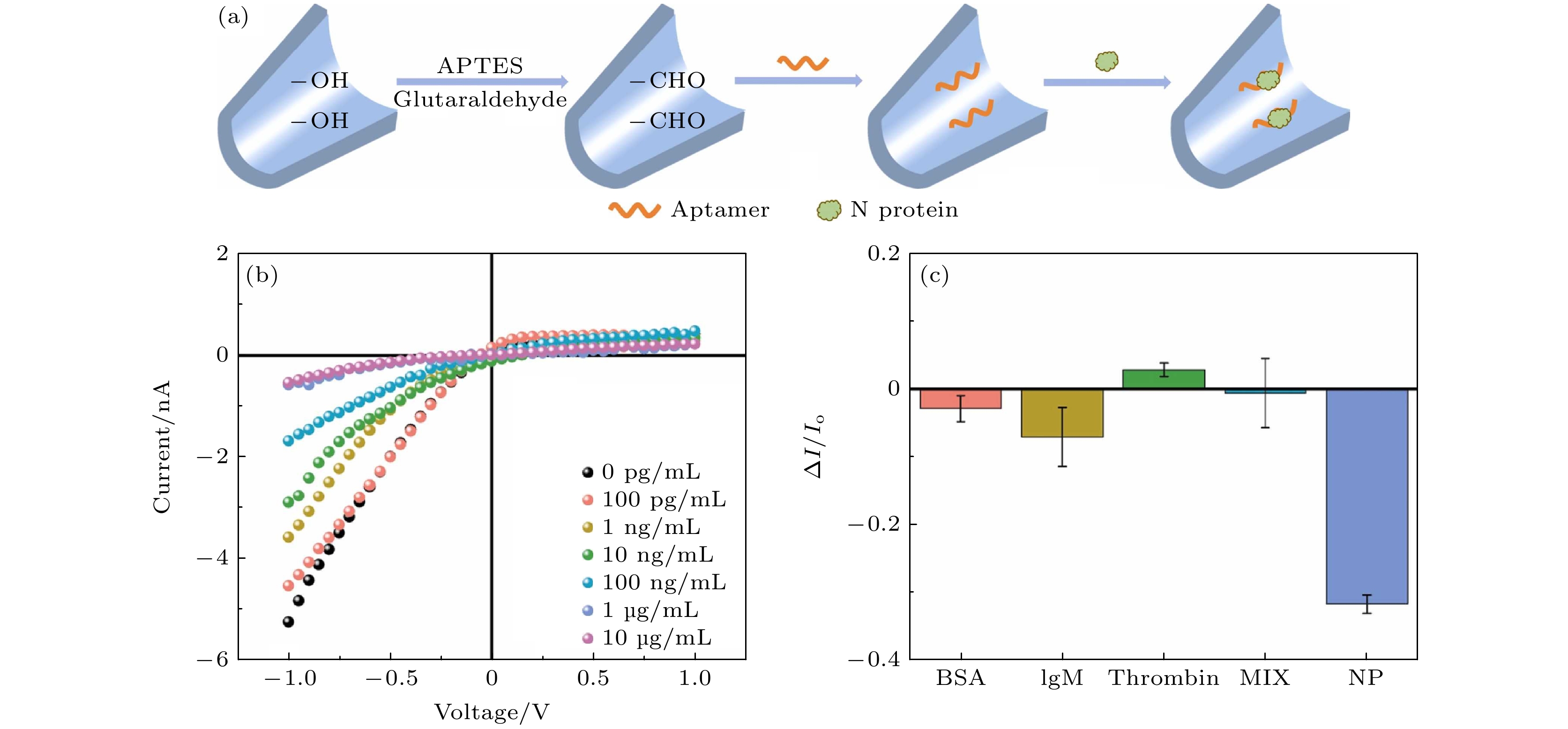
图 8 (a)纳米移液器共价修饰流程示意图; (b)检测不同浓度N蛋白的纳米移液器的I-V曲线; (c)不同蛋白质引起的归一化离子电流变化[47]
Figure 8. (a) Schematic description of the covalent modification procedures of nanopipettes; (b) I-V curves of the nanopipettes for detecting different concentrations of the N protein; (c) normalized ionic current changes caused by different proteins[47].
图 9 (a)未与配体结合(开放)和与配体结合(闭合)状态下的GBP示意图; (b)不同半乳糖浓度下的电流迹线(左), 闭合 (L1) 和开放 (L2) 构象在 30 s内的分布图(右)[49]
Figure 9. (a) Schematic diagram of GBP in the ligand-free (open) and ligand-bound (closed) states; (b) current trace at different galactose concentrations (left), histogram of the closed (L1) and open (L2) conformations in 30 s (right)[49].
图 10 (a)通过DNA链杂交将Ty1固定在ClyA纳米孔上的方法示意图; (b)添加不同浓度的刺突蛋白前后 ClyA-f-Ty1 的代表性电流迹线[50]
Figure 10. (a) Schematic diagram of the method of fixing Ty1 on ClyA nanopore by DNA strand hybridization; (b) the representative current trace of ClyA-f-Ty1 before and after the addition of different concentrations of spike protein[50].
图 14 (a) PNRSS结构示意图, 孔隙限制处的反应部分(棕色)被苯硼酸(PBA)修饰. PBA 作为固定反应物, 结合目标分析物(如儿茶酚胺)并报告传感事件, 蓝色、绿色和黄色区域分别代表 PNRSS 链的延伸部分、牵引部分和系绳部位; (b)振幅 SD 与阻塞百分比Ib的散点图, 蓝色 (L-N)、橙色 (D-N)、黄色 (L-E) 和紫色 (D-E)[61]
Figure 14. (a) PNRSS structure diagram, the reactive portion (brown) at the pore limit is modified by phenylboric acid (PBA). PBA acts as a fixed reactant, binds to target analytes (such as catecholamine) and reports sensing events, the blue, green, and yellow areas represent the PNRSS chain extension, traction, and tether, respectively; (b) scatter plot of amplitude SD versus percentage blockade Ib, blue (L-N), orange (D-N), yellow (L-E), and purple (D-E)[61].
图 16 (a)固态纳米孔用于CRP检测示意图, 右上图为15 nm孔径的纳米孔TEM图像; (b) CRP、适配体和 CRP-适配体复合物通过纳米孔的阻塞电流比以及过孔时间的散点图与直方图[72]
Figure 16. (a) Schematic diagram of solid-state nanopores used for CRP detection, the up right is a TEM image of an aperture of 15 nm in diameter; (b) scatter plots and histograms of blocking current ratios and times of translocation of CRP, aptamer, and CRP-aptamer complexes through nanopore[72].
图 18 (a)石英纳米移液管选择性检测CEA分子示意图, 左边的分子代表Apt-MNPs, 右边的分子代表CEA-Apt-MNP复合物; (b)在+400 mV下1 mol/L KCl的CEA和Apt-MNP混合溶液的CEA-Apt-MNPs(左)和Apt-MNP(右)相应阻塞信号的电流迹线; (c)使用适配体官能化MNPs分离CEA的策略[84]
Figure 18. (a) Schematic illustration of selective detection of CEA molecules with a quartz nanopipette, the left molecule represents the Apt-MNPs, and the right one refers to CEA-Apt-MNP complexes; (b) current-time trace for the presence of a mixed solution of CEA and Apt–MNPs with 1 mol/L KCl at +400 mV, the arrow points to the current traces of the corresponding blockage signals of CEA-Apt-MNPs (left) and Apt-MNPs (right); (c) the strategy for separation of CEA using aptamer functionalized MNPs[84].
图 20 (a)载体制备示意图; (b) α-Syn低聚物与DNA载体结合示意图. (c) DNA载体与不同α-Syn低聚物结合的统计结果 (i) 每个样品的典型电流迹线, 峰值电流随着α-Syn低聚物聚合时间的延长而逐渐增大; (ii) 每个样品的3个代表性信号[93]
Figure 20. (a) Schematic diagram of carrier preparation; (b) schematic diagram of α-Syn oligomer binding to DNA carrier; (c) statistical results of binding of DNA vectors to different α-Syn oligomers: (i) typical current trace for each sample; the peak current increases gradually with the extension of polymerization time of α-Syn oligomer; (ii) three representative signals for each sample[93].
图 21 (a) DNA载体的2D示意图, 具有凝血酶和AChE的特异性探针; (b)在–200 mV下记录的电流时间迹线, 并在5 kHz下滤波, 清楚显示3个电平, 第1个与DNA载体相关, 第2个与凝血酶相关, 第3个与AChE相关[65]
Figure 21. (a) 2D schematic of the λ-DNA carrier with two independent aptamer probes specific to thrombin and AChE; (b) typical current-time trace recorded at –200 mV and re-filtered at 5 kHz clearly showing three levels, the 1st associated with the DNA carrier, 2nd with thrombin and 3rd from AChE[65].
图 22 (a)带有分子信标的DNA载体制备及其与相应miRNA结合示意图; (b)存在miRNA (let-7a, miR-375-3p和miR-141-3p)的情况下, DNA载体过孔的代表性光子和电流时间迹线, 代表 5.6, 10 和 38.5 kbp DNA 片段长度的过孔事件的3个电信号用实心圆、方形和星号标记, 光信号中相应同步事用圆圈标记[95]
Figure 22. (a) Schematic representation of the preparation of size-coded DNA probes and their binding to respective miRNA targets; (b) representative photon and current time traces of DNA carrier translocations in the presence of miRNAs (let-7a, miR-375-3p, and miR-141-3p), three electrical signals representing translocation events of 5.6, 10, and 38.5 kbp DNA fragment lengths are marked with a filled circle, square, and asterisk, the corresponding synchronization in the optical signal is marked with a circle[95].
图 24 (a) opTET2 和 opTET2/SA 的原理图, 红色T代表碱基的生物素修饰; (b) opTET结构的原子力显微镜(AFM) 图像; (c) DNA 四面体opTET2 和 opTET2/SA 通过 30 nm 纳米孔过孔事件的散点图与直方图[102]
Figure 24. (a) Schematic diagram of opTET2 and opTET2/SA, and the red T representing biotin modification of the base; (b) AFM images of opTET structures; (c) scatter plots and histograms of DNA tetrahedron opTET2 and opTET2/SA translocation events through 30 nm nanopore[102].
表 1 目标生物分子对应的特异性增强技术以及检测极限
Table 1. Specific enhancement techniques and detection limits corresponding to target biomolecules.
特异性
增强技术目标生物分子 纳米孔类型 检测极限 表面官能化 α-thrombin
γ-thrombinSiNx N/A[39] miRNA-21 Quartz 0.1 pmol/L[42] HAdV
SARS-CoV-2PET 6 pfu/mL[43]
104 copies/mL[43]N protein Nanopipette 73.204 pg/mL[47] Neuraminidase ClyA 38 nmol/L[49] SARS-CoV-2 Spike Protein ClyA 2.3 nmol/L[50] Barstar t-FhuA 12.6 nmol/L[51] Streptavidin PET N/A[52] L-His, D-His PET N/A[58] L-Trp PI N/A[59] norepinephrine epinephrine MspA 1 μmol/L[61] ribonucleotide MspA N/A[62] 分子探针 miR155 SiNx 10 nmol/L[68] C-reactive protein SiNx 0.3 ng/μL[72] alpha-1 antitrypsin
Tau 381
BACE1Aerolysin 77.9 fmol/L[73]
6.79 fmol/L[73]
86.4 fmol/L[73]miRNA-21 Glass 5 nmol/L[83] Carcinoembryonic-antigen Quartz 0.01 nmol/L[84] alpha fetoprotein α-HL 1 fmol/L[85] ESAT-6/CFP-10 α-HL 10 amol/L[86] α-Syn Quartz 2.2 pmol/L[93] Thrombin
AChEQuartz N/A[65] miR-375-3p
miR-141-3pSiNx 8 fmol/L[95]
5 fmol/L[95]E. coli DH5α 16 SrRNA
Salmonella 16 SrRNA
A. Baumanii 16 SrRNAGlass N/A[98] Streptavidin SiNx N/A[102] -
[1] Yamazaki H, Hu R, Henley R Y, Halman J, Afonin K A, Yu D, Zhao Q, Wanunu M 2017 Nano Lett. 17 7067
 Google Scholar
[2] Kim J D, Lee Y G 2014 Biomed. Opt. Express 5 2471
 Google Scholar
[3] Verschueren D, Shi X, Dekker C 2019 Small Methods 3 1800465
 Google Scholar
[4] Fologea D, Gershow M, Ledden B, McNabb D S, Golovchenko J A, Li J 2005 Nano Lett. 5 1905
 Google Scholar
[5] Xue L, Yamazaki H, Ren R, Wanunu M, Ivanov A P, Edel J B 2020 Nat. Rev. Mater. 5 931
 Google Scholar
[6] Elaguech M A, Bahri M, Djebbi K, Zhou D, Shi B, Liang L, Komarova N, Kuznetsov A, Tlili C, Wang D 2022 Food Chem. 389 133051
 Google Scholar
[7] Beamish E, Tabard-Cossa V, Godin M 2019 ACS Sens. 4 2458
 Google Scholar
[8] Wang L, Han Y J, Zhou S, Guan X Y 2014 Biosens. Bioelectron. 62 158
 Google Scholar
[9] Oh S, Lee M K, Chi S W 2019 ACS Sens. 4 2849
 Google Scholar
[10] He L, Tessier D R, Briggs K, Tsangaris M, Charron M, McConnell E M, Lomovtsev D, Tabard-Cossa V 2021 Nat. Commun. 12 5348
 Google Scholar
[11] Chen X H, Zhou S, Wang Y J, Zheng L, Guan S, Wang D Q, Wang L, Guan X Y 2023 TrAC Trends Anal. Chem. 162 117060
 Google Scholar
[12] Ying Y L, Zhang J, Gao R, Long Y T 2013 Angew. Chem. Int. Ed. 52 13154
 Google Scholar
[13] Haque F, Li J H, Wu H C, Liang X J, Guo P X 2013 Nano Today 8 56
 Google Scholar
[14] Li S J, Xia N, Yuan B Q, Du W M, Sun Z F, Zhou B B 2015 Electrochim. Acta 159 234
 Google Scholar
[15] Ali M, Neumann R, Ensinger W 2010 ACS Nano 4 7267
 Google Scholar
[16] Ding D F, Gao P C, Ma Q, Wang D G, Xia F 2019 Small 15 1804878
 Google Scholar
[17] Wang H Y, Gu Z, Cao C, Wang J, Long Y T 2013 Anal. Chem. 85 8254
 Google Scholar
[18] Piguet F, Ouldali H, Pastoriza-Gallego M, Manivet P, Pelta J, Oukhaled A 2018 Nat. Commun. 9 966
 Google Scholar
[19] Liu Y, Zhang S Y, Wang Y Q, Wang L Y, Cao Z Y, Sun W, Fan P P, Zhang P K, Chen H Y, Huang S 2022 J. Am. Chem. Soc. 144 13717
 Google Scholar
[20] Hou G L, Zhang H C, Xie G H, Xiao K, Wen L P, Li S H, Tian Y, Jiang L 2014 J. Mater. Chem. A 2 19131
 Google Scholar
[21] Guo L P, Liu Y C, Zeng H O, Zhang S P, Song R Y, Yang J, Han X, Wang Y, Wang L D 2024 Adv. Mater. 36 2307242
 Google Scholar
[22] Ying Y L, Hu Z L, Zhang S L, Qing Y J, Fragasso A, Maglia G, Meller A, Bayley H, Dekker C, Long Y T 2022 Nat. Nanotechnol. 17 1136
 Google Scholar
[23] Mayer S F, Cao C, Dal Peraro M 2022 iScience 25 104145
 Google Scholar
[24] Lee K, Park K B, Kim H J, Yu J S, Chae H, Kim H M, Kim K B 2018 Adv. Mater. 30 1704680
 Google Scholar
[25] Wanunu M, Meller A 2007 Nano Lett. 7 1580
 Google Scholar
[26] Anderson B N, Muthukumar M, Meller A 2013 ACS Nano 7 1408
 Google Scholar
[27] Soni N, Chandra Verma N, Talor N, Meller A 2023 Nano Lett. 23 4609
 Google Scholar
[28] Tang Z P, Lu B, Zhao Q, Wang J J, Luo K F, Yu D P 2014 Small 10 4332
 Google Scholar
[29] Schneider G F, Xu Q, Hage S, Luik S, Spoor J N H, Malladi S, Zandbergen H, Dekker C 2013 Nat. Commun. 4 2619
 Google Scholar
[30] Yusko E C, Prangkio P, Sept D, Rollings R C, Li J, Mayer M 2012 ACS Nano 6 5909
 Google Scholar
[31] Li Q, Ying Y L, Liu S C, Lin Y, Long Y T 2019 ACS Sens. 4 1185
 Google Scholar
[32] Feng S L, Chen C T, Wang W, Que L 2018 Biosens. Bioelectron. 105 36
 Google Scholar
[33] Wilson D S, Szostak J W 1999 Annu. Rev. Biochem. 68 611
 Google Scholar
[34] Zhou J, Rossi J 2016 Nat. Rev. Drug Discovery 16 181
[35] Negrier C, Shima M, Hoffman M 2019 Blood Rev. 38 100582
 Google Scholar
[36] Jaberi N, Soleimani A, Pashirzad M, Abdeahad H, Mohammadi F, Khoshakhlagh M, Khazaei M, Ferns G A, Avan A, Hassanian S M 2018 J. Cell. Biochem. 120 4757
[37] Bock L C, Griffin L C, Latham J A, Vermaas E H, Toole J J 1992 Nature 355 564
 Google Scholar
[38] Rotem D, Jayasinghe L, Salichou M, Bayley H 2012 J. Am. Chem. Soc. 134 2781
 Google Scholar
[39] Reynaud L, Bouchet-Spinelli A, Janot J M, Buhot A, Balme S, Raillon C 2021 Anal. Chem. 93 7889
 Google Scholar
[40] Cao M Y, Zhang L J, Tang H R, Qiu X, Li Y X 2022 Anal. Chem. 94 17405
 Google Scholar
[41] Chou J, Shahi P, Werb Z 2014 Cell Cycle 12 3262
[42] Qiu X, Dong J Y, Dai Q S, Huang M M, Li Y X 2023 Biosens. Bioelectron. 240 115594
 Google Scholar
[43] Peinetti A S, Lake R J, Cong W, Cooper L, Wu Y, Ma Y, Pawel G T, Toimil-Molares M E, Trautmann C, Rong L, Mariñas B, Azzaroni O, Lu Y 2021 Sci. Adv. 7 eabh2848
 Google Scholar
[44] Wu D, Wu T T, Liu Q, Yang Z C 2020 Int. J. Infect. Dis. 94 44
 Google Scholar
[45] Wu F, Zhao S, Yu B, Chen Y M, Wang W, Song Z G, Hu Y, Tao Z W, Tian J H, Pei Y Y, Yuan M L, Zhang Y L, Dai F H, Liu Y, Wang Q M, Zheng J J, Xu L, Holmes E C, Zhang Y Z 2020 Nature 579 265
 Google Scholar
[46] Kim D, Lee J Y, Yang J S, Kim J W, Kim V N, Chang H 2020 Cell 181 914
 Google Scholar
[47] Ma W H, Xie W Y, Tian R, Zeng X Q, Liang L Y, Hou C J, Huo D Q, Wang D Q 2023 Sens. Actuators B 377 133075
 Google Scholar
[48] Albrecht C, Kuznetsov A S, Appert-Collin A, Dhaideh Z, Callewaert M, Bershatsky Y V, Urban A S, Bocharov E V, Bagnard D, Baud S, Blaise S, Romier-Crouzet B, Efremov R G, Dauchez M, Duca L, Gueroult M, Maurice P, Bennasroune A 2020 Front. Cell Dev. Biol. 8 611121
[49] Kwak D K, Kim J S, Lee M K, Ryu K S, Chi S W 2020 Anal. Chem. 92 14303
 Google Scholar
[50] Zhang X, Galenkamp N S, van der Heide N J, Moreno J, Maglia G, Kjems J 2023 ACS Nano 17 9167
 Google Scholar
[51] Thakur A K, Movileanu L 2018 Nat. Biotechnol. 37 96
[52] Ali M, Yameen B, Neumann R, Ensinger W, Knoll W, Azzaroni O 2008 J. Am. Chem. Soc. 130 16351
 Google Scholar
[53] Liu Y, Xuan W M, Cui Y 2010 Adv. Mater. 22 4112
 Google Scholar
[54] Fortuna A, Alves G, Falcão A 2013 Biomed. Chromatogr. 28 27
[55] Wang J, Prajapati J D, Gao F, Ying Y L, Kleinekathöfer U, Winterhalter M, Long Y T 2022 J. Am. Chem. Soc. 144 15072
 Google Scholar
[56] Ramirez J, He F, Lebrilla C B 1998 J. Am. Chem. Soc. 120 7387
 Google Scholar
[57] Kim B Y, Yang J, Gong M, Flachsbart B R, Shannon M A, Bohn P W, Sweedler J V 2009 Anal. Chem. 81 2715
 Google Scholar
[58] Han C P, Hou X, Zhang H C, Guo W, Li H B, Jiang L 2011 J. Am. Chem. Soc. 133 7644
 Google Scholar
[59] Xie G H, Tian W, Wen L P, Xiao K, Zhang Z, Liu Q, Hou G L, Li P, Tian Y, Jiang L 2015 Chem. Commun. 51 3135
 Google Scholar
[60] Jia W D, Hu C Z, Wang Y Q, Gu Y M, Qian G, Du X Y, Wang L, Liu Y, Cao J, Zhang S Y, Yan S, Zhang P K, Ma J, Chen H Y, Huang S 2021 Nat. Commun. 12 5811
 Google Scholar
[61] Jia W D, Hu C Z, Wang Y Q, Liu Y, Wang L Y, Zhang S Y, Zhu Q, Gu Y M, Zhang P K, Ma J, Chen H Y, Huang S 2022 ACS Nano 16 6615
 Google Scholar
[62] Wang Y Q, Fan P P, Zhang S Y, Wang L Y, Li X Y, Jia W D, Liu Y, Wang K F, Du X Y, Zhang P K, Huang S 2022 ACS Nano 16 21356
 Google Scholar
[63] Talaga D S, Li J 2009 J. Am. Chem. Soc. 131 9287
 Google Scholar
[64] Firnkes M, Pedone D, Knezevic J, Döblinger M, Rant U 2010 Nano Lett. 10 2162
 Google Scholar
[65] Sze J Y Y, Ivanov A P, Cass A E G, Edel J B 2017 Nat. Commun. 8 1552
 Google Scholar
[66] Wanunu M, Dadosh T, Ray V, Jin J, McReynolds L, Drndić M 2010 Nat. Nanotechnol. 5 807
 Google Scholar
[67] Carlsen A T, Zahid O K, Ruzicka J A, Taylor E W, Hall A R 2014 Nano Lett. 14 5488
 Google Scholar
[68] Zahid O K, Wang F, Ruzicka J A, Taylor E W, Hall A R 2016 Nano Lett. 16 2033
 Google Scholar
[69] Ilktac A, Kalkan S, Caliskan S 2020 Int. J. Clin. Pract. 75 e13935
[70] Mosquera-Sulbaran J A, Pedreañez A, Carrero Y, Callejas D 2021 Rev. Med. Virol. 31 e2221
 Google Scholar
[71] Tatsuoka T, Okuyama T, Takeshita E, Oi H, Noro T, Mitsui T, Yoshitomi H, Oya M 2020 Surgery Today 51 397
[72] Wu J, Liang L Y, Zhang M K, Zhu R, Wang Z, Yin Y J, Yin B H, Weng T, Fang S X, Xie W Y, Wang L, Wang D Q 2022 ACS Appl. Mater. Interfaces 14 12077
 Google Scholar
[73] Zou Z, Yang H, Yan Q, Qi P, Qing Z H, Zheng J, Xu X, Zhang L H, Feng F, Yang R H 2019 Chem. Commun. 55 6433
 Google Scholar
[74] Saha K, Agasti S S, Kim C, Li X, Rotello V M 2012 Chem. Rev. 112 2739
 Google Scholar
[75] Wang H, Yang R H, Yang L, Tan W H 2009 ACS Nano 3 2451
 Google Scholar
[76] Billinge E R, Broom M, Platt M 2013 Anal. Chem. 86 1030
[77] Blundell E L C J, Vogel R, Platt M 2016 Langmuir 32 1082
 Google Scholar
[78] Hernández-Neuta I, Pereiro I, Ahlford A, Ferraro D, Zhang Q, Viovy J L, Descroix S, Nilsson M 2018 Biosens. Bioelectron. 102 531
 Google Scholar
[79] Kühnemund M, Nilsson M 2015 Biosens. Bioelectron. 67 11
 Google Scholar
[80] Rosen C B, Rodriguez-Larrea D, Bayley H 2014 Nat. Biotechnol. 32 179
 Google Scholar
[81] Zhang Z H, Wang X Q, Wei X J, Zheng S W, Lenhart B J, Xu P S, Li J, Pan J, Albrecht H, Liu C 2021 Biosens. Bioelectron. 181 113134
 Google Scholar
[82] Karhanek M, Kemp J T, Pourmand N, Davis R W, Webb C D 2005 Nano Lett. 5 403
 Google Scholar
[83] Wang H, Tang H R, Yang C, Li Y X 2019 Anal. Chem. 91 7965
 Google Scholar
[84] Tang H R, Wang H, Yang C, Zhao D D, Qian Y Y, Li Y X 2020 Anal. Chem. 92 3042
 Google Scholar
[85] Zhang Z H, Li T, Sheng Y Y, Liu L, Wu H C 2018 Small 15 1804078
[86] Wang X Q, Wei X J, van der Zalm M M, Zhang Z H, Subramanian N, Demers A M, Ghimenton Walters E, Hesseling A, Liu C 2023 ACS Nano 17 21093
 Google Scholar
[87] Japrung D, Bahrami A, Nadzeyka A, Peto L, Bauerdick S, Edel J B, Albrecht T 2014 J. Phys. Chem. B 118 11605
 Google Scholar
[88] Bell N A W, Keyser U F 2015 J. Am. Chem. Soc. 137 2035
 Google Scholar
[89] Squires A, Atas E, Meller A 2015 Sci. Rep. 5 11643
 Google Scholar
[90] Plesa C, Ruitenberg J W, Witteveen M J, Dekker C 2015 Nano Lett. 15 3153
 Google Scholar
[91] Kulenkampff K, Wolf Perez A-M, Sormanni P, Habchi J, Vendruscolo M 2021 Nat. Rev. Chem. 5 277
 Google Scholar
[92] van Steenoven I, Majbour N K, Vaikath N N, Berendse H W, van der Flier W M, van de Berg W D J, Teunissen C E, Lemstra A W, El-Agnaf O M A 2018 Movement Disorders 33 1724
 Google Scholar
[93] Liu Y X, Wang X Y, Campolo G, Teng X Y, Ying L M, Edel J B, Ivanov A P 2023 ACS Nano 17 22999
 Google Scholar
[94] Ivankin A, Henley R Y, Larkin J, Carson S, Toscano M L, Wanunu M 2014 ACS Nano 8 10774
 Google Scholar
[95] Cai S, Pataillot-Meakin T, Shibakawa A, Ren R, Bevan C L, Ladame S, Ivanov A P, Edel J B 2021 Nat. Commun. 12 3515
 Google Scholar
[96] Chandrasekaran A R, MacIsaac M, Vilcapoma J, Hansen C H, Yang D, Wong W P, Halvorsen K 2021 Nano Lett. 21 469
 Google Scholar
[97] Koussa M A, Halvorsen K, Ward A, Wong W P 2015 Nat. Methods 12 123
 Google Scholar
[98] Zhu J B, Tivony R, Bošković F, Pereira-Dias J, Sandler S E, Baker S, Keyser U F 2023 J. Am. Chem. Soc. 145 12115
 Google Scholar
[99] Al-Zarah H, Serag M F, Abadi M, Habuchi S 2023 ACS Appl. Nano Mater. 6 9515
 Google Scholar
[100] Ding T L, Yang J, Wang J, Pan V, Lu Z H, Ke Y G, Zhang C 2022 Biosens. Bioelectron. 195 113658
 Google Scholar
[101] Kim S H, Kim K R, Ahn D R, Lee J E, Yang E G, Kim S Y 2017 ACS Nano 11 9352
 Google Scholar
[102] Yang J, Wang J, Liu X, Chen Y M, Liang Y, Wang Q, Jiang S X, Zhang C 2023 Small 19 2303715
 Google Scholar
Catalog
Metrics
- Abstract views: 9873
- PDF Downloads: 213
- Cited By: 0


 DownLoad:


