


-
高压极端条件是实现和调控新奇物态的重要途径, 磁共振技术是材料微观磁结构和磁性表征的重要方法, 两者的融合为物质科学前沿研究提供了新的机遇. 然而, 传统磁共振技术受限于自旋极化度低、信号探测效率差等因素, 难以实现超高压极端条件下微米级小样品的原位测量. 近年来, 以金刚石氮空位中心为代表的色心量子传感迅速发展, 为高压极端条件下的磁共振和原位量子传感提供了全新解决方案. 本文总结了高压极端条件对金刚石氮空位中心自旋和光学性质的影响, 梳理了高压下色心磁共振的基本现象和规律. 同时, 以高压下微区磁成像、压强探测、超导迈斯纳效应测量等应用为例, 本文还介绍了高压下色心量子传感的近期研究进展.High-pressure extreme conditions are crucial for realizing novel states and regulating material properties, while magnetic resonance technology is a widely used method to characterize microscopic magnetic structures and magnetic properties. The integration of these two fields offers new opportunities for cutting-edge research in condensed matter physics and materials science. However, conventional magnetic resonance is limited by several factors, such as low spin polarization and low signal detection efficiency, which makes in-situ measurement of micrometer-sized samples under ultra-high pressure a challenge. Recent advances in quantum sensing with color centers in solids, in particular, the development of quantum sensors based on nitrogen vacancy (NV) centers in diamond, provide an innovative solution for magnetic resonance and in-situ quantum sensing under high pressure. This article summarizes the effects of high-pressure conditions on the spin and optical properties, as well as on the magnetic resonance of diamond NV centers. In addition, this article reviews recent advances in high-pressure quantum sensing through applications such as magnetic imaging, pressure detection, and the study of the superconducting Meissner effect under high pressure.
[1] Wang H, Tse J S, Tanaka K, Iitaka T, Ma Y M 2012 PNAS 109 6463
 Google Scholar
[2] Drozdov A P, Eremets M I, Troyan I A, Ksenofontov V, Shylin S I 2015 Nature 525 73
Google Scholar
[3] Drozdov A P, Kong P P, Minkov V S, Besedin S P, Kuzovnikov M A, Mozaffari S, Balicas L, Balakirev F F, Graf D E, Prakapenka V B, Greenberg E, Knyazev D A, Tkacz M, Eremets M I 2019 Nature 569 528
Google Scholar
[4] Somayazulu M, Ahart M, Mishra A K, Geballe Z M, Baldini M, Meng Y, Struzhkin V V, Hemley R J 2019 Phys. Rev. Lett. 122 027001
Google Scholar
[5] Zhang L J, Wang Y C, Lv J, Ma Y M 2017 Nat. Rev. Mater. 2 17005
Google Scholar
[6] Ekimov E A, Sidorov V A, Bauer E D, Mel'nik N N, Curro N J, Thompson J D, Stishov S M 2004 Nature 428 542
Google Scholar
[7] Hirose K, Fei Y, Ma Y, Mao H K 1999 Nature 397 53
Google Scholar
[8] Hu Q Y, Kim D Y, Yang W G, Yang L X, Meng Y, Zhang L, Mao H K 2016 Nature 534 241
Google Scholar
[9] Meier T 2018 Annu. Rep. NMR Spectrosc. 93 1
[10] Meier T, Trybel F, Khandarkhaeva S, Steinle-Neumann G, Chariton S, Fedotenko T, Petitgirard S, Hanfland M, Glazyrin K, Dubrovinskaia N, Dubrovinsky L 2019 Phys. Rev. X 9 031008
[11] Dai J H, Shang Y X, Yu Y H, Xu Y, Yu H, Hong F, Yu X H, Pan X Y, Liu G Q 2022 Chin. Phys. Lett. 39 117601
Google Scholar
[12] Hilberer A, Toraille L, Dailledouze C, Adam M P, Hanlon L, Weck G, Schmidt M, Loubeyre P, Roch J F 2023 Phys. Rev. B 107 L220102
Google Scholar
[13] Bhattacharyya P, Chen W, Huang X, Chatterjee S, Huang B, Kobrin B, Lyu Y, Smart T J, Block M, Wang E, Wang Z, Wu W, Hsieh S, Ma H, Mandyam S, Chen B, Davis E, Geballe Z M, Zu C, Struzhkin V, Jeanloz R, Moore J E, Cui T, Galli G, Halperin B I, Laumann C R, Yao N Y 2024 Nature 627 73
Google Scholar
[14] Wang M Q, Wang Y, Liu Z X, Xu G Y, Yang B, Yu P, Sun H Y, Ye X Y, Zhou J W, Goncharov A F, Wang Y, Du J F 2024 Nat. Commun. 15 8843
Google Scholar
[15] Liu G Q, Feng X, Wang N, Li Q, Liu R B 2019 Nat. Commun. 10 1344
Google Scholar
[16] Fan J W, Guo S W, Lin C, Wang N, Liu G Q, Li Q, Liu R B 2024 Nano Lett. 24 14806
Google Scholar
[17] Fortman B, Mugica-Sanchez L, Tischler N, Selco C, Hang Y X, Holczer K, Takahashi C 2021 J Appl. Phys. 130 083901
Google Scholar
[18] 刘刚钦 2022 71 066101
Google Scholar
Liu G Q 2022 Acta Phys. Sin. 71 066101
Google Scholar
[19] Rondin L, Tetienne J P, Hingant T, Roch J F, Maletinsky P, Jacques V 2014 Rep. Prog. Phys. 77 056503
Google Scholar
[20] Schirhagl R, Chang K, Loretz M, Degen C L 2014 Annu. Rev. Phys. Chem. 65 83
Google Scholar
[21] Degen C L, Reinhard F, Cappellaro P 2017 Rev. Mod. Phys. 89 035002
Google Scholar
[22] Casola F, Van Der Sar T, Yacoby A 2018 Nat. Rev. Mater. 3 17088
Google Scholar
[23] Liu G Q, Liu R B, Li Q 2022 Acc. Chem. Res. 56 95
[24] 刘刚钦, 邢健, 潘新宇 2019 68 120302
Google Scholar
Liu G Q, Xing J, Pan X Y 2019 Acta Phys. Sin. 68 120302
Google Scholar
[25] 董杨, 杜博, 张少春, 陈向东, 孙方稳 2018 67 160301
Google Scholar
Dong Y, Du B, Zhang S C, Chen X D, Sun F W 2018 Acta Phys. Sin. 67 160301
Google Scholar
[26] 彭世杰, 刘颖, 马文超, 石发展, 杜江峰 2018 67 167601
Google Scholar
Peng S J, Liu Y, Ma W C, Shi F Z, Du J F 2018 Acta Phys. Sin. 67 167601
Google Scholar
[27] Aslam N, Zhou H Y, Urbach E K, Turner M J, Walsworth R L, Lukin M D, Park H 2023 Nat. Rev. Phys. 5 157
Google Scholar
[28] Wu Y, Weil T 2022 Adv. Sci. 9 2200059
Google Scholar
[29] Barry J F, Schloss J M, Bauch E, Turner M J, Hart C A, Pham L A, Walsworth R L 2020 Rev. Mod. Phys. 92 015004
Google Scholar
[30] Lawson A W, Tang T Y 1950 Rev. Sci. Instrum. 21 815
[31] Jayaraman A 1983 Rev. Mod. Phys. 55 65
Google Scholar
[32] Gruber A, Dräbenstedt A, Tietz C, Fleury L, Wrachtrup J, von Borczyskowski C 1997 Science 276 2012
Google Scholar
[33] Doherty M W, Manson N B, Delaney P, Jelezko F, Wrachtrup J, Hollenberg L C L 2013 Phys. Rep. 528 1
Google Scholar
[34] Doherty M W, Struzhkin V V, Simpson D A, McGuinness L P, Meng Y F, Stacey A, Karle T J, Hemley R J, Manson N B, Hollenberg L C L, Prawer S 2014 Phys. Rev. Lett. 112 047601
Google Scholar
[35] Shang Y X, Hong F, Dai H J, Yu H, Lu Y N, Liu E K, Yu X H, Liu G Q, Pan X Y 2019 Chin. Phys. Lett. 36 086201
Google Scholar
[36] Yip K Y, Ho K O, Yu K Y, Chen Y, Zhang W, Kasahara S, Mizukami Y, Shibauchi T, Matsuda Y, Goh S K, Yang S 2019 Science 366 1355
Google Scholar
[37] Shelton D P, Cabriales W, Salamat A 2024 Rev. Sci. Instrum. 95 083901
Google Scholar
[38] Hsieh S, Bhattacharyya P, Zu C, Mittiga T, Smart T J, Machado F, Kobrin B, Höhn T O, Rui N Z, Kamrani M, Chatterjee S, Choi S, Zaletel M, Struzhkin V V, Moore J E, Levitas V I, Jeanloz R, Yao N Y 2019 Science 366 1349
Google Scholar
[39] Lesik M, Plisson T, Toraille L, Renaud J, Occelli F, Schmidt M, Salord O, Delobbe A, Debuisschert T, Rondin L, Loubeyre P, Roch J F 2019 Science 366 1359
Google Scholar
[40] Wen J Y, Xu Y, Wang G, He Z X, Chen Y, Wang N N, Lu T L, Ma X L, Jin F, Chen L C, Liu M, Fan J W, Liu X B, Pan X Y, Liu G Q, Cheng J G, Yu X H 2024 arXiv: 2410.10275 https://doi.org/10.48550/arXiv.2410.10275
[41] Meijer J, Burchard B, Domhan M, Wittmann C, Gaebel T, Popa I, Jelezko F, Wrachtrup J 2005 Appl. Phys. Lett. 87 261909
Google Scholar
[42] Lyapin S G, Ilichev I D, Novikov A P, Davydov V A, Agafonov V N 2018 Nanosystems: Physics, Chemistry, Mathematics 9 55
[43] Shang Y X, Hong F, Dai J H, Lu Y N, Yu H, Yu Y H, Yu X H, Pan X Y, Liu G Q 2022 arXiv: 2203.10511 https://doi.org/10.48550/arXiv.2203.10511
[44] Jacques V, Neumann P, Beck J, Markham M, Twitchen D, Meijer J, Kaiser F, Balasubramanian G, Jelezko F, Wrachtrup J 2009 Phys. Rev. Lett. 102 57403
Google Scholar
[45] London P, Scheuer J, Cai J M, Schwarz I, Retzker A, Plenio M B, Katagiri M, Teraji T, Koizumi S, Isoya J, Fischer R, McGuinness L P, Naydenov B, Jelezko F 2013 Phys. Rev. Lett. 111 067601
Google Scholar
[46] Liu G Q, Jiang Q Q, Chang Y C, Liu D Q, Li W X, Gu C Z, Po H C, Zhang W X, Zhao N, Pan X Y 2014 Nanoscale 6 10134
Google Scholar
[47] Zhang G, Cheng Y, Chou J P, Gali A 2020 Appl. Phys. Rev. 7 031308
Google Scholar
[48] Wang J F, Liu L, Liu X D, Li Q, Cui J M, Zhou D F, Zhou J Y, Wei Y, Xu H A, Xu W, Lin W X, Yan J W, He Z X, Liu Z H, Hao Z H, Li H O, Liu W, Xu J S, Gregoryanz E, Li C F 2023 Nat. Mater. 22 489
Google Scholar
[49] Liu L, Wang J F, Liu X D, Xu H A, Cui J M, Li Q, Zhou J Y, Lin W X, He Z X, Xu W, Wei Y, Liu Z H, Wang P, Hao Z H, Ding J F, Li H O, Liu W, Li H, You L X, Xu J S, Gregoryanz E, Li C F, Guo G C 2022 Nano Lett. 22 9943
Google Scholar
[50] He G H, Gong R T, Wang Z P, Liu Z W, Hong J, Zhang T X, Riofrio A L, Rehfuss Z, Chen M F, Yao C Y, Poirier T, Ye B T, Wang X, Ran S, Edgar J H, Zhang S X, Yao N Y, Zu C 2025 arXiv: 2501.03319
[51] Zhong C, Wang Y P, Mai D, Ye C H, Li X D, Wang H, Dai R C, Wang Z P, Sun X Y, Zhang Z M 2024 Nano Lett. 24 4993
[52] Mao H K 2024 Natl. Sci. Rev. 11 nwae004
Google Scholar
[53] Eremets M I 2024 Natl. Sci. Rev. 11 nwae047
Google Scholar
[54] Hamlin J J 2019 Nature 569 491
Google Scholar
[55] Sun H L, Huo M W, Hu X W, Li J Y, Liu Z J, Han Y F, Tang L Y, Mao Z Q, Yang P T, Wang B S, Cheng J G, Yao D X, Zhang G M, Wang M 2023 Nature 621 493
Google Scholar
[56] Wang N N, Wang G, Shen X L, Hou J, Luo J, Ma X P, Yang H X, Shi L F, Dou J, Feng J, Yang J, Shi Y Q, Ren Z A, Ma H M, Yang P T, Liu Z Y, Liu Y, Zhang H, Dong X L, Wang Y X, Jiang K, Hu J P, Nagasaki S, Kitagawa K, Calder S, Yan J Q, Sun J P, Wang B S, Zhou R, Uwatoko Y, Cheng J G 2024 Nature 634 579
Google Scholar
[57] Zhou Y Z, Guo J, Cai S, Sun H L, Li C Y, Zhao J Y, Wang P Y, Han J Y, Chen X T, Chen Y J, Wu Q, Ding Y, Xiang T, Mao H K, Sun L L 2025 Matter Radiat. Extremes 10 027801
Google Scholar
-
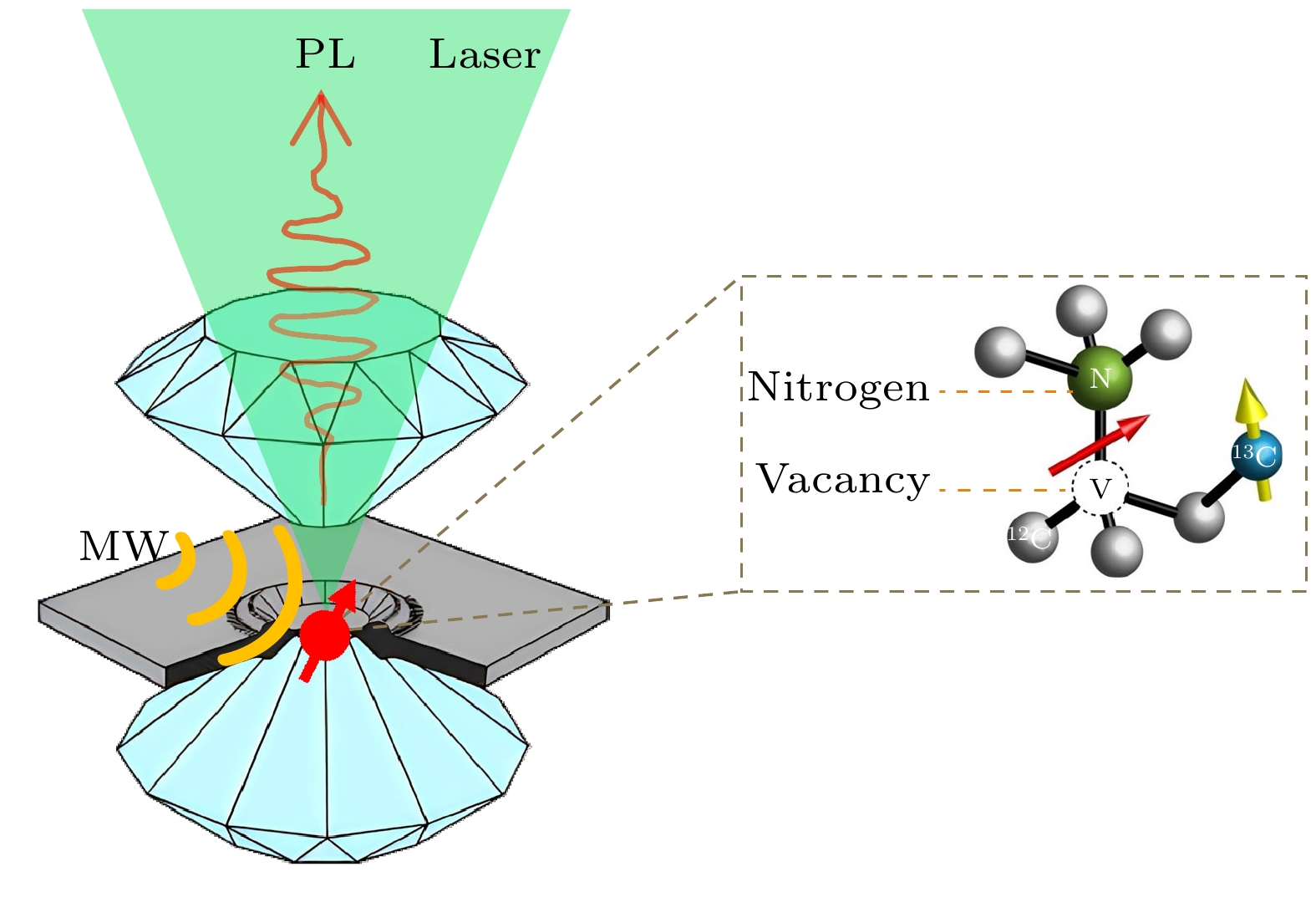
图 1 高压下的金刚石色心量子传感. 左图为金刚石对顶砧的基本结构, 由两块特殊切割的金刚石和金属垫片构成, 垫片中心孔内装载样品并填充传压介质, 通过上下两片金刚石的挤压给样品施加高压. 右图为金刚石氮空位(nitrogen-vacancy, NV)中心的物理结构——由一个替代位氮原子和一个近邻空位构成. NV中心自旋状态可通过光学方法高效地极化、操控和读出, 可作为灵敏的纳米尺度量子传感器. 基于NV中心的量子传感完全兼容于金刚石对顶砧压机, 为高压极端条件下的磁共振和微区磁测量提供了全新的方案
Fig. 1. Diamond NV center-based quantum sensing under high pressure. The figure on the left illustrates the basic structure of a diamond anvil cell, which consists of two specially cut diamonds and a metal seal. The sample is loaded into the central hole of the gasket, which is filled with a pressure-transmitting medium. High pressure is applied to the sample by compressing the upper and lower diamond anvils. The diagram on the right shows the physical structure of a nitrogen-vacancy (NV) center in diamond, which consists of a substituted nitrogen atom and an adjacent vacancy. The spin state of NV centers can be efficiently polarized, controlled and read out using optical methods, enabling sensitive quantum sensing at the nanoscale. NV-based quantum sensing is compatible with diamond anvil cells and provides a novel method to realize magnetic resonance and magnetic measurements under high pressure conditions.
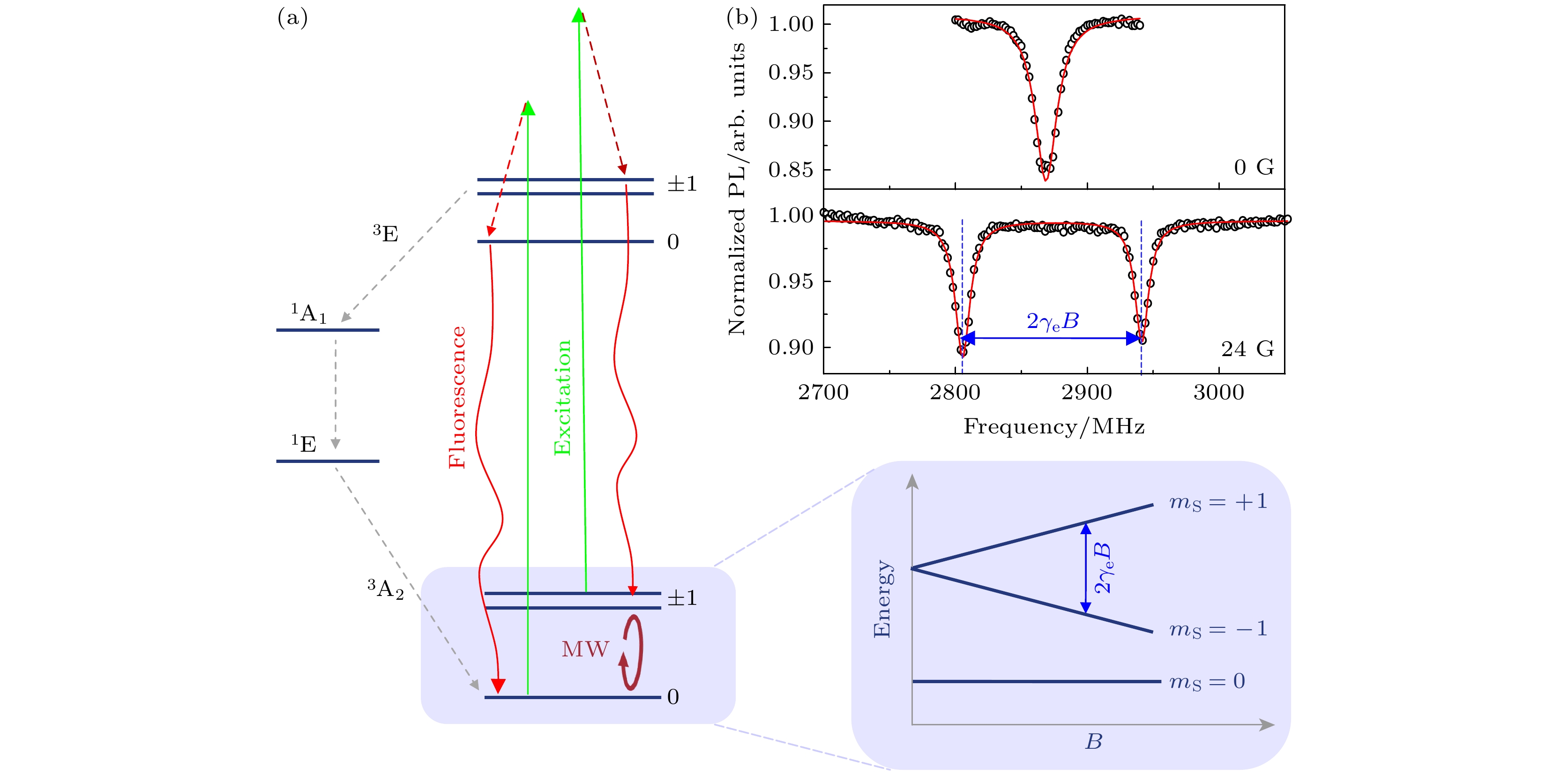
图 2 金刚石NV中心量子传感工作原理 (a) 金刚石NV中心自旋能级结构和光学跃迁, 右侧为基态能级随外磁场的变化规律(塞曼效应); (b) 典型的光探磁共振(ODMR)谱线, 上图为零场ODMR谱线, 下图为外加24 G (1 G = 10–4 T)磁场的结果; 通过拟合共振频率, 可以得到NV中心所处位置的磁场信息
Fig. 2. Working principle of diamond quantum sensing: (a) The energy level structure and the optical transitions of NV centers in diamond; the right diagram shows the ground states of an NV center under different external magnetic fields (Zeeman effect); (b) typical optically detected magnetic resonance (ODMR) spectra. Top: ODMR spectrum at zero-field. Bottom: ODMR spectrum under an external magnetic field of 24 G (1 G = 10–4 T). By fitting the resonance frequency of the ODMR spectra, we can determine the strength and orientation of the magnetic field.
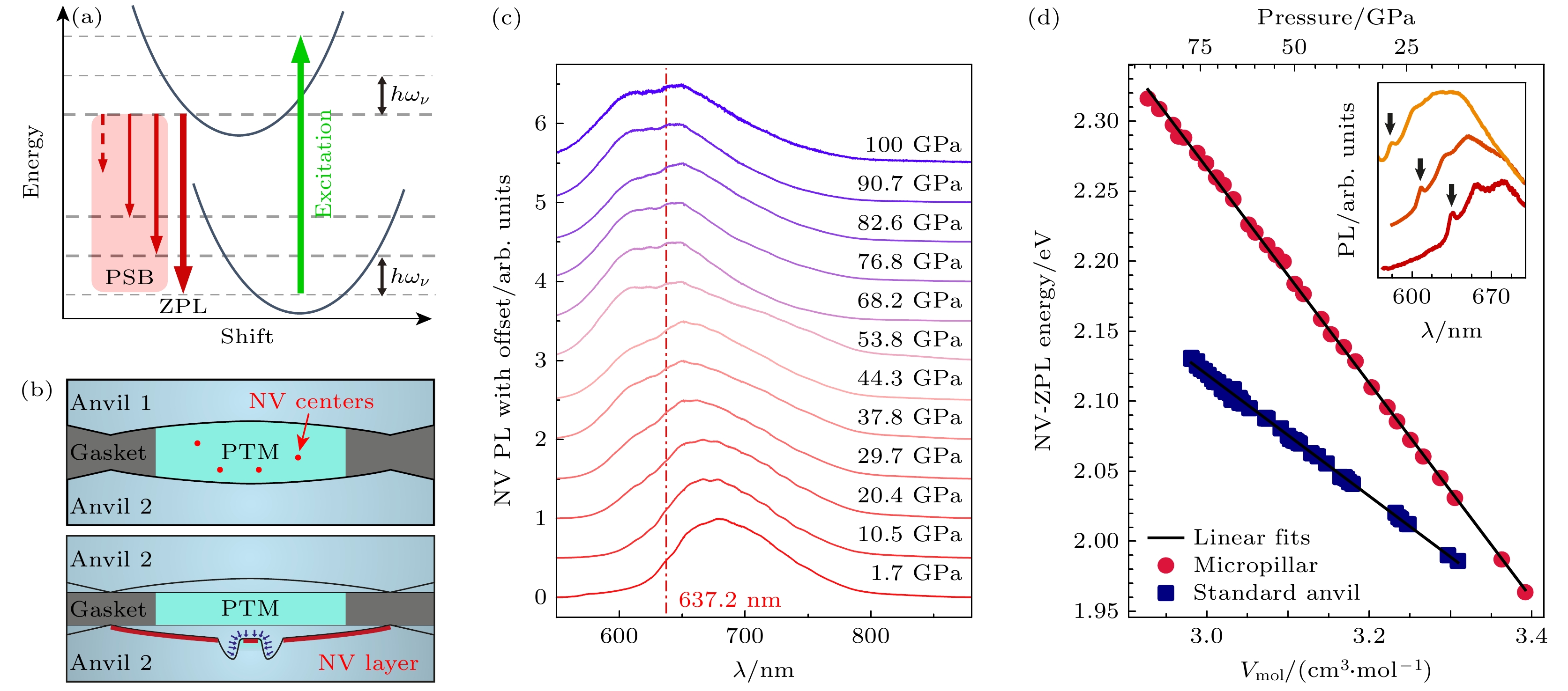
图 3 高压极端条件对金刚石NV中心光学性质的影响 (a) 零声线(ZPL)和声子边带(PSB)的示意图[11]; (b) 金刚石对顶砧高压腔中引入NV中心的两种方法, 上图为在传压介质中放置包含NV中心的金刚石颗粒, 下图为在对顶砧砧面上制备NV中心[12]; (c)不同压强下金刚石NV中心的荧光光谱, 激发光源为532 nm激光, 该实验使用的NV中心来自微米金刚石颗粒[11]; (d) 金刚石NV中心ZPL随压强变化规律, 该实验使用了砧面上的NV中心, 结果显示静水压条件对于实现高压ODMR至关重要[12]
Fig. 3. The influence of pressure on the optical properties of NV centers: (a) Schematic representation of the zero-phonon line (ZPL) and the phonon sideband (PSB) [11]; (b) two methods for placing NV centers in the DAC high-pressure chamber, the top diagram shows placement of diamond particles with NV centers in the pressure-transmitting medium, and the bottom diagram shows fabrication of shallow NV centers on the diamond culet [12]; (c) PL spectra of NV centers under different pressures, the experiment is performed with 532 nm laser excitation and NV centers in microdiamond [11]; (d) pressure dependence of the ZPL, the experiment is performed with shallow NV centers on the culet, the results emphasize the importance of the hydrostatic environment for ODMR at high pressure [12].
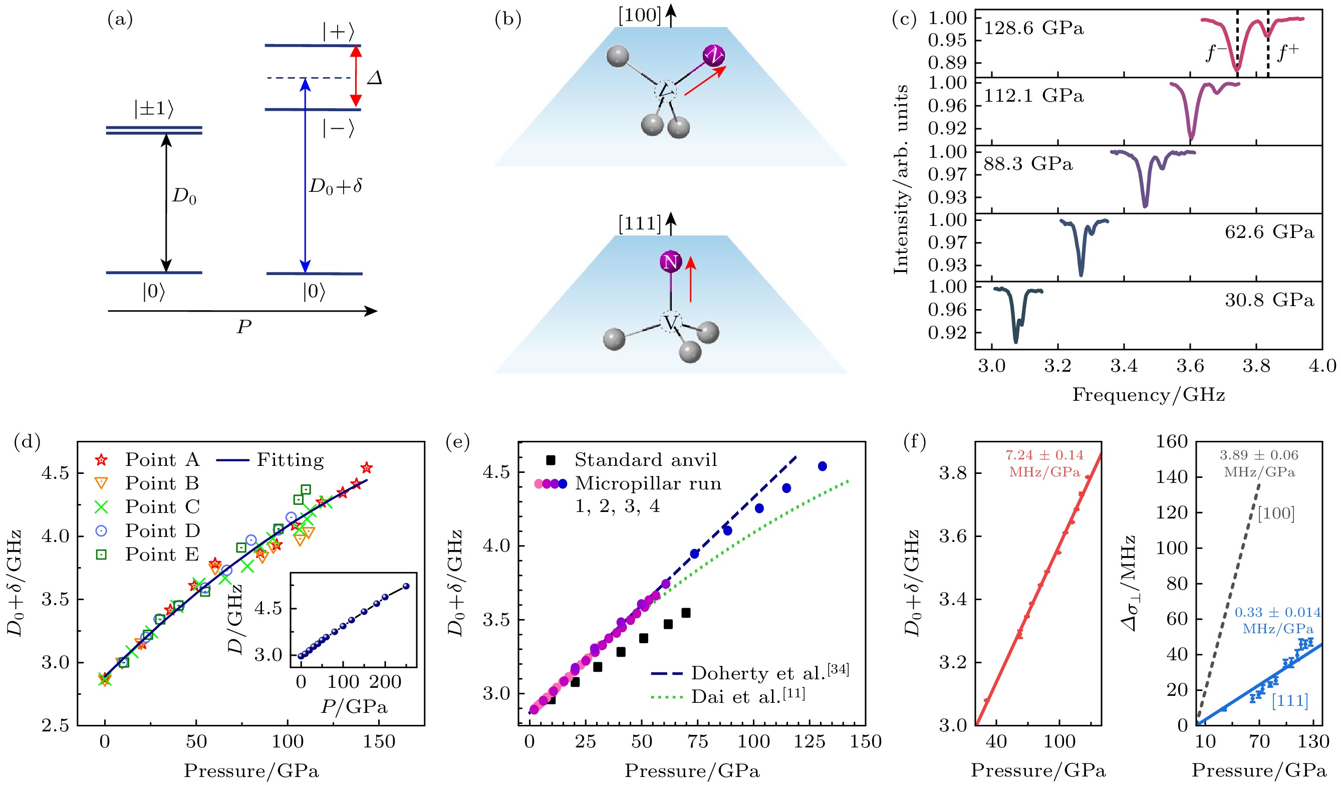
图 4 高压极端条件对金刚石NV中心自旋性质的影响 (a) 压强对NV中心基态能级的影响; (b) 沿(100)和(111)晶向切割的金刚石对顶砧砧面上NV中心的受力示意; (c) 不同压强下的NV中心零场ODMR谱线, 数据来自(111)切割的对顶砧砧面浅层NV中心[14]; (d), (e) 实验测量的NV中心零场劈裂值D随压强变化规律, 其中(d)图来自传压介质中的微米金刚石[11]; (e)图来自(100)切割的砧面浅层NV中心[12]; (f)图来自(111)切割的砧面浅层NV中心[11,14]
Fig. 4. The influence of pressure on the spin properties of NV centers: (a) Ground states of NV centers with and without external pressure; (b) schematic representation of NV orientation and diamond cut direction; (c) ODMR spectra of NV centers under different pressures, the experiment is performed with shallow NV centers in (111) cut diamond [14]; (d), (e) pressure dependence of zero-field splitting, D; data are acquired with (d) NV centers in microdiamond [11], (e) shallow NV centers in (100) cut diamond [12], and (f) shallow NV centers in (111) cut diamond[11,14].
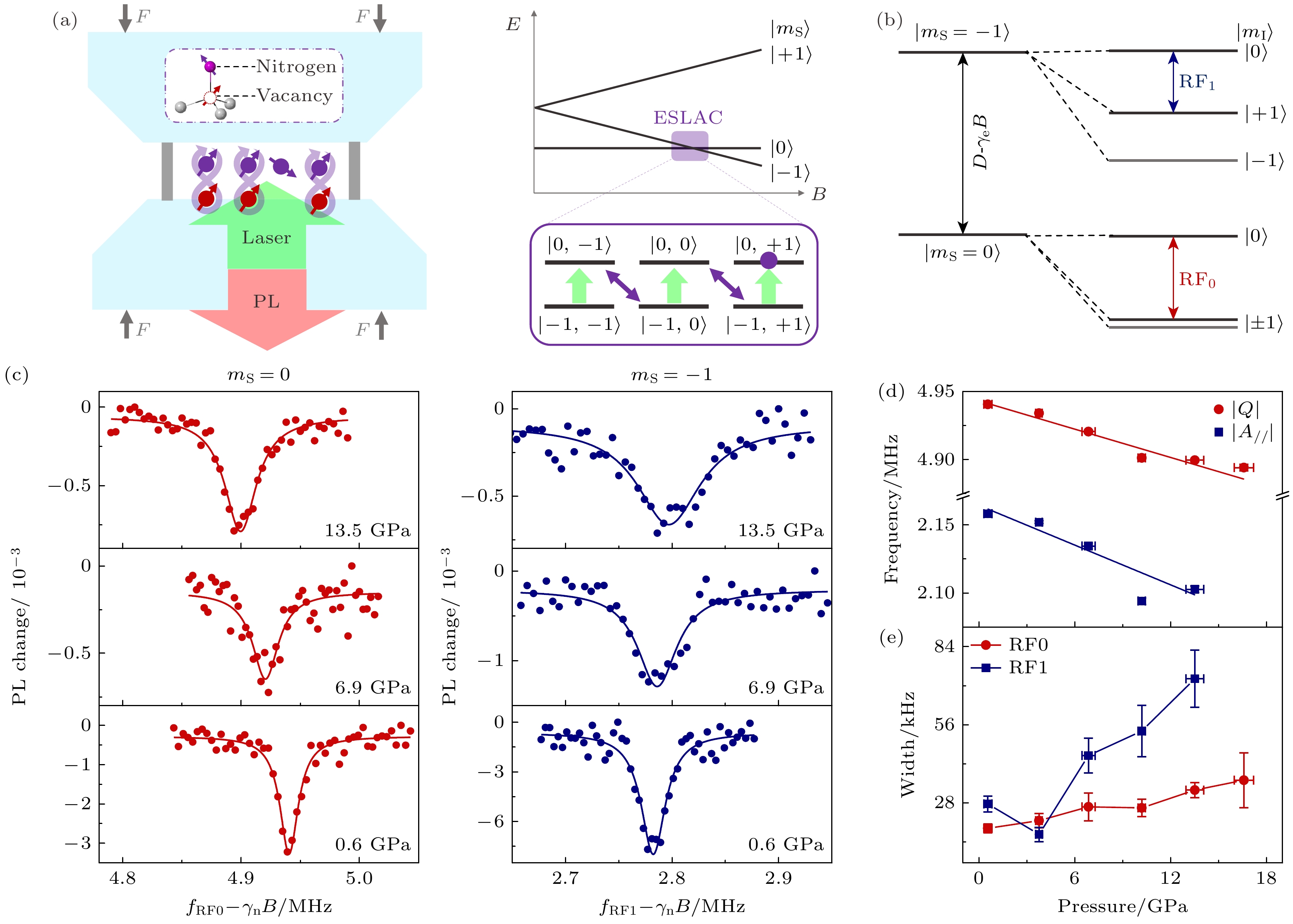
图 6 高压下金刚石中14N核磁共振[43] (a)基于金刚石NV中心的高压磁共振示意图(左), 在激发态能级交叉点附近实现14N核自旋的动态极化(右); (b) 电子自旋-核自旋耦合系统的能级结构, 磁共振探测的频率用箭头标出; (c) 典型的金刚石内14N核磁共振谱线, 左侧数据NV中心处于$ |{m}_{{\mathrm{s}}}=0\rangle $态, 右侧数据NV中心处于$ |{m}_{{\mathrm{s}}}=-1\rangle $态; (d) 14N核自旋四极矩项Q和超精细耦合参数A//随压强变化规律; (e) NMR谱线线宽随压强依赖关系
Fig. 6. NMR of 14N spin ensemble under high pressure[43]: (a) Schematic representation of high-pressure NMR enabled by NV centers(left), dynamical nuclear spin polarization of 14N nuclear spin at ESLAC(right); (b) energy levels of the coupled electron and nuclear spin system, with the transitions of the NMR measurements labeled; (c) typical NMR spectra of 14N spin ensemble under different pressures, NV electron spins in the $ |{m}_{{\mathrm{s}}}=0\rangle $ state(left), NV electron spins in the $ |{m}_{{\mathrm{s}}}=-1\rangle $ state(right); (d) absolute value of Q (red) and A// (blue) as a function of pressure; (e) pressure dependence of the width of 14N NMR spectra.
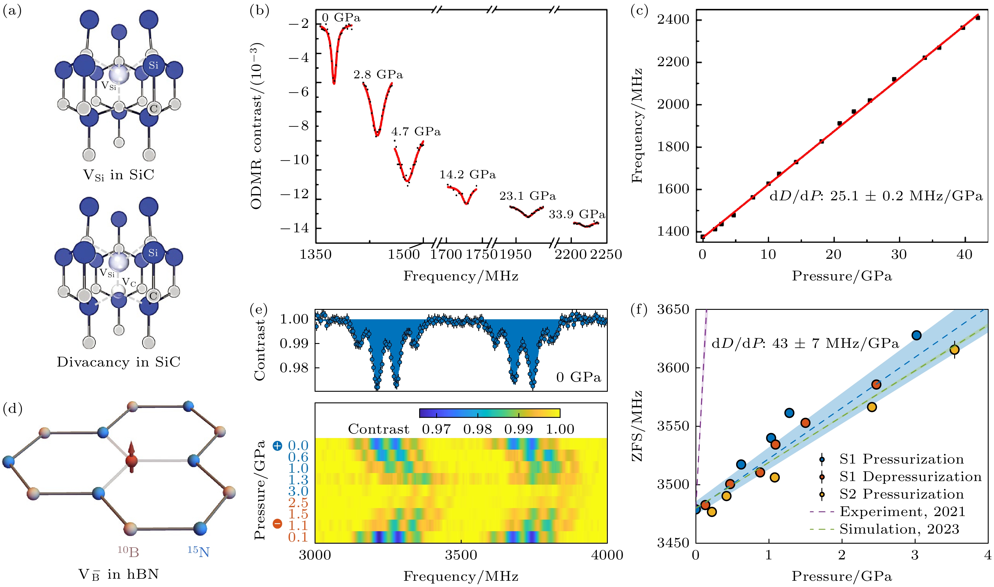
图 7 基于SiC和hBN中色心的高压量子传感 (a) 4H-SiC中的Si空位($ {{\mathrm{V}}}_{{\mathrm{S}}{\mathrm{i}}} $)色心(左)和双空位色心(右)物理结构[47]; (b), (c) 双空位色心PL5@SiC的ODMR谱线和对应零场劈裂值随压强变化规律[49]; (d) hBN中带负电的B空位$ {{\mathrm{V}}}_{{\mathrm{B}}}^{-} $物理结构; (e), (f) $ {{\mathrm{V}}}_{{\mathrm{B}}}^{-} $@hBN在不同压强下ODMR谱线和对应零场劈裂值随压强变化规律[50]
Fig. 7. High-pressure quantum sensing with color centers in SiC and hBN: (a) The physical structure of Si vacancy and divacancy in 4H-SiC [47]; (b), (c) typical ODMR spectra of the PL5@SiC divacancy center and the pressure dependence of its zero-field splitting[49]; (d) the physical structure of B vacancy in hBN, $ {{\mathrm{V}}}_{{\mathrm{B}}}^{-} $; (e), (f) typical ODMR spectra of $ {{\mathrm{V}}}_{{\mathrm{B}}}^{-}@{\mathrm{h}}{\mathrm{B}}{\mathrm{N}} $ and the pressure dependence of its zero-field splitting[50].
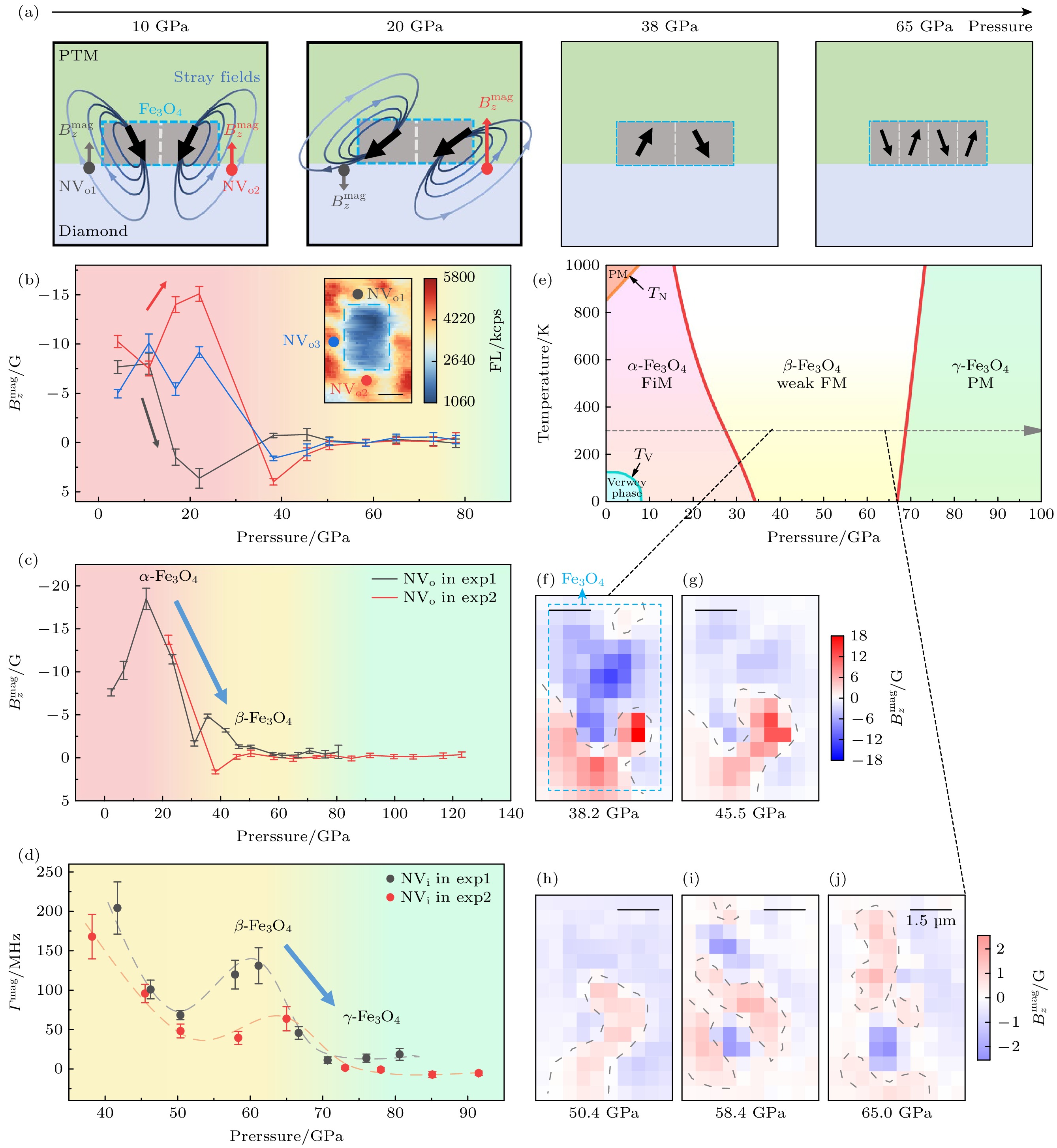
图 8 兆巴高压下的Fe3O4颗粒磁成像和磁相变过程测量[14] (a) 磁畴和近邻磁场随压强的演化示意图; (b), (c) 对顶砧选定位置(见图中标记)处的磁场$ {B}_{z}^{{\mathrm{m}}{\mathrm{a}}{\mathrm{g}}} $随压强变化规律; (d) ODMR线宽$ {\varGamma }^{{\mathrm{m}}{\mathrm{a}}{\mathrm{g}}} $随压强变化规律; (e) Fe3O4的相图, FM代表铁磁, FiM代表亚铁磁, PM代表顺磁; (f)—(j) 在压强为38.2, 45.5, 50.4, 58.4和65.0 GPa下的局部磁成像, 外加磁场约为240 G下对exp2中Fe3O4表面的磁场成像; 蓝色点划线标记了样品边界, 灰色点划线标记了磁畴边界
Fig. 8. Magnetism evolution of magnetite to megabar pressures[14]: (a) Schematic diagram of the evolution of magnetic domains and their stray magnetic fields in magnetite with pressure; (b), (c) pressure dependence of the magnetite magnetic field $ {B}_{z}^{{\mathrm{m}}{\mathrm{a}}{\mathrm{g}}} $ at the selected positions; (d) pressure dependence of the linewidth broadening $ {\varGamma }^{{\mathrm{m}}{\mathrm{a}}{\mathrm{g}}} $; (e) the phase diagram of Fe3O4, FM stands for ferromagnetic, FiM stands for ferrimagnetic, and PM stands for paramagnetic; (f)–(j) magnetic field imaging of the surface of Fe3O4 in exp2 with an external magnetic field of ~240 G at pressures of 38.2, 45.5, 50.4, 58.4 and 65.0 GPa, respectively. The dashed blue line in (e) marks the Fe3O4 sample and dashed gray lines mark the magnetic domain wall.
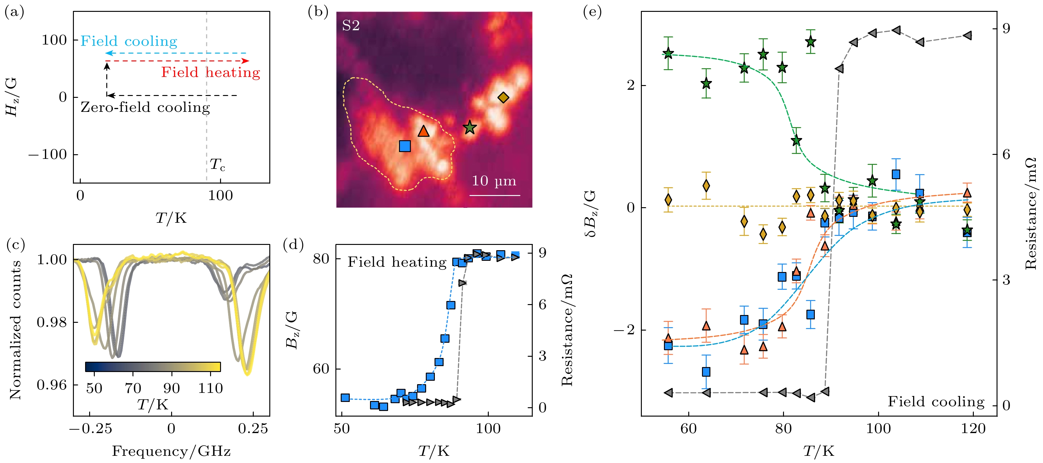
图 9 高压下CeH9超导迈斯纳效应的实验测量[13] (a) 测量抗磁现象的实验序列; (b) 样品及近邻区域的共聚焦荧光图; (c) 样品上蓝色标记位置在不同温度下的ODMR谱线, 该数据对应外加磁场为$ {H}_{{{z}}} $= 79 G, 且在零场降温后采集, 随着温度升高和靠近$ {T}_{{\mathrm{C}}} $, ODMR劈裂逐渐增加; (d) 局部磁场(左侧Y轴)和四电极法测量电阻(右侧Y轴)随温度变化, 零场降温后加场升温测试; (e) 在$ {H}_{{{z}}} $= 79 G磁场下, 降温过程中局部磁场和电阻随温度变温规律, 电阻在$ {T}_{{\mathrm{C}}}\approx $ 91 K时出现明显转变
Fig. 9. Imaging the Meissner effect in CeH9 under high pressure[13]: (a) The experimental sequence for probing local diamagnetism; (b) confocal fluorescence image of the sample, ODMR spectroscopy is performed at the labeled points; (c) NV ODMR spectra collected at the blue spatial point in (b) on field heating at $ {H}_{{{z}}} $= 79 G (following zero-field cooling), the ODMR splitting increases as T is increased across $ {T}_{{\mathrm{C}}} $; (d) the local field, Bz (left y-axis) and the four-point resistance (right y-axis) as a function of temperature; (e) simultaneous measurements of four-point resistance (right y-axis) and the change in the local field, $ \delta {B}_{{{z}}} $ (left y-axis) on field cooling with $ {H}_{{{z}}} $= 79 G, the measured resistance identifies a clear transition at $ {T}_{{\mathrm{C}}}\approx $ 91 K.
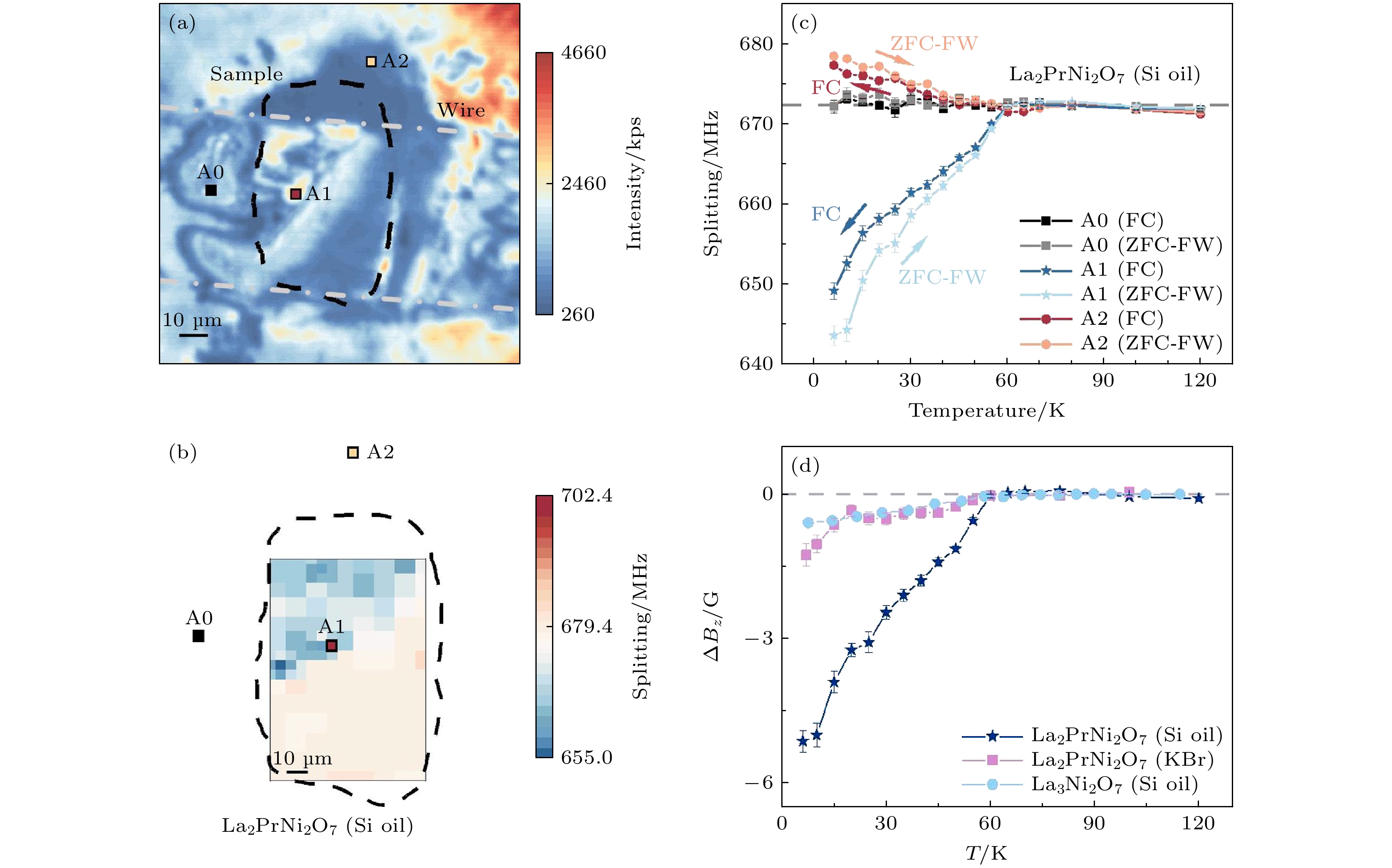
图 10 高压下La3Ni2O7-δ和La2PrNi2O7超导迈斯纳效应的原位测量[40] (a) La2PrNi2O7样品的共聚焦荧光扫描图, 传压介质为硅油; (b) 零场冷却到7 K后外加120 G磁场时的磁成像, 其中蓝色区域磁场小于外加磁场, 是典型的抗磁区域; (c) 典型位置的NV中心测得磁场随温度变温规律. 其中A0远离样品, 为测量参考点; A1在样品上, 低温区呈现出明显的抗磁现象; A2区在样品边沿, 低温下观测到了局部磁场增强, 在零场冷却加场升温(ZFC-FW)和场冷(FC)实验中观测到了相似现象, 外加磁场大小为120 G; (d) 不同样品和传压介质产生的超导抗磁信号对比, 外加磁场均为120 G
Fig. 10. Probing the Meissner effect in pressurized bilayer nickelate La3Ni2O7-δ and La2PrNi2O7 [40]: (a) Fluorescence image of sample A (La2PrNi2O7 in silicon oil); (b) magnetic field imaging under an external magnetic field of HZ = 120 G after zero-field cooling of the sample to 7 K, the blue area shows clear diamagnetism; (c) ODMR splitting of three selected points under ZFC-FW and FC measurements, point A0 is far away from the sample and serves as a reference, point A1 is on the sample and local demagnetization is observed at low temperature, point A2 is located at the sample edge and a local enhancement of magnetic field is observed, similar phenomena are observed in the ZFC-FW and FC measurements, (d) comparison of the diamagnetism effect of the three samples during the ZFW-FW measurement, the external magnetic field is about 120 G.
-
[1] Wang H, Tse J S, Tanaka K, Iitaka T, Ma Y M 2012 PNAS 109 6463
Google Scholar
[2] Drozdov A P, Eremets M I, Troyan I A, Ksenofontov V, Shylin S I 2015 Nature 525 73
Google Scholar
[3] Drozdov A P, Kong P P, Minkov V S, Besedin S P, Kuzovnikov M A, Mozaffari S, Balicas L, Balakirev F F, Graf D E, Prakapenka V B, Greenberg E, Knyazev D A, Tkacz M, Eremets M I 2019 Nature 569 528
Google Scholar
[4] Somayazulu M, Ahart M, Mishra A K, Geballe Z M, Baldini M, Meng Y, Struzhkin V V, Hemley R J 2019 Phys. Rev. Lett. 122 027001
Google Scholar
[5] Zhang L J, Wang Y C, Lv J, Ma Y M 2017 Nat. Rev. Mater. 2 17005
Google Scholar
[6] Ekimov E A, Sidorov V A, Bauer E D, Mel'nik N N, Curro N J, Thompson J D, Stishov S M 2004 Nature 428 542
Google Scholar
[7] Hirose K, Fei Y, Ma Y, Mao H K 1999 Nature 397 53
Google Scholar
[8] Hu Q Y, Kim D Y, Yang W G, Yang L X, Meng Y, Zhang L, Mao H K 2016 Nature 534 241
Google Scholar
[9] Meier T 2018 Annu. Rep. NMR Spectrosc. 93 1
[10] Meier T, Trybel F, Khandarkhaeva S, Steinle-Neumann G, Chariton S, Fedotenko T, Petitgirard S, Hanfland M, Glazyrin K, Dubrovinskaia N, Dubrovinsky L 2019 Phys. Rev. X 9 031008
[11] Dai J H, Shang Y X, Yu Y H, Xu Y, Yu H, Hong F, Yu X H, Pan X Y, Liu G Q 2022 Chin. Phys. Lett. 39 117601
Google Scholar
[12] Hilberer A, Toraille L, Dailledouze C, Adam M P, Hanlon L, Weck G, Schmidt M, Loubeyre P, Roch J F 2023 Phys. Rev. B 107 L220102
Google Scholar
[13] Bhattacharyya P, Chen W, Huang X, Chatterjee S, Huang B, Kobrin B, Lyu Y, Smart T J, Block M, Wang E, Wang Z, Wu W, Hsieh S, Ma H, Mandyam S, Chen B, Davis E, Geballe Z M, Zu C, Struzhkin V, Jeanloz R, Moore J E, Cui T, Galli G, Halperin B I, Laumann C R, Yao N Y 2024 Nature 627 73
Google Scholar
[14] Wang M Q, Wang Y, Liu Z X, Xu G Y, Yang B, Yu P, Sun H Y, Ye X Y, Zhou J W, Goncharov A F, Wang Y, Du J F 2024 Nat. Commun. 15 8843
Google Scholar
[15] Liu G Q, Feng X, Wang N, Li Q, Liu R B 2019 Nat. Commun. 10 1344
Google Scholar
[16] Fan J W, Guo S W, Lin C, Wang N, Liu G Q, Li Q, Liu R B 2024 Nano Lett. 24 14806
Google Scholar
[17] Fortman B, Mugica-Sanchez L, Tischler N, Selco C, Hang Y X, Holczer K, Takahashi C 2021 J Appl. Phys. 130 083901
Google Scholar
[18] 刘刚钦 2022 71 066101
Google Scholar
Liu G Q 2022 Acta Phys. Sin. 71 066101
Google Scholar
[19] Rondin L, Tetienne J P, Hingant T, Roch J F, Maletinsky P, Jacques V 2014 Rep. Prog. Phys. 77 056503
Google Scholar
[20] Schirhagl R, Chang K, Loretz M, Degen C L 2014 Annu. Rev. Phys. Chem. 65 83
Google Scholar
[21] Degen C L, Reinhard F, Cappellaro P 2017 Rev. Mod. Phys. 89 035002
Google Scholar
[22] Casola F, Van Der Sar T, Yacoby A 2018 Nat. Rev. Mater. 3 17088
Google Scholar
[23] Liu G Q, Liu R B, Li Q 2022 Acc. Chem. Res. 56 95
[24] 刘刚钦, 邢健, 潘新宇 2019 68 120302
Google Scholar
Liu G Q, Xing J, Pan X Y 2019 Acta Phys. Sin. 68 120302
Google Scholar
[25] 董杨, 杜博, 张少春, 陈向东, 孙方稳 2018 67 160301
Google Scholar
Dong Y, Du B, Zhang S C, Chen X D, Sun F W 2018 Acta Phys. Sin. 67 160301
Google Scholar
[26] 彭世杰, 刘颖, 马文超, 石发展, 杜江峰 2018 67 167601
Google Scholar
Peng S J, Liu Y, Ma W C, Shi F Z, Du J F 2018 Acta Phys. Sin. 67 167601
Google Scholar
[27] Aslam N, Zhou H Y, Urbach E K, Turner M J, Walsworth R L, Lukin M D, Park H 2023 Nat. Rev. Phys. 5 157
Google Scholar
[28] Wu Y, Weil T 2022 Adv. Sci. 9 2200059
Google Scholar
[29] Barry J F, Schloss J M, Bauch E, Turner M J, Hart C A, Pham L A, Walsworth R L 2020 Rev. Mod. Phys. 92 015004
Google Scholar
[30] Lawson A W, Tang T Y 1950 Rev. Sci. Instrum. 21 815
[31] Jayaraman A 1983 Rev. Mod. Phys. 55 65
Google Scholar
[32] Gruber A, Dräbenstedt A, Tietz C, Fleury L, Wrachtrup J, von Borczyskowski C 1997 Science 276 2012
Google Scholar
[33] Doherty M W, Manson N B, Delaney P, Jelezko F, Wrachtrup J, Hollenberg L C L 2013 Phys. Rep. 528 1
Google Scholar
[34] Doherty M W, Struzhkin V V, Simpson D A, McGuinness L P, Meng Y F, Stacey A, Karle T J, Hemley R J, Manson N B, Hollenberg L C L, Prawer S 2014 Phys. Rev. Lett. 112 047601
Google Scholar
[35] Shang Y X, Hong F, Dai H J, Yu H, Lu Y N, Liu E K, Yu X H, Liu G Q, Pan X Y 2019 Chin. Phys. Lett. 36 086201
Google Scholar
[36] Yip K Y, Ho K O, Yu K Y, Chen Y, Zhang W, Kasahara S, Mizukami Y, Shibauchi T, Matsuda Y, Goh S K, Yang S 2019 Science 366 1355
Google Scholar
[37] Shelton D P, Cabriales W, Salamat A 2024 Rev. Sci. Instrum. 95 083901
Google Scholar
[38] Hsieh S, Bhattacharyya P, Zu C, Mittiga T, Smart T J, Machado F, Kobrin B, Höhn T O, Rui N Z, Kamrani M, Chatterjee S, Choi S, Zaletel M, Struzhkin V V, Moore J E, Levitas V I, Jeanloz R, Yao N Y 2019 Science 366 1349
Google Scholar
[39] Lesik M, Plisson T, Toraille L, Renaud J, Occelli F, Schmidt M, Salord O, Delobbe A, Debuisschert T, Rondin L, Loubeyre P, Roch J F 2019 Science 366 1359
Google Scholar
[40] Wen J Y, Xu Y, Wang G, He Z X, Chen Y, Wang N N, Lu T L, Ma X L, Jin F, Chen L C, Liu M, Fan J W, Liu X B, Pan X Y, Liu G Q, Cheng J G, Yu X H 2024 arXiv: 2410.10275 https://doi.org/10.48550/arXiv.2410.10275
[41] Meijer J, Burchard B, Domhan M, Wittmann C, Gaebel T, Popa I, Jelezko F, Wrachtrup J 2005 Appl. Phys. Lett. 87 261909
Google Scholar
[42] Lyapin S G, Ilichev I D, Novikov A P, Davydov V A, Agafonov V N 2018 Nanosystems: Physics, Chemistry, Mathematics 9 55
[43] Shang Y X, Hong F, Dai J H, Lu Y N, Yu H, Yu Y H, Yu X H, Pan X Y, Liu G Q 2022 arXiv: 2203.10511 https://doi.org/10.48550/arXiv.2203.10511
[44] Jacques V, Neumann P, Beck J, Markham M, Twitchen D, Meijer J, Kaiser F, Balasubramanian G, Jelezko F, Wrachtrup J 2009 Phys. Rev. Lett. 102 57403
Google Scholar
[45] London P, Scheuer J, Cai J M, Schwarz I, Retzker A, Plenio M B, Katagiri M, Teraji T, Koizumi S, Isoya J, Fischer R, McGuinness L P, Naydenov B, Jelezko F 2013 Phys. Rev. Lett. 111 067601
Google Scholar
[46] Liu G Q, Jiang Q Q, Chang Y C, Liu D Q, Li W X, Gu C Z, Po H C, Zhang W X, Zhao N, Pan X Y 2014 Nanoscale 6 10134
Google Scholar
[47] Zhang G, Cheng Y, Chou J P, Gali A 2020 Appl. Phys. Rev. 7 031308
Google Scholar
[48] Wang J F, Liu L, Liu X D, Li Q, Cui J M, Zhou D F, Zhou J Y, Wei Y, Xu H A, Xu W, Lin W X, Yan J W, He Z X, Liu Z H, Hao Z H, Li H O, Liu W, Xu J S, Gregoryanz E, Li C F 2023 Nat. Mater. 22 489
Google Scholar
[49] Liu L, Wang J F, Liu X D, Xu H A, Cui J M, Li Q, Zhou J Y, Lin W X, He Z X, Xu W, Wei Y, Liu Z H, Wang P, Hao Z H, Ding J F, Li H O, Liu W, Li H, You L X, Xu J S, Gregoryanz E, Li C F, Guo G C 2022 Nano Lett. 22 9943
Google Scholar
[50] He G H, Gong R T, Wang Z P, Liu Z W, Hong J, Zhang T X, Riofrio A L, Rehfuss Z, Chen M F, Yao C Y, Poirier T, Ye B T, Wang X, Ran S, Edgar J H, Zhang S X, Yao N Y, Zu C 2025 arXiv: 2501.03319
[51] Zhong C, Wang Y P, Mai D, Ye C H, Li X D, Wang H, Dai R C, Wang Z P, Sun X Y, Zhang Z M 2024 Nano Lett. 24 4993
[52] Mao H K 2024 Natl. Sci. Rev. 11 nwae004
Google Scholar
[53] Eremets M I 2024 Natl. Sci. Rev. 11 nwae047
Google Scholar
[54] Hamlin J J 2019 Nature 569 491
Google Scholar
[55] Sun H L, Huo M W, Hu X W, Li J Y, Liu Z J, Han Y F, Tang L Y, Mao Z Q, Yang P T, Wang B S, Cheng J G, Yao D X, Zhang G M, Wang M 2023 Nature 621 493
Google Scholar
[56] Wang N N, Wang G, Shen X L, Hou J, Luo J, Ma X P, Yang H X, Shi L F, Dou J, Feng J, Yang J, Shi Y Q, Ren Z A, Ma H M, Yang P T, Liu Z Y, Liu Y, Zhang H, Dong X L, Wang Y X, Jiang K, Hu J P, Nagasaki S, Kitagawa K, Calder S, Yan J Q, Sun J P, Wang B S, Zhou R, Uwatoko Y, Cheng J G 2024 Nature 634 579
Google Scholar
[57] Zhou Y Z, Guo J, Cai S, Sun H L, Li C Y, Zhao J Y, Wang P Y, Han J Y, Chen X T, Chen Y J, Wu Q, Ding Y, Xiang T, Mao H K, Sun L L 2025 Matter Radiat. Extremes 10 027801
Google Scholar


 下载:
下载:


计量
- 文章访问数: 1188
- PDF下载量: 91
- 被引次数: 0
