


-
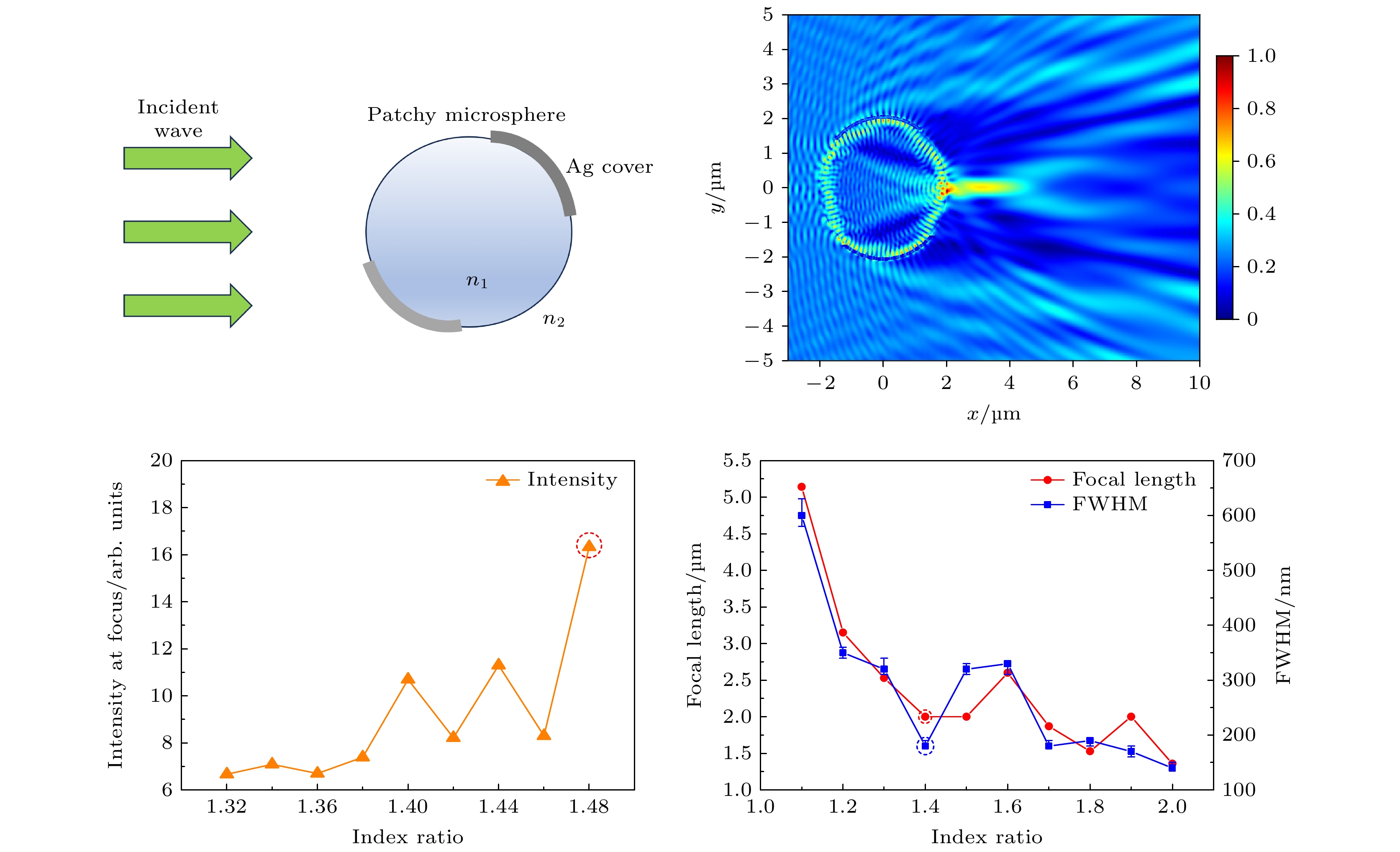
Photonic nanojet (PNJ) has gradually attracted the attention of researchers in the recent years. PNJ has unique properties, such as high intensity, high localization and subwavelength scale focusing ability, making it a narrow beam with wavelength scale. The full-width at half maximum (FWHM) of PNJ at the focus can exceed the diffraction limit while maintaining high intensity with a long distance, which can significantly enhance the imaging resolution. In this work, the characteristics of PNJ are explored through numerical simulation, with a focus on studying the patchy microspheres under various conditions, including coverage area, incident angle, and the refractive index of the immersion medium. The findings reveal that when the microsphere size is fixed and the coverage area accounts for 69%, the performance of PNJ is optimal. Under this condition, adjusting the incident angle to –5.74° can accurately position the PNJ focal point on the microsphere surface. Furthermore, at this specific angle, the patchy microspheres can generated PNJ with “S”-typed and “Y”-typed field intensity distribution, and the FWHM is reduced to 180 nm, significantly exceeding the traditional diffraction limit. This optimization strategy not only facilitates super-resolution focusing, but also greatly enhances both the intensity and efficiency of the PNJ. Additionally, this study demonstrates that the PNJ performance improves when the refractive index ratio between the patchy microsphere and the immersion medium approaches 1.4. Notably, a resonance effect occurs when the refractive index ratio reaches 1.48, resulting in enhanced PNJ performance. In this case, the PNJ focal point remains on the surface of the microsphere, with an FWHM of 180 nm, while the light intensity is further amplified to approximately three times the intensity of the PNJ generated by the microspheres without resonance effect. This research provides theoretical support for the application of patchy microspheres in fields such as super-resolution imaging.
[1] 周锐, 吴梦雪, 沈飞, 洪明辉 2017 66 140702
 Google Scholar
Zhou R, Wu M X, Shen F, Hong M H 2017 Acta Phys. Sin. 66 140702
Google Scholar
[2] 王淑莹, 章海军, 张冬仙 2013 62 034207
Google Scholar
Wang S Y, Zhang H J, Zhang D X 2013 Acta Phys. Sin. 62 034207
Google Scholar
[3] 宋扬, 杨西斌, 闫冰, 王驰, 孙建美, 熊大曦 2020 69 134201
Google Scholar
Song Y, Yang X B, Yan B, Wang C, Sun J M, Xiong D X 2020 Acta Phys. Sin. 69 134201
Google Scholar
[4] Chen Z, Taflove A, Backman V 2004 Opt. Express 12 1214
Google Scholar
[5] Lee J Y, Hong B H, Kim W Y, Min S K, Kim Y, Jouravlev M V, Bose R, Kim K S, Hwang I C, Kaufman L J, Wong C W, Kim P, Kim K S 2009 Nature 460 498
Google Scholar
[6] Wang Z B, Guo W, Li L, Luk’yanchuk B, Khan A, Liu Z, Chen Z C, Hong M H 2011 Nat. Commun. 2 218
Google Scholar
[7] Hao X, Kuang C F, Liu X, Zhang H J, Li Y H 2011 Appl. Phys. Lett. 99 203102
Google Scholar
[8] Lee S, Li L, Wang Z B, Guo W, Yan Y Z, Wang T 2013 Appl. Opt. 52 7265
Google Scholar
[9] Allen K W, Farahi N, Li Y, Limberopoulos N I, Walker D E, Urbas A M, Astratov V N 2015 Opt. Express 23 24484
Google Scholar
[10] Liu C Y, Lo W C 2017 Opt. Commun. 399 104
Google Scholar
[11] Yang S L, Cao Y R, Shi Q F, Wang X Q, Chen T, Wang J G, Ye Y H 2019 J. Phys. Chem. C 123 28353
Google Scholar
[12] Cao Y R, Yang S L, Wang J G, Shi Q F, Ye Y H 2020 J. Appl. Phys. 127 233103
Google Scholar
[13] Liu X, Hu S, Tang Y 2020 Photonics 7 84
Google Scholar
[14] Xu C, Yang T, Zou P, Ye R 2022 Proc. SPIE 12316 1231602
Google Scholar
[15] Tam W G, Corriveau R 1978 J. Opt. Soc. Am. 68 763
Google Scholar
[16] 董哲, 杨洗陈 2009 光学学报 29 1296
Google Scholar
Dong Z, Yang X C 2009 Acta Opt. Sin. 29 1296
Google Scholar
[17] Devilez A, Stout B, Bonod N, Popov E 2008 Opt. Express 16 14200
Google Scholar
[18] Ritchie R H, Eldridge H B 1962 Phys. Rev. 126 1935
Google Scholar
[19] Shin Y B, Kim H M, Jung Y, Chung B H 2010 Sens. Actuators B Chem. 150 1
Google Scholar
[20] Shi L P, Chong T C, Yao H B, Tan P K, Miao X S 2002 J. Appl. Phys. 91 10209
Google Scholar
[21] Luo X, Ishihara T 2004 Appl. Phys. Lett. 84 4780
Google Scholar
[22] Bonaccorso F, Sun Z, Hasan T, Ferrari A C 2010 Nat. Photonics 4 611
Google Scholar
[23] Peterson A W, Halter M, Tona A, Plant A L 2014 BMC Cell Biol. 15 35
Google Scholar
[24] Wei F F, Lu D, Shen H, Wan W W, Ponsetto J L, Huang E, Liu Z W 2014 Nano Lett. 14 4634
Google Scholar
[25] Sun T, Chen H Y, Yang S, Hu J P, Wang C H 2018 Opt. Laser Technol. 108 551
Google Scholar
-
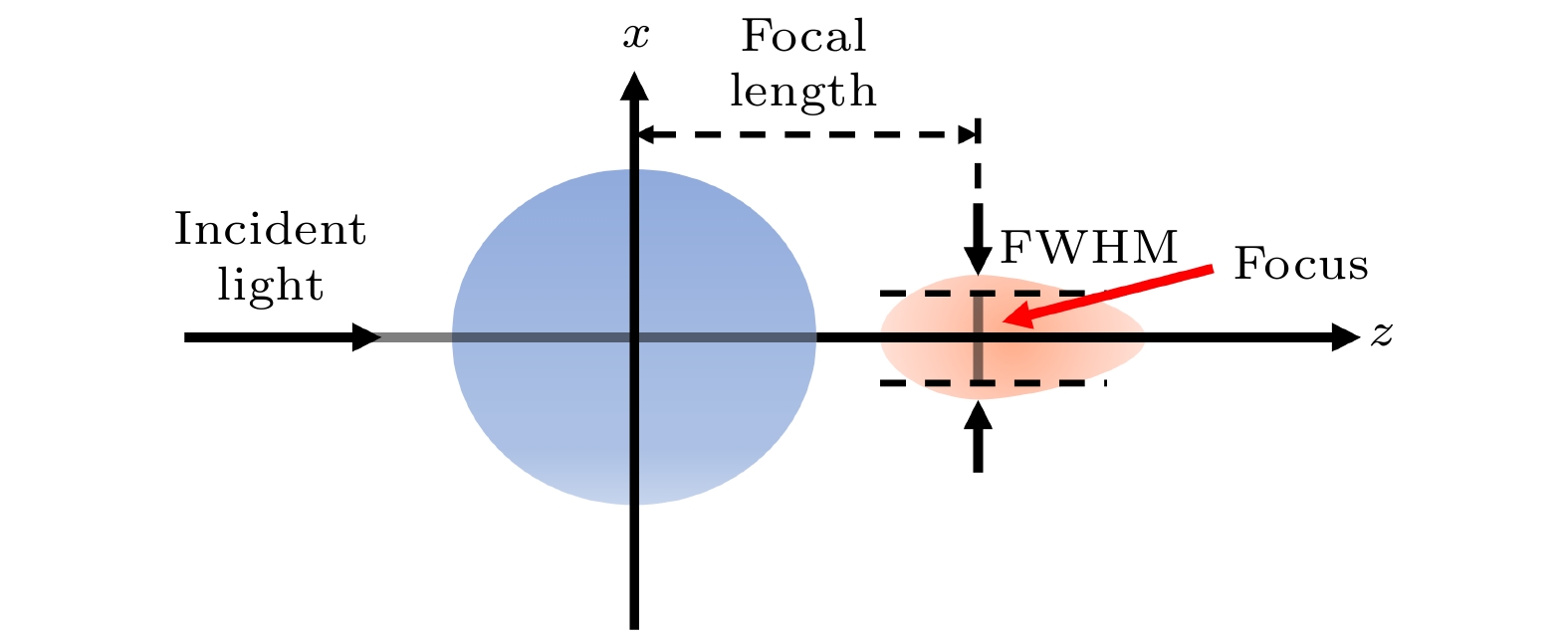
图 1 PNJ的特征
Figure 1. Characteristics of PNJ.
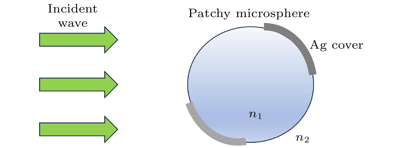
图 2 非均匀镀膜微球仿真示意图
Figure 2. Simulation diagram of patchy microspheres.
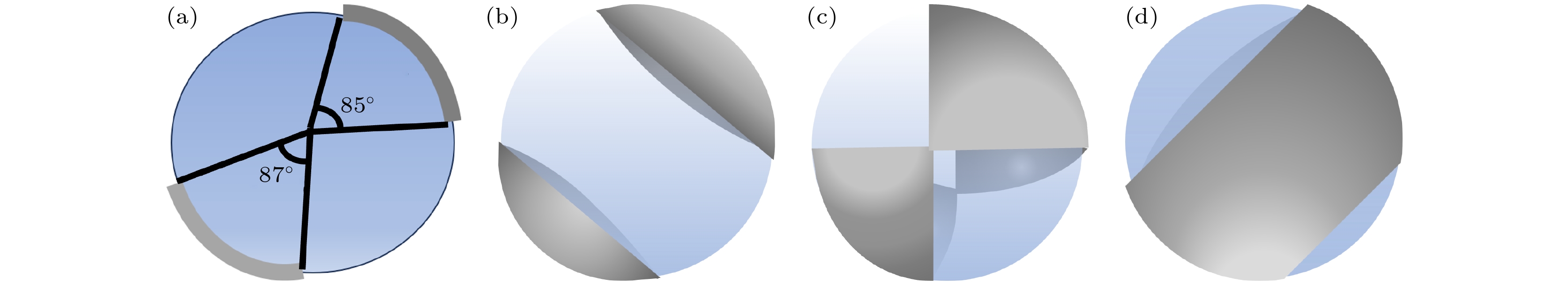
图 3 非均匀镀膜微球示意图 (a)横截面图; (b)镀膜面积占比为27%; (c)镀膜面积占比为48%; (d)镀膜面积占比为69%
Figure 3. Schematic diagram of patchy microspheres: (a) Cross sectional view; (b) the patchy area is 27%; (c) the patchy area is 48%; (d) the patchy area is 69%.
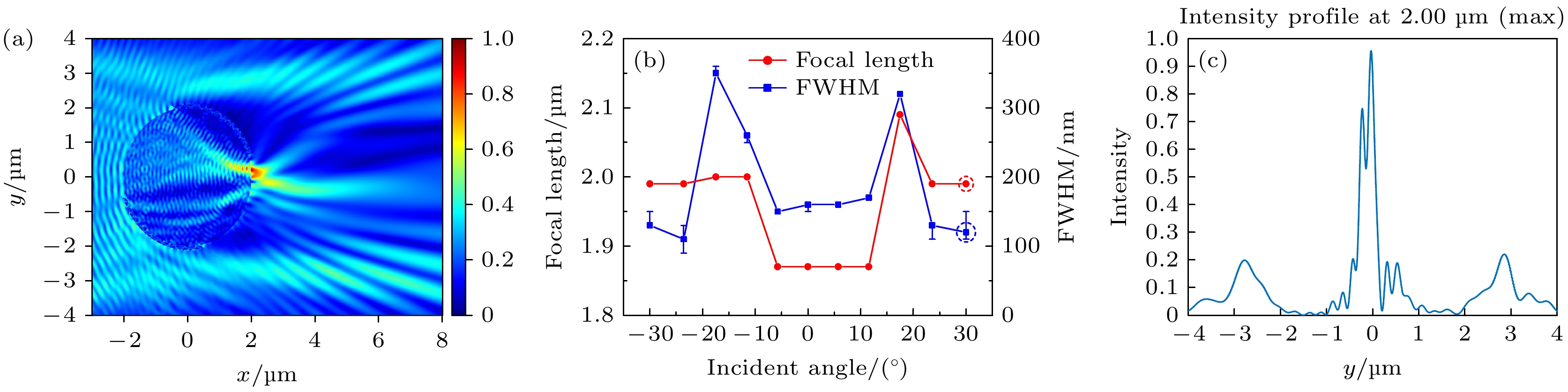
图 4 非均匀镀膜微球镀膜面积占比为27%时PNJ的特征 (a)倾斜照明角度为30°时的电场强度横截面图; (b) PNJ的焦距和FWHM随倾斜照明角度的变化; (c)倾斜照明角度为–17.46°时焦点处强度分布随y的变化
Figure 4. Characteristics of PNJ when the coating area ratio is 27%: (a) Cross-sectional view of electric field intensity when the angle of oblique illumination is 30°; (b) the variety of the focal length and FWHM of PNJ with the change of oblique illumination angle; (c) intensity distribution at focal point with y when oblique illumination is –17.46°.
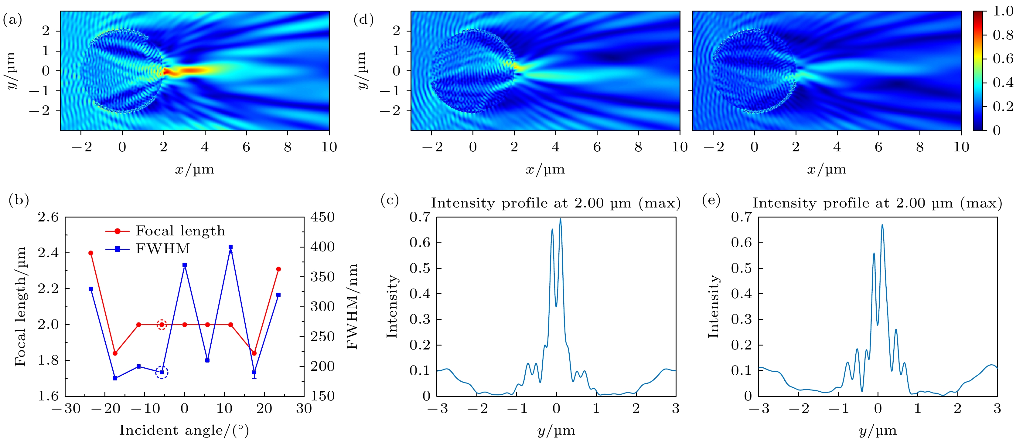
图 5 非均匀镀膜微球镀膜面积占比为48%时PNJ的特征 (a)倾斜照明角度为–5.74°时的电场强度横截面图; (b) PNJ的焦距和FWHM随倾斜照明角度的变化; (c)倾斜照明角度为0°时焦点处强度分布随y的变化; (d)倾斜照明角度为30°和–30°时的电场强度横截面图; (e)倾斜照明角度为11.54°时焦点处强度分布随y的变化
Figure 5. Characteristics of PNJ when the coating area ratio is 48%: (a) Cross-sectional view of electric field intensity when the oblique illumination angle is –5.74°; (b) the variation of the focal length and FWHM with the oblique illumination angle; (c) the intensity distribution at focal point with y when the oblique illumination angle is –0°; (d) the cross-sectional view of electric field intensity when the oblique illumination angle is 30° and –30°; (e) the intensity distribution at focal point with y when the oblique illumination angle is 11.54°.
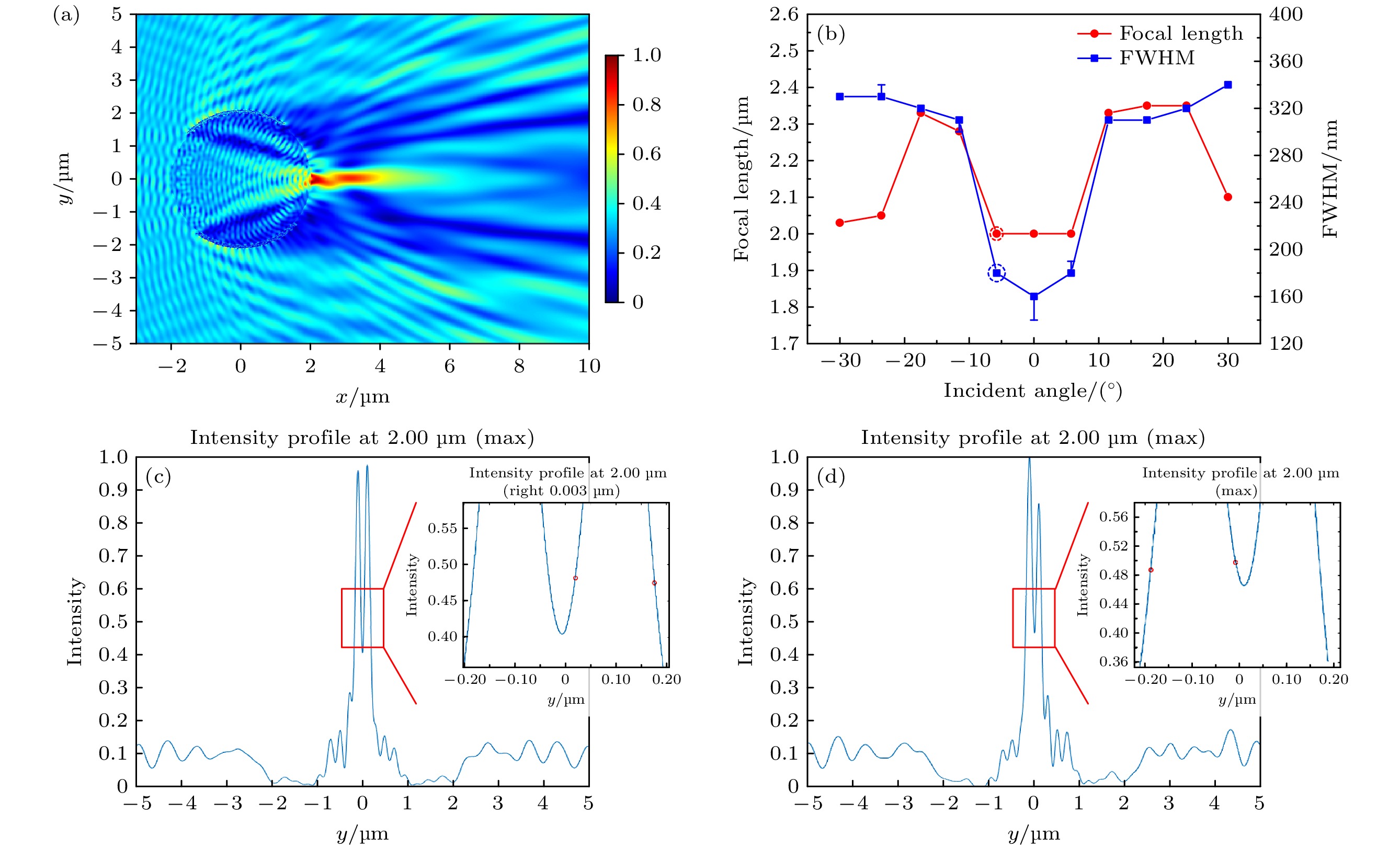
图 6 非均匀镀膜微球镀膜面积占比为69%时PNJ的特征 (a) –5.74°时的电场强度横截面图; (b) PNJ的焦距和FWHM随倾斜照明角度的变化; (c)倾斜照明角度为0°时焦点处强度分布随y的变化; (d)倾斜照明角度为–5.74°时焦点处强度分布随y的变化
Figure 6. Characteristics of PNJ when the coating area ratio is 48%: (a) Cross-sectional view of electric field intensity when the oblique illumination angle is –5.74°; (b) the variation of the focal length and FWHM with the oblique illumination angle; (c) the intensity distribution at focal point with y when the oblique illumination angle is 0°; (d) the intensity distribution at focal point with y when the oblique illumination angle is –5.74°.

图 7 浸没介质与非均匀镀膜微球的折射率比示意图
Figure 7. Schematic diagram of the refractive index ratio between immersion medium and patchy microspheres.
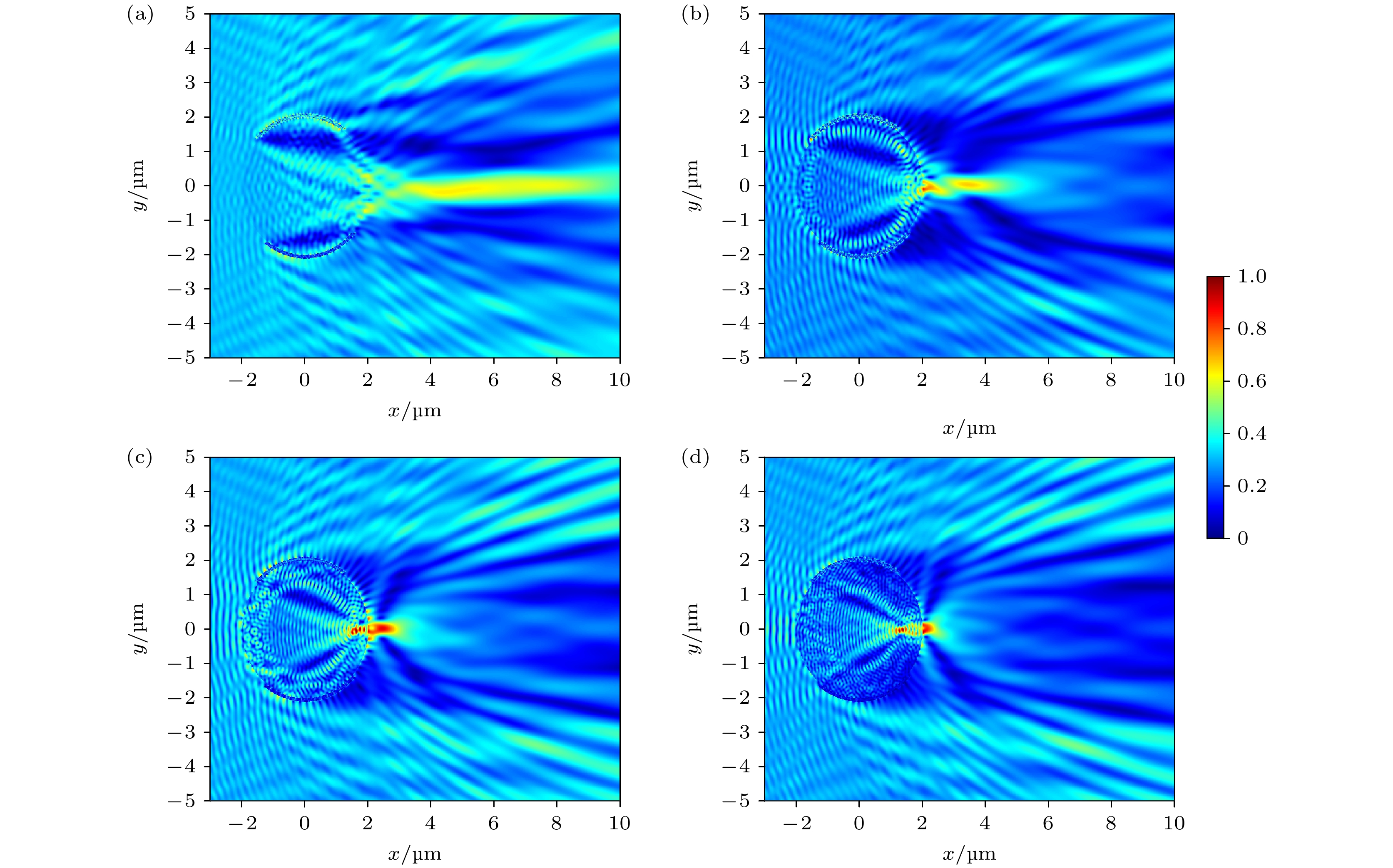
图 8 电场强度横截面图 (a)折射率比为1.1; (b)折射率比为1.4; (c)折射率比为1.7; (d)折射率比为2.0
Figure 8. Electric field intensity cross-section diagram: (a) The refractive index ratio is 1.1; (b) the refractive index ratio is 1.4; (c) the refractive index ratio is 1.7; (d) the refractive index ratio is 2.0.
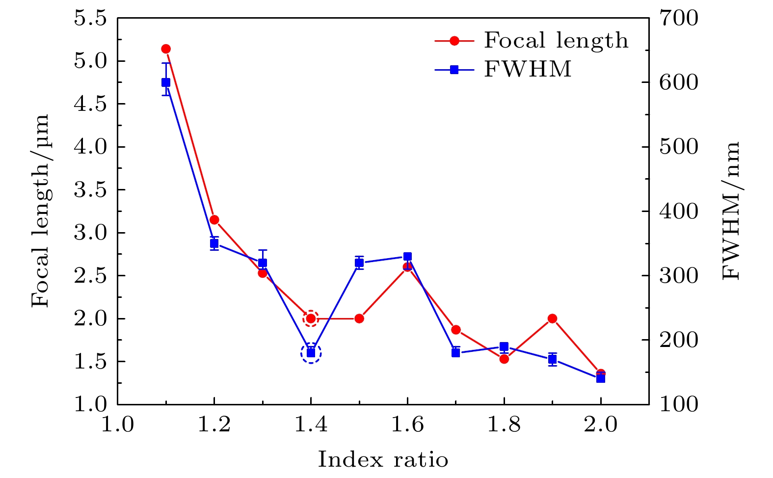
图 9 PNJ焦距和FWHM随折射率比的变化
Figure 9. Variation of PNJ’s focal length and FWHM with the refractive index ratio.
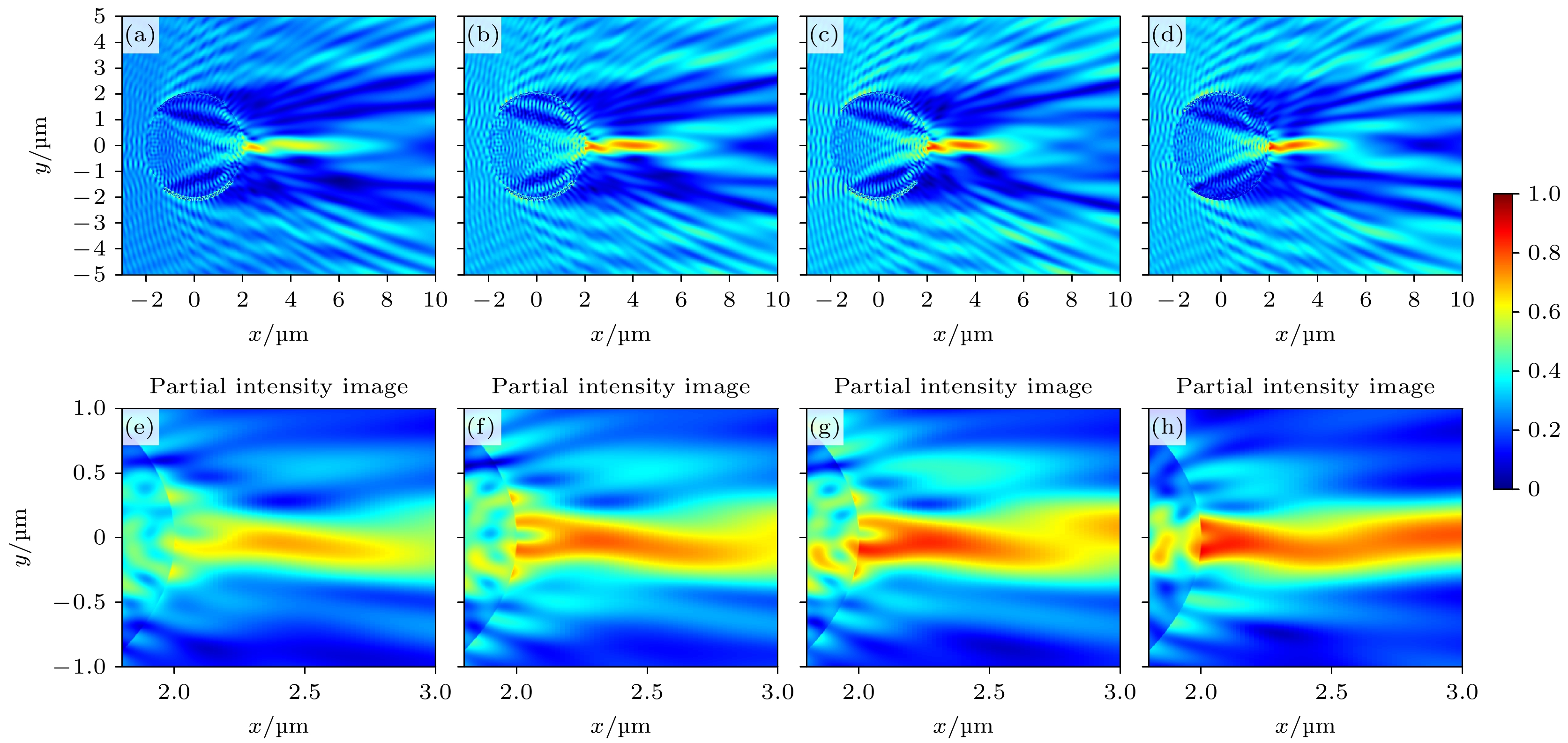
图 10 光子纳米喷流电场强度分布图 (a)折射率比为1.34时场强分布; (b)折射率比为1.36时场强分布; (c)折射率比为1.38时场强分布; (d)折射率比为1.44时场强分布; (e)折射率比为1.34时局部放大图; (f)折射率比为1.36时局部放大图; (g)折射率比为1.38时局部放大图; (h)折射率比为1.44时局部放大图
Figure 10. PNJ’s electric field intensity distribution: (a) Field intensity distribution when the refractive index ratio is 1.34; (b) field intensity distribution when the refractive index ratio is 1.36; (c) field intensity distribution when the refractive index ratio is 1.38; (d) field intensity distribution when the refractive index ratio is 1.44; (e) partial enlargement when the refractive index ratio is 1.34; (f) partial enlargement when the refractive index ratio is 1.36; (g) partial enlargement when the refractive index ratio is 1.38; (h) partial enlargement when the refractive index ratio is 1.44.
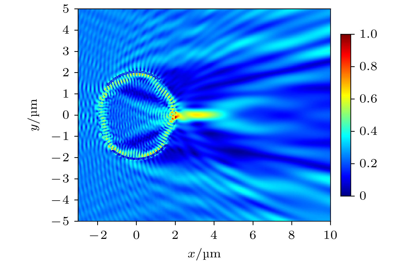
图 11 折射率比为1.48时的谐振现象
Figure 11. Resonance phenomenon when the refractive index ratio is 1.48.
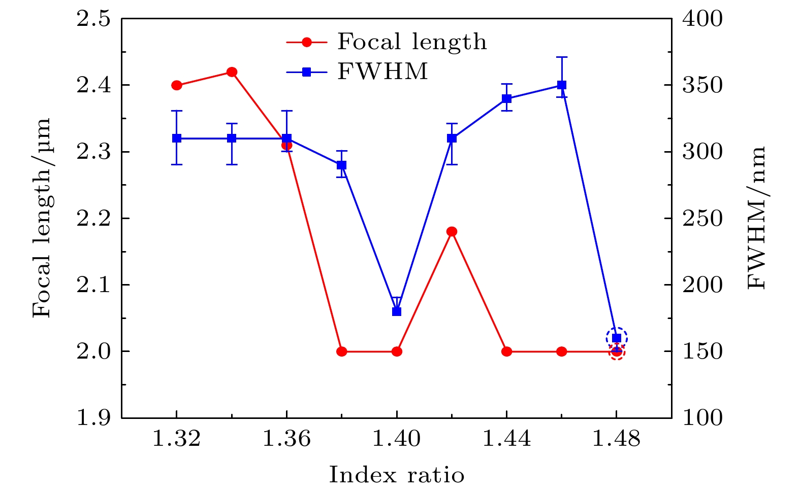
图 12 PNJ焦距和FWHM随折射率比的变化
Figure 12. Variation of PNJ’s focal length and FWHM with the refractive index ratio.
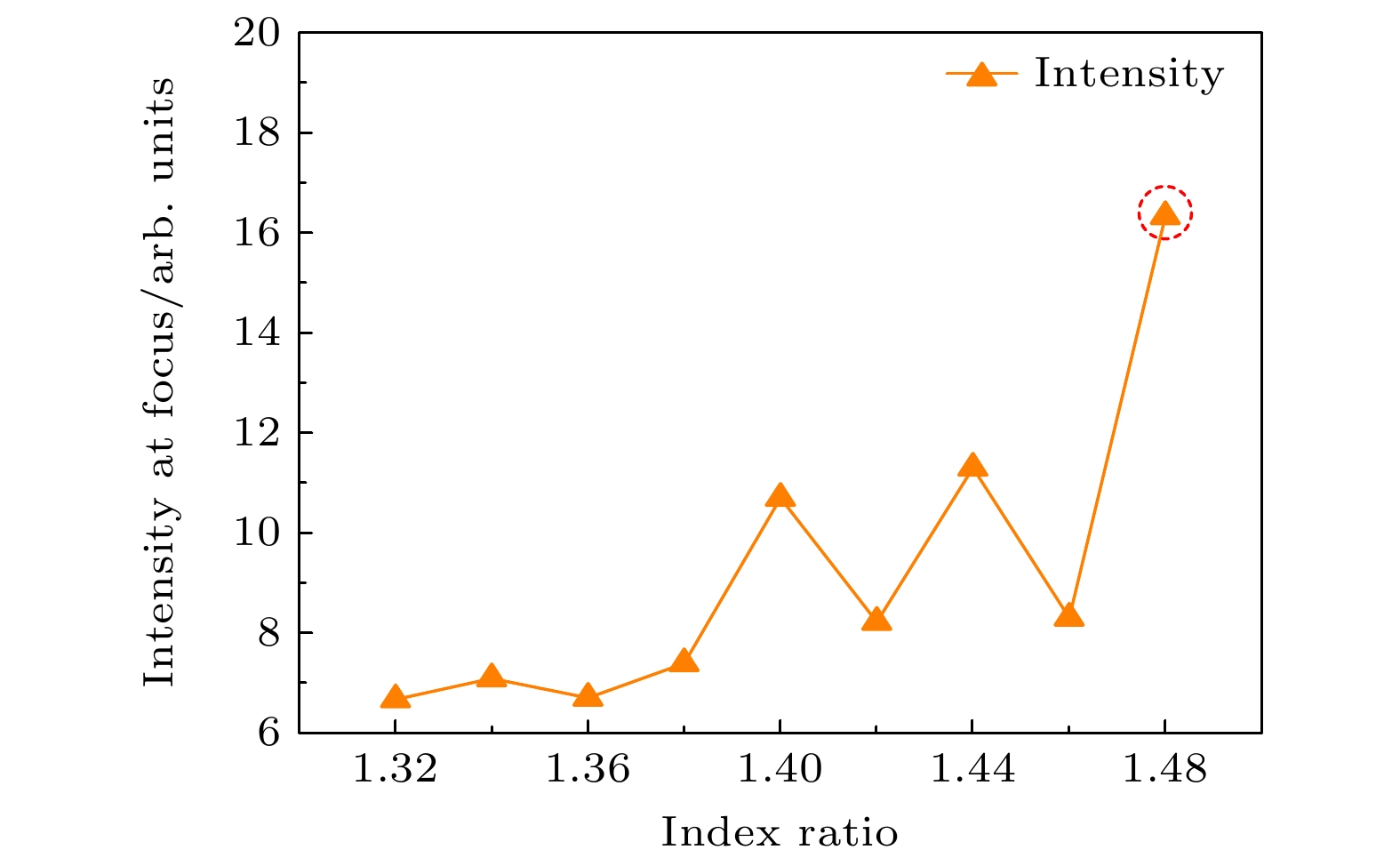
图 13 PNJ焦点处强度随折射率变化
Figure 13. Variation of PNJ’s intensity at the focus with the refractive index ratio.
表 1 不同镀膜模型比较
Table 1. Comparison of different patchy models.
微球模型 文献 类型 优点 缺陷 应用意义 PDMS包覆BaTiO3
玻璃微球[9] 均匀镀膜 通过异丙醇挥发调节距离 蒸发速率不稳定, 表面湿润性控制差, 无法长时间动态成像 为超分辨率显微成像提供了一种新的途径 核壳微纤维 [10] 均匀镀膜 大面积超分辨成像, 等离子体
效应增强聚焦, 高强度聚焦成像方向受限, 纤芯折射率变化可导致散射 展示了通过结构设计来增强成像分辨率的可能性 金属-介电纳米结构 [12] 非均匀镀膜 增强的近场效应, 有效的等离子体-微球相互作用 PDMS聚合物耦合方式复杂, 环境影响大 增强的近场电场效应提供了一种新的超分辨率成像技术 PS涂层BTG微球 [13] 均匀镀膜 增强PNJ强度, 改善聚焦效果, 提升超分辨成像效果 仅能在液体介质中进行实验 在液体介质中实现了更高的分辨率成像 AI薄膜包覆介电微球 [14] 非均匀镀膜 光子钩提升成像效果, 提供新的PNJ应用 未系统研究谐振现象 为非均匀镀膜微球的应用提供了新的可能性 非均匀镀膜微球 本文 非均匀镀膜 特性优化的光子钩, 提升超分辨成像性能, 谐振现象提升聚焦强度 为优化PNJ设计提供了重要依据, 将谐振现象引入PNJ特性分析  DownLoad: CSV
-
[1] 周锐, 吴梦雪, 沈飞, 洪明辉 2017 66 140702
Google Scholar
Zhou R, Wu M X, Shen F, Hong M H 2017 Acta Phys. Sin. 66 140702
Google Scholar
[2] 王淑莹, 章海军, 张冬仙 2013 62 034207
Google Scholar
Wang S Y, Zhang H J, Zhang D X 2013 Acta Phys. Sin. 62 034207
Google Scholar
[3] 宋扬, 杨西斌, 闫冰, 王驰, 孙建美, 熊大曦 2020 69 134201
Google Scholar
Song Y, Yang X B, Yan B, Wang C, Sun J M, Xiong D X 2020 Acta Phys. Sin. 69 134201
Google Scholar
[4] Chen Z, Taflove A, Backman V 2004 Opt. Express 12 1214
Google Scholar
[5] Lee J Y, Hong B H, Kim W Y, Min S K, Kim Y, Jouravlev M V, Bose R, Kim K S, Hwang I C, Kaufman L J, Wong C W, Kim P, Kim K S 2009 Nature 460 498
Google Scholar
[6] Wang Z B, Guo W, Li L, Luk’yanchuk B, Khan A, Liu Z, Chen Z C, Hong M H 2011 Nat. Commun. 2 218
Google Scholar
[7] Hao X, Kuang C F, Liu X, Zhang H J, Li Y H 2011 Appl. Phys. Lett. 99 203102
Google Scholar
[8] Lee S, Li L, Wang Z B, Guo W, Yan Y Z, Wang T 2013 Appl. Opt. 52 7265
Google Scholar
[9] Allen K W, Farahi N, Li Y, Limberopoulos N I, Walker D E, Urbas A M, Astratov V N 2015 Opt. Express 23 24484
Google Scholar
[10] Liu C Y, Lo W C 2017 Opt. Commun. 399 104
Google Scholar
[11] Yang S L, Cao Y R, Shi Q F, Wang X Q, Chen T, Wang J G, Ye Y H 2019 J. Phys. Chem. C 123 28353
Google Scholar
[12] Cao Y R, Yang S L, Wang J G, Shi Q F, Ye Y H 2020 J. Appl. Phys. 127 233103
Google Scholar
[13] Liu X, Hu S, Tang Y 2020 Photonics 7 84
Google Scholar
[14] Xu C, Yang T, Zou P, Ye R 2022 Proc. SPIE 12316 1231602
Google Scholar
[15] Tam W G, Corriveau R 1978 J. Opt. Soc. Am. 68 763
Google Scholar
[16] 董哲, 杨洗陈 2009 光学学报 29 1296
Google Scholar
Dong Z, Yang X C 2009 Acta Opt. Sin. 29 1296
Google Scholar
[17] Devilez A, Stout B, Bonod N, Popov E 2008 Opt. Express 16 14200
Google Scholar
[18] Ritchie R H, Eldridge H B 1962 Phys. Rev. 126 1935
Google Scholar
[19] Shin Y B, Kim H M, Jung Y, Chung B H 2010 Sens. Actuators B Chem. 150 1
Google Scholar
[20] Shi L P, Chong T C, Yao H B, Tan P K, Miao X S 2002 J. Appl. Phys. 91 10209
Google Scholar
[21] Luo X, Ishihara T 2004 Appl. Phys. Lett. 84 4780
Google Scholar
[22] Bonaccorso F, Sun Z, Hasan T, Ferrari A C 2010 Nat. Photonics 4 611
Google Scholar
[23] Peterson A W, Halter M, Tona A, Plant A L 2014 BMC Cell Biol. 15 35
Google Scholar
[24] Wei F F, Lu D, Shen H, Wan W W, Ponsetto J L, Huang E, Liu Z W 2014 Nano Lett. 14 4634
Google Scholar
[25] Sun T, Chen H Y, Yang S, Hu J P, Wang C H 2018 Opt. Laser Technol. 108 551
Google Scholar


 DownLoad:


Catalog
Metrics
- Abstract views: 1123
- PDF Downloads: 45
- Cited By: 0
