


-
High precision measurement of uranium isotope ratio (235U/238U) has important application in the field of nuclear energy safety. In this paper, based on high sensitivity tunable absorption spectroscopy technology, combined with the sample processing method of pulsed laser ablation plasma, high-precision measurement of uranium 235U/238U isotope ratio in solid material is realized. In the experimental measurement, transitions near 394.4884 nm/394.4930 nm (vacuum) are selected as the 235U/238U analytical lines. The influence of buffer gas and its pressure on the persistence time of uranium atom in laser ablated plasma are studied in detail. The experimental results show that different buffer gases have different ability to restrict the movement of particles in the plasma, which leads to different longitudinal expansion velocity of the plasma (perpendicular to the surface of the sample), and increases the persistence time of uranium atoms in the laser beam. The effect of pressure change on plasma evolution can be reduced by adding buffer gas. When helium is used as the buffer gas, the persistence time of uranium atoms in the plasma is longer, which can improve the selection space of data acquisition delay. In the ablation environment with helium, the electron number density of laser ablated plasma is relatively low, which can reduce the influence of Stark broadening effect and obtain narrower absorption lines, which is more conducive to the measurement of uranium atomic absorption spectrum. In order to reduce the influence of Doppler shift effect on absorption spectrum measurement and avoid misjudgment in spectrum analysis, it is more appropriate to carry out experimental measurement after 3μs sampling delay. Through experiments, the optimal conditions for measuring atomic absorption spectrum of uranium are obtained. Under these conditions, five different samples with 235U content of 4.95%, 4.10%, 3.00%, 1.10% and 0.25% respectively are measured, and the high-resolution absorption spectrum signals of 235U and 238U are obtained. The absorption spectra of samples with different content are measured and statistically analyzed, the 235U absorption signal has high linearity, the fitting correlation coefficient can reach 0.989, and the limit of detection is 0.033% (3σ). The stability test of absorption spectrum signal shows that the relative standard deviation of 238U, 235U and 235U / 238U signals are 2.054%, 2.152% and 0.524% respectively. The wavelength scanning mode is superior to the fixed wavelength spectrum measurement, and the influence of the energy fluctuation between different ablation pulses on the spectrum measurement is weakened by the wavelength scanning mode to a certain extent. The results show that laser ablation combined with absorption spectroscopy technology is suitable for uranium isotope ratio analysis and has great potential applications in rapid isotope analysis of nuclear fuel. -
Keywords:
- laser ablation /
- plasma /
- atomic absorption spectroscopy /
- isotopic ratio analysis
[1] Russo R E 1995 Appl. Spectrosc. 49 14
 Google Scholar
[2] Chichkov B N, Momma C, Nolte S, Alvensleben F, Tünnermann A 1996 Appl. Phys. A 63 109
Google Scholar
[3] Russo R E, Mao X, Liu H, Gonzalez J, Mao S S 2002 Talanta 57 425
Google Scholar
[4] Harilal S S, Brumfield B E, LaHaye N L, Hartig K C, Phillips M C 2018 Appl. Phys. Rev. 5 021301
Google Scholar
[5] Miziolek A W, Palleschi V, Schechter I 2006 Crit. Rev. Anal. Chem. 27 257
Google Scholar
[6] Harilal S S, Lahaye N L, Phillips M C 2017 Opt. Express. 25 2312
Google Scholar
[7] Skrodzki P J, Shah N P, Taylor N, Hartig K C, Lahaye N L, Brumfield B E, Jovanovic I, Phillips M C, Harilal S S 2016 Spectrochim. Acta, Part B 122 112
Google Scholar
[8] Smith C A, Martinez M A, Veirs D K, Cremers D A 2000 Spectrochim. Acta, Part B 57 929
Google Scholar
[9] Cremers D A, Beddingfield A, Smithwick R, Chinni R C, Jones C R, Beardsley B, Karch L 2012 Appl. Spectrosc. 66 250
Google Scholar
[10] Chan C Y, Choi I, Mao X, Zorba V, Lam O P, Shuh D K, Russo R E 2016 Spectrochim. Acta, Part B 122 31
Google Scholar
[11] Phillips M C, Brumfield B E, Lahaye N, Harilal S S, Hartig K C, Jovanovic I 2017 Scie. Rep 7 3784
Google Scholar
[12] Quentmeier A, Bolshov M, Niemax K 2001 Spectrochim. Acta, Part B 56 45
Google Scholar
[13] Liu H, Quentmeier A, Niemax K 2002 Spectrochim. Acta, Part B 57 1611
Google Scholar
[14] Miyabe M, Oba M, Iimura H, Akaoka K, Maruyama Y, Ohba H 2013 Appl. Phys. A 112 87
Google Scholar
[15] Miyabe M, Oba M, Jung K, Iimura H, Akaokaa K, Katoa M, Otobeb H, Khumaeni A, Wakaida I 2017 Spectrochim. Acta, Part B 134 42
Google Scholar
[16] Taylor N R, Phillips M C 2014 Opt. lett. 39 594
Google Scholar
[17] 叶浩, 张骏昕, 梅海平, 黄尧, 袁子豪, 曹振松, 黄印博 2020 中国激光 47 299
Google Scholar
Ye H, Zhang J X, Mei H P, Huang Y, Yuan Z H, Cao Z S, Huang Y B 2020 Chin. J. Lasers 47 299
Google Scholar
[18] Miyabe M, Oba M, Iimura H, Akaoka K, Maruyama Y, Wakaida I 2010 Appl. Phys. A 101 65
Google Scholar
[19] Yan P, Luo W, Zhang J, Wang L 1992 Chin. J. Lasers 5 27
[20] Kramida Y, Ralchenko J, Reader N A NIST Atomic Spectra Database, National Institute of Standards and Technology http://physics.nist.gov/asd [2021-01-25]
[21] Miyabe M, Oba M, Iimura H, Akaoka K, Maruyama Y, Wakaida I, Watanabe K 2009 4th international conference on laser probing Nagoya, Japan, October 6–10, 2008 p30
[22] Man B Y, Wang X T, Liu A H 1998 J. Appl. Phys. 83 3509
Google Scholar
[23] 张树东, 陈冠英, 刘亚楠, 董晨钟 2002 原子核物理评论 19 206
Google Scholar
Zhang S D, Chen G Y, Liu Y N, Dong C Z 2002 Nucl. Phys. Rev. 19 206
Google Scholar
-

图 1 LAAS测量原理示意图
Figure 1. Principle of LAAS.
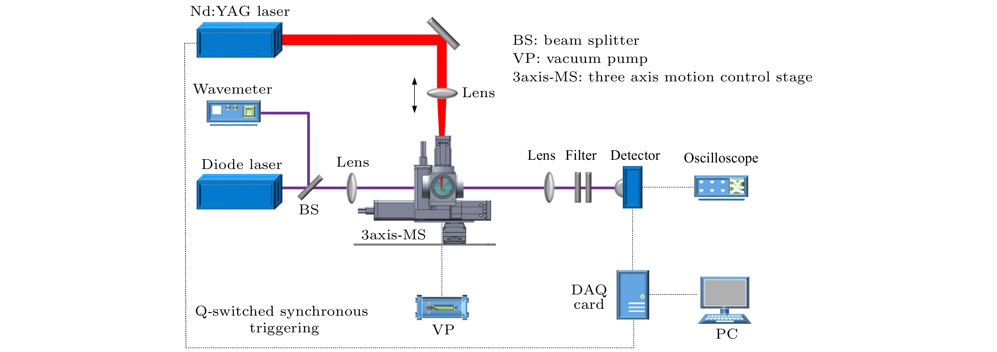
图 2 LAAS实验装置简图
Figure 2. Schematic diagram of the experimental setup of LAAS.
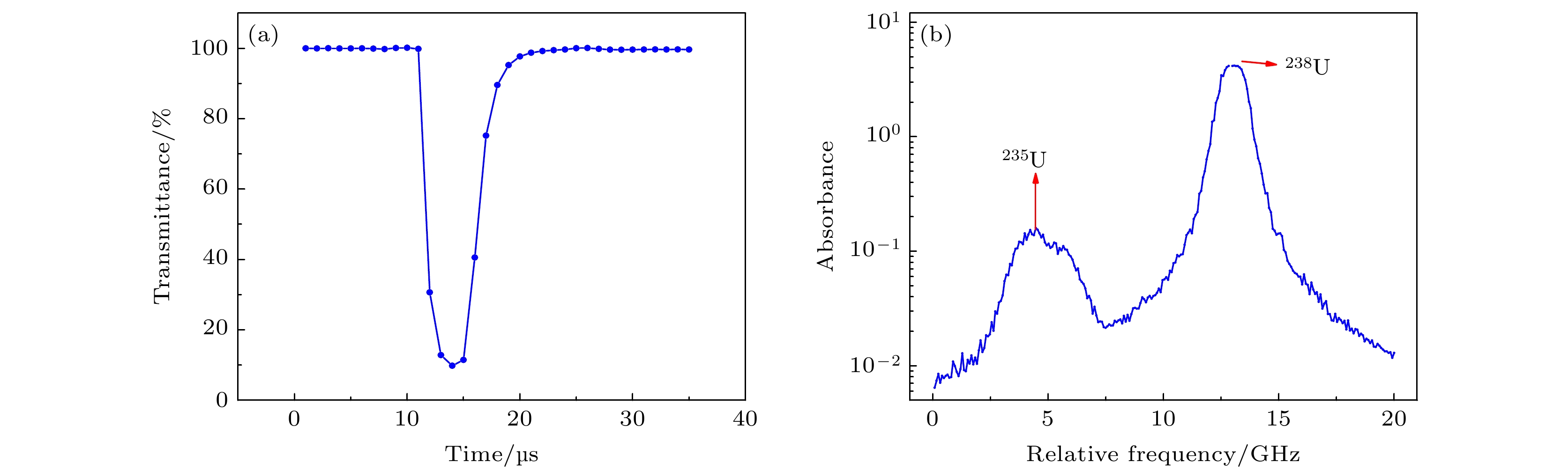
图 3 (a)等离子体透过率测量; (b)实验测量的235U/ 238U吸收光谱
Figure 3. (a) Plasma transmittance measurement; (b) measured absorption spectrum of 235U/ 238U.
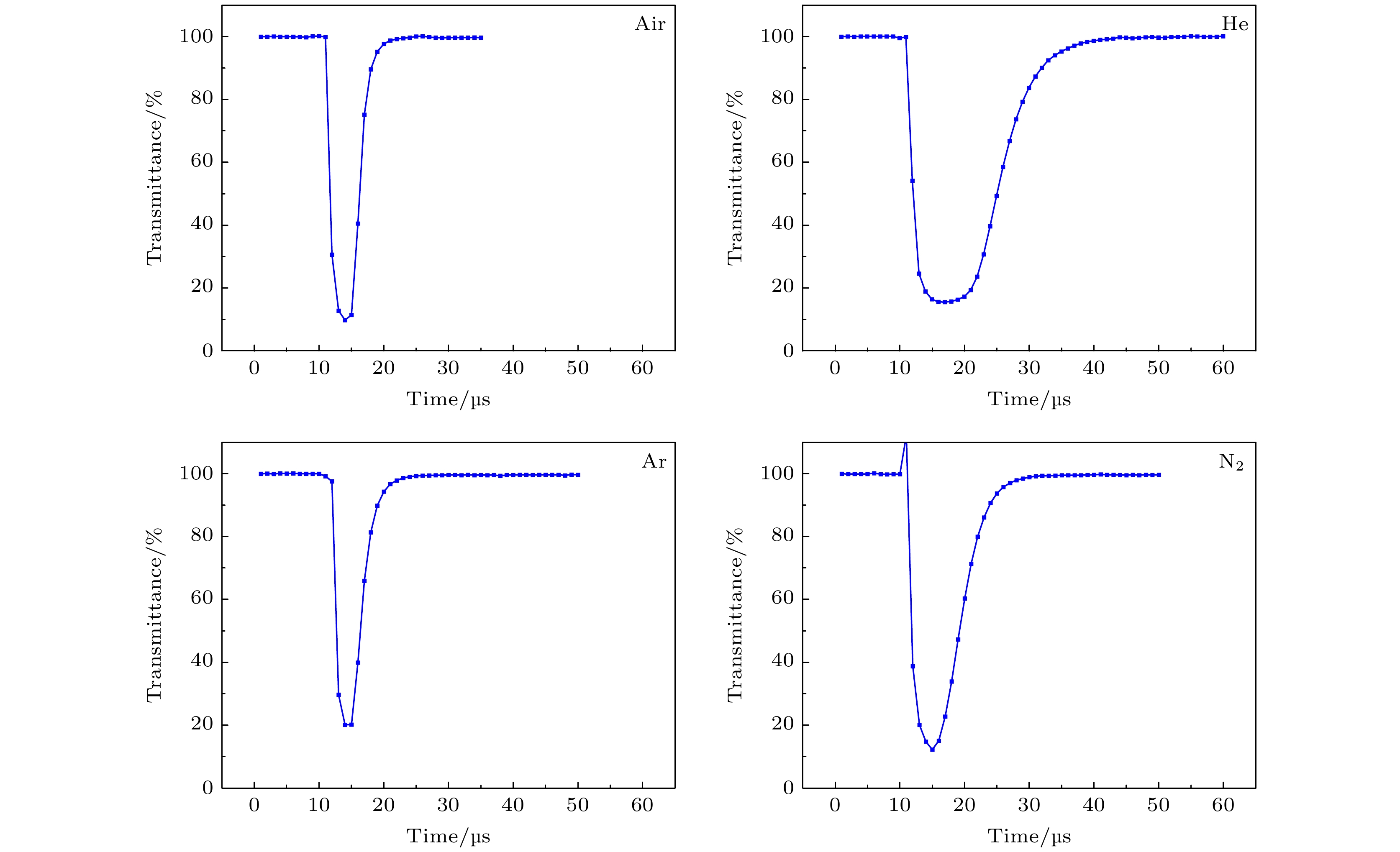
图 4 不同环境气体下测量结果比较(Air, He, Ar, N2)
Figure 4. Comparison of measurement results under different ambient gases (Air, He, Ar, N2).

图 5 不同烧蚀环境下等离子体持续时间随样品池内压力的变化
Figure 5. The persistence of ablation plasma changes with pressure.
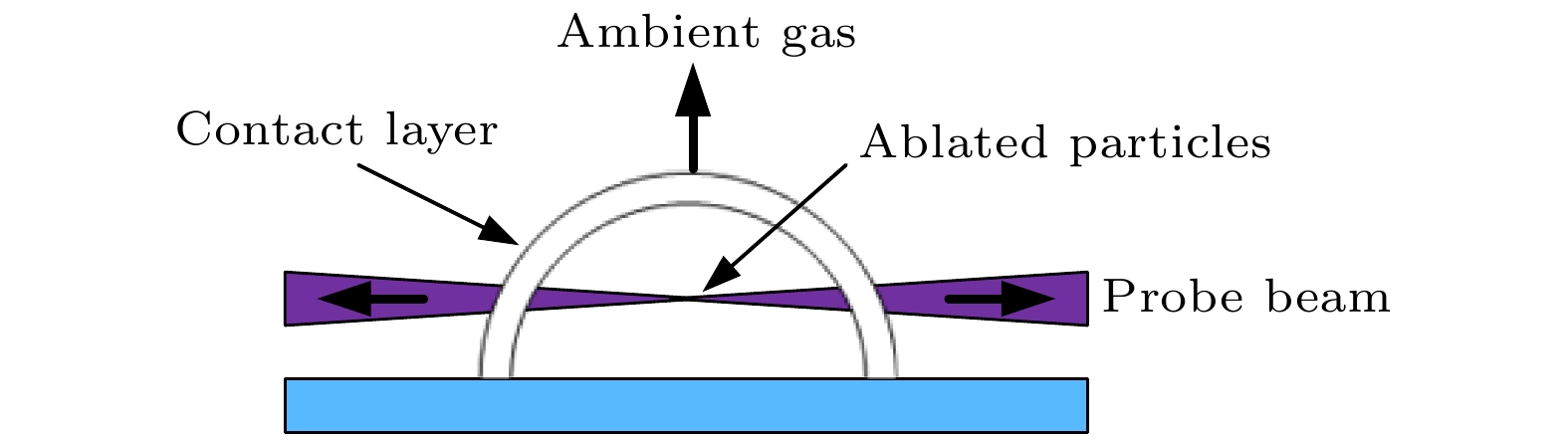
图 6 等离子体膨胀简易模型示意图
Figure 6. Simple model of plasma expansion.

图 7 不同采样延时的238U吸收光谱
Figure 7. Absorption spectra with different sampling delays.
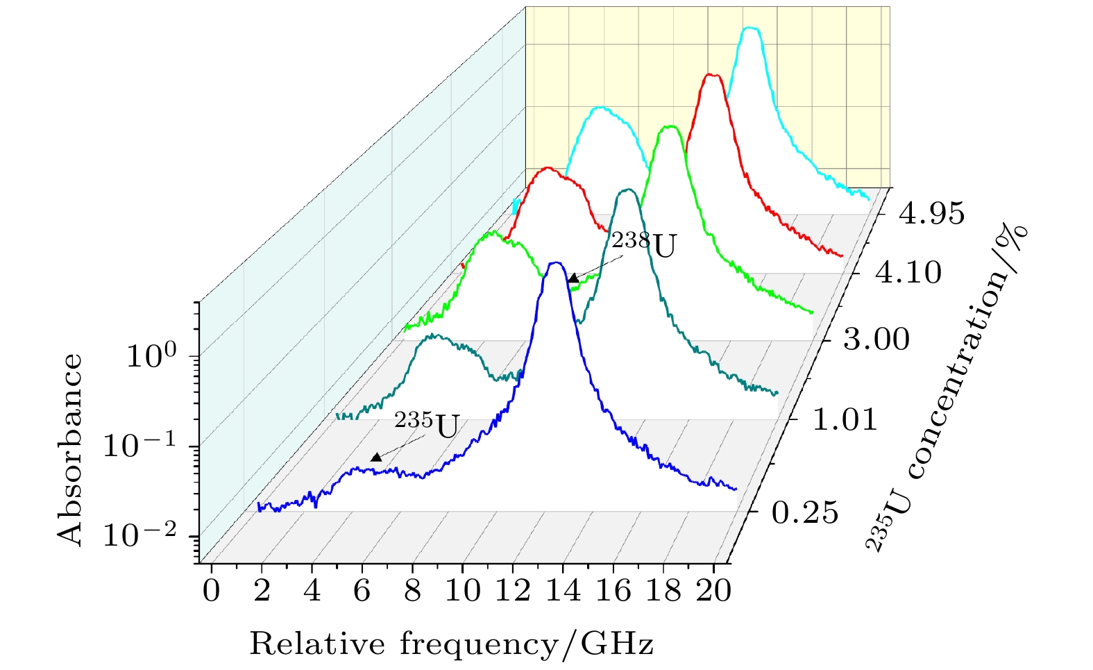
图 8 不同含量样品235U/238U吸收光谱
Figure 8. 235U/238U absorption spectra of samples with different concentration.

图 9 不同含量样品235U/238U吸收光谱及Voigt线型拟合光谱, 拟合曲线下部为拟合残差图
Figure 9. 235U/238U absorption spectra and Voigt fitting spectra of samples with different concentration, the lower part of the fitted curve is the fitted residual graph.
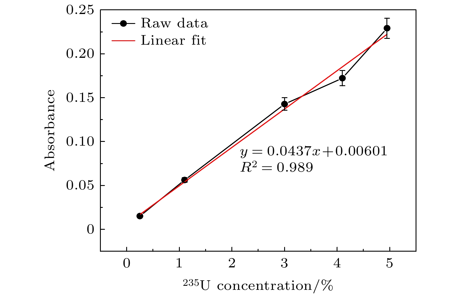
图 10 235U丰度与吸收强度的定标曲线
Figure 10. Calibration curve of 235U abundance and absorption intensity.

图 11 1.10%样品235U/238U吸收光谱及Voigt线型拟合光谱
Figure 11. 235U/238U absorption spectrum and Voigt fitting spectrum of 1.10% sample.
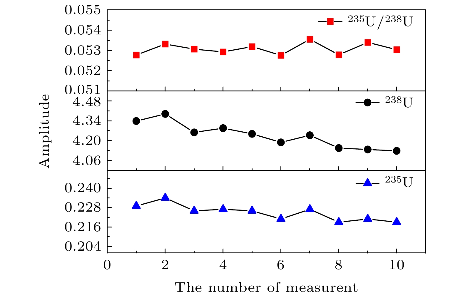
图 12 235U/238U吸收光谱信号稳定性研究
Figure 12. Study on the stability of 235U/238U absorption spectrum signal.
表 1 LAAS实验装置关键器件参数
Table 1. Key device parameters of LAAS experimental device.
实验装置关键器件 参数 探测激光器 线宽100 kHz 烧蚀激光器 波长1064 nm, 脉宽8 ns, 重复频率1—20 Hz, 单脉冲能量最大为200 mJ, 能量稳定性 ≤ 1% 带通滤光片 Semrock, 中心波长λ = 395 nm, 带宽Δλ = 11 nm 陷波滤光片 Thorlabs, 中心波长λ = 1064 nm, 带宽Δλ = 44 nm 光电探测器 Thorlabs, 探测带宽150 MHz  DownLoad: CSV
表 2 实验参数设置
Table 2. experimental parameter setting
实验
参数烧蚀激光
能量/ mJ采样延
时/μs缓冲
气体压力/kPa 扫描时
间/s数值 40 4 He 4 50
DownLoad: CSV
-
[1] Russo R E 1995 Appl. Spectrosc. 49 14
Google Scholar
[2] Chichkov B N, Momma C, Nolte S, Alvensleben F, Tünnermann A 1996 Appl. Phys. A 63 109
Google Scholar
[3] Russo R E, Mao X, Liu H, Gonzalez J, Mao S S 2002 Talanta 57 425
Google Scholar
[4] Harilal S S, Brumfield B E, LaHaye N L, Hartig K C, Phillips M C 2018 Appl. Phys. Rev. 5 021301
Google Scholar
[5] Miziolek A W, Palleschi V, Schechter I 2006 Crit. Rev. Anal. Chem. 27 257
Google Scholar
[6] Harilal S S, Lahaye N L, Phillips M C 2017 Opt. Express. 25 2312
Google Scholar
[7] Skrodzki P J, Shah N P, Taylor N, Hartig K C, Lahaye N L, Brumfield B E, Jovanovic I, Phillips M C, Harilal S S 2016 Spectrochim. Acta, Part B 122 112
Google Scholar
[8] Smith C A, Martinez M A, Veirs D K, Cremers D A 2000 Spectrochim. Acta, Part B 57 929
Google Scholar
[9] Cremers D A, Beddingfield A, Smithwick R, Chinni R C, Jones C R, Beardsley B, Karch L 2012 Appl. Spectrosc. 66 250
Google Scholar
[10] Chan C Y, Choi I, Mao X, Zorba V, Lam O P, Shuh D K, Russo R E 2016 Spectrochim. Acta, Part B 122 31
Google Scholar
[11] Phillips M C, Brumfield B E, Lahaye N, Harilal S S, Hartig K C, Jovanovic I 2017 Scie. Rep 7 3784
Google Scholar
[12] Quentmeier A, Bolshov M, Niemax K 2001 Spectrochim. Acta, Part B 56 45
Google Scholar
[13] Liu H, Quentmeier A, Niemax K 2002 Spectrochim. Acta, Part B 57 1611
Google Scholar
[14] Miyabe M, Oba M, Iimura H, Akaoka K, Maruyama Y, Ohba H 2013 Appl. Phys. A 112 87
Google Scholar
[15] Miyabe M, Oba M, Jung K, Iimura H, Akaokaa K, Katoa M, Otobeb H, Khumaeni A, Wakaida I 2017 Spectrochim. Acta, Part B 134 42
Google Scholar
[16] Taylor N R, Phillips M C 2014 Opt. lett. 39 594
Google Scholar
[17] 叶浩, 张骏昕, 梅海平, 黄尧, 袁子豪, 曹振松, 黄印博 2020 中国激光 47 299
Google Scholar
Ye H, Zhang J X, Mei H P, Huang Y, Yuan Z H, Cao Z S, Huang Y B 2020 Chin. J. Lasers 47 299
Google Scholar
[18] Miyabe M, Oba M, Iimura H, Akaoka K, Maruyama Y, Wakaida I 2010 Appl. Phys. A 101 65
Google Scholar
[19] Yan P, Luo W, Zhang J, Wang L 1992 Chin. J. Lasers 5 27
[20] Kramida Y, Ralchenko J, Reader N A NIST Atomic Spectra Database, National Institute of Standards and Technology http://physics.nist.gov/asd [2021-01-25]
[21] Miyabe M, Oba M, Iimura H, Akaoka K, Maruyama Y, Wakaida I, Watanabe K 2009 4th international conference on laser probing Nagoya, Japan, October 6–10, 2008 p30
[22] Man B Y, Wang X T, Liu A H 1998 J. Appl. Phys. 83 3509
Google Scholar
[23] 张树东, 陈冠英, 刘亚楠, 董晨钟 2002 原子核物理评论 19 206
Google Scholar
Zhang S D, Chen G Y, Liu Y N, Dong C Z 2002 Nucl. Phys. Rev. 19 206
Google Scholar


 DownLoad:


Catalog
Metrics
- Abstract views: 7241
- PDF Downloads: 148
- Cited By: 0
