-
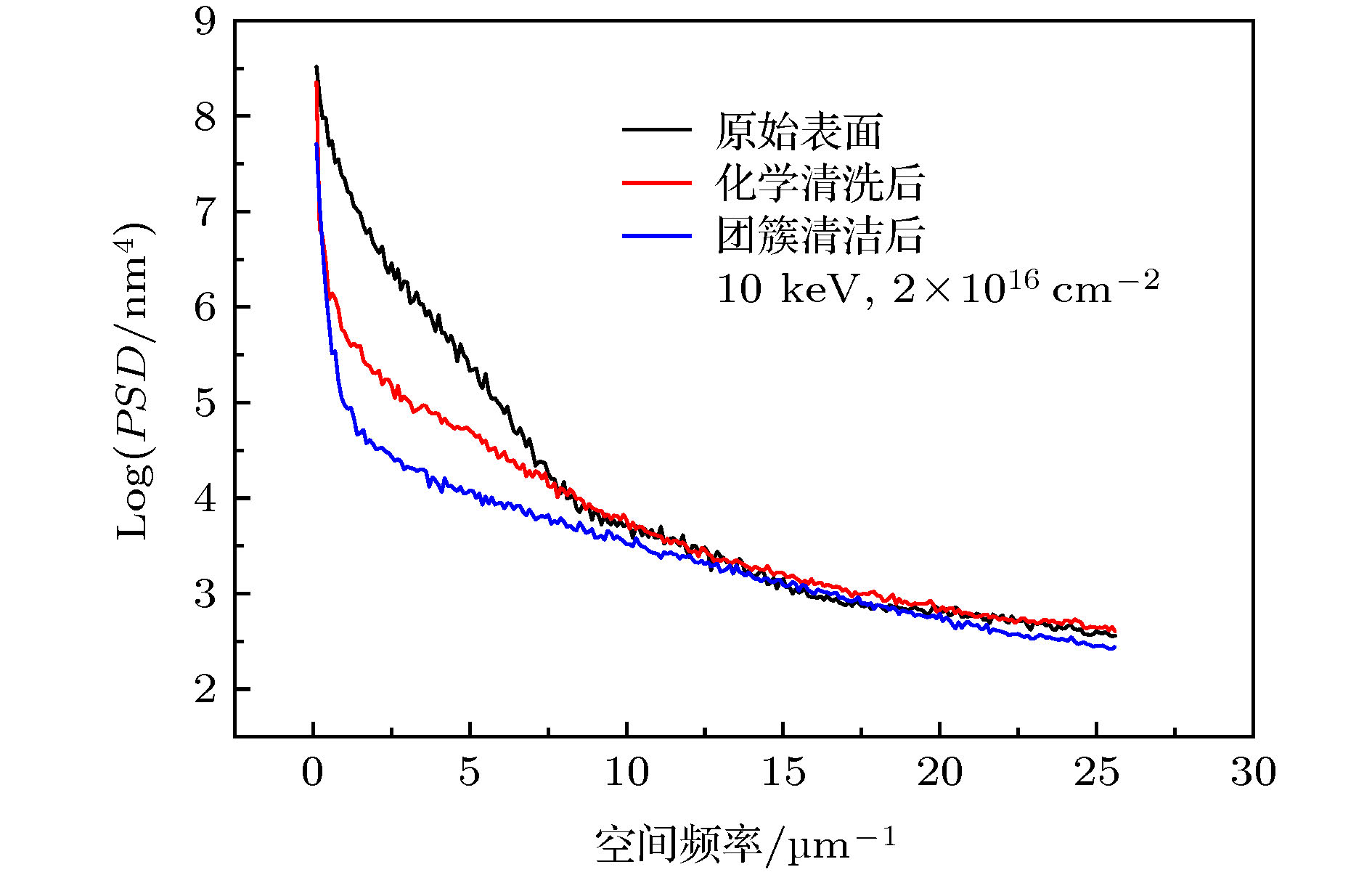
A custom-built gas cluster ion source with energy up to 50 keV is constructed, and Ar, CO2, N2, and O2 are used as the working gases. The clusters are formed by a metal supersonic conical nozzle with critical diameter in a range of 65–135 μm and a cone angle of 14°. The nozzle is powered in the pulsed mode, which improves the pumping conditions, and also makes it possible to increase the gas pressure in the stagnation zone to 15 atm and thereby obtain larger clusters. Based on the principle of ultrasonic expansion, gas cluster ions with an average size of 3000 atoms are obtained. The cluster beam current of 50 μA is obtained. The Ar cluster beam, which is less reactive, is used for treating surface, namely, surface smoothing and formation of self-assembled nanostructures. The Ar cluster bombardment perpendicular to the surface of the substrate is used to demonstrate the smoothing of the surface of Si wafers, Ti coating, and Au film. For the initial Si wafer, its root-mean-square (RMS) roughness of 1.92 nm decreases down to 0.5 nm after cluster beam treatment. The cleaning effect of the cluster beam is also observed very well. The one-dimensional (1D) isotropic power spectral density of the Si surface topography before and after smoothing are also discussed. The off-normal irradiation Ar cluster beam is also used to form self-assembled surface nanoripple arrays on the surface of flat ZnO single crystal substrates. The ripple formation is observed when the incident angle of the cluster beam is in a range of 30°–60°. The process of nanoripple fabrication is significantly governed by the cluster beam incident angle, energy and dose. The nano-ripples formed on the flat substrates remain eolian sand ripples and their formation starts at the incident angle of 30°. The most developed nanoripples are observed at the incident angle within a range of 45°–60°. The surface morphology and characteristic distribution of the nano-structures on the flat ZnO substrate are also analyzed by the two-dimensional (2D) power spectral density function. Next, Ar cluster beam is used for irradiating the ZnO nanorod arrays grown on the Si substrate. Due to various angles between the nanorod’s axis and the substrate normal, the conditions of the ripple formation on the nanorod facets are also realized. The dependence of wavelength on the accelerating voltage of the cluster ions and the dose are studied. Similar dependence of wavelength on accelerating voltage and dose are found for nanorods. Comparing with the flat ZnO surface, nanoripples on the ZnO nanorod faces at high irradiation doses demonstrate an ordering effect, and morphology of the ripples resembles that of more parallel steps rather than eolian ripples.
-
Keywords:
- gas cluster ion beam /
- surface smoothing /
- self-assembled nanostructures /
- power spectral density function
[1] Yamada I, Matsuo J, Toyoda N, Kirkpatrick A 2001 Mater. Sci. Eng., R 34 231
 Google Scholar
[2] 郭尔夫, 韩纪锋, 李永青, 杨朝文, 周荣 2014 63 103601
 Google Scholar
Guo E F, Han J F, Li Y Q, Yang C W, Zhou R 2014 Acta Phys. Sin. 63 103601
 Google Scholar
[3] Becker E W, Bier K, Henkes W 1956 Z. Phys. 146 333
 Google Scholar
[4] Matsuo J, Abe H, Takaoka G H, Yamada I 1995 Nucl. Instrum. Meth. B 99 244
 Google Scholar
[5] Yamada I, Matsuo J, Insepov Z, Takeuchi D, Akizuki M, Toyoda N 1996 J. Vac. Sci. Technol., A 14 781
 Google Scholar
[6] Yamada I, Matsuo J 1998 Mater. Sci. Semicond. Process. 1 27
 Google Scholar
[7] Song J H, Kwon S N, Choi D K, Choi W K 2001 Nucl. Instrum. Meth. B 179 568
 Google Scholar
[8] Andreev A A, Chernysh V S, Ermakov Yu A, Ieshkin A E 2013 Vacuum 91 47
 Google Scholar
[9] Kirkpatrick A, Greer J 1999 AIP Conf. Proc. 475 405
[10] Qin W, Howson R P, Akizuki M, Matsuo J, Takaoka G, Yamada I 1998 Mater. Chem. Phys. 54 258
 Google Scholar
[11] Nose T, Inoue S, Toyoda N, Mochiji K, Mitamura T, Yamada I 2005 Nucl. Instrum. Meth. B 241 626
 Google Scholar
[12] Greer J A, Fenner D B, Hautala J, Allen L P, DiFilippo V, Toyoda N, Yamada I, Matsuo J, Minami E, Katsumata H 2000 Surf. Coat. Technol. 133 273
 Google Scholar
[13] Toyoda N, Yamada I 2008 Nucl. Instrum. Methods B 266 2529
 Google Scholar
[14] Lozano O, Chen Q Y, Tilakaratne B P, Seo H W, Wang X M, Wadekar P V, Chinta P V, Tu LW, Ho N J, Wijesundera D, Chu W K 2013 AIP Adv. 3 062107
 Google Scholar
[15] Saleem I, Tilakaratne B P, Li Y, Bao J, Wijesundera D N, Chu W K 2016 Nucl. Instrum. Methods B 380 20
 Google Scholar
[16] Bradley R M, Harper J M E 1988 J. Vac. Sci. Technol., A 6 2390
 Google Scholar
[17] Motta F C, Shipman P D, Bradley R M 2012 J. Phys. D: Appl. Phys. 45 122001
 Google Scholar
[18] Chao L C, Li Y K, Chang W C 2011 Mater. Lett. 65 1615
 Google Scholar
[19] Babonneau D, Camelio S, Simonot L, Pailloux F, Guerin P, Lamongie B, Lyon O 2011 EPL 93 26005
 Google Scholar
[20] Gkogkou D, Schreiber B, Shaykhutdinov T, Ly H K, Kuhlmann U, Gernert U, Facsko S, Hildebrandt P, Esser N, Hinrichs K, Weidinger I M, Oates T W H 2016 ACS Sens. 1 318
 Google Scholar
[21] Arranz M A, Colino J M, Palomares F J 2014 J. Appl. Phys. 115 183906
 Google Scholar
[22] Zhang K, Uhrmacher M, Hofsäss H, Krauser J 2008 J. Appl. Phys. 103 083507
 Google Scholar
[23] Zeng X M, Pelenovich V, Liu C S, Fu D J 2017 Chin. Phys. C 41 087003
 Google Scholar
[24] Zeng X M, Pelenovich V, Wang Z S, Zuo W B, Belykh S, Tolstogouzov A, Fu D J, Xiao X H 2019 Beilstein J. Nanotechnol. 10 135
 Google Scholar
[25] 瓦西里·帕里诺维奇, 曾晓梅, 付德君 2017 ZL 2017 1 0664910.3
Pelenovich V, Zeng X M, Fu D J 2017 ZL2017 1 0664910.3 (in Chinese)
[26] Pelenovich V, Zeng X M, Ieshkin A, Zuo W B, Chernysh V S, Tolstogouzov A B, Yang B, Fu D J 2019 Nucl. Instrum. Methods B 450 131
 Google Scholar
[27] Pelenovich V O, Zeng X M, Ieshkin A E, Chernysh V S, Tolstogouzov A B, Yang B, Fu D J 2019 J. Surf. Invest. 13 344
 Google Scholar
[28] Korobeishchikov N G, Nikolaev I V, Roenko M A 2019 Nucl. Instrum. Methods B 438 1
 Google Scholar
[29] Ieshkin A, Kireev D, Chernysh V, Molchanov A, Serebryakov A, Chirkin M 2019 Surf. Topogr. Metrol. Prop. 7 025016
 Google Scholar
[30] Insepov Z, Yamada I 1999 Nucl. Instrum. Methods B 148 121
 Google Scholar
[31] Toyoda N, Hagiwara N, Matsuo J, Yamada I 2000 Nucl. Instrum. Methods B 161 980
 Google Scholar
[32] Wang L, Kang Y, Liu X, Zhang S, Huang W, Wang S 2012 Sens. Actuators, B 162 237
 Google Scholar
[33] Shinde S D, Patil G E, Kajale D D, Gaikwad V B, Jain G H 2012 J. Alloys Compd. 528 109
 Google Scholar
[34] Inoue S, Toyoda N, Tsubakino H, Yamada I 2005 Nucl. Instrum. Methods B 237 449
 Google Scholar
[35] Sumie K, Toyoda N, Yamada I 2013 Nucl. Instrum. Methods B 307 290
 Google Scholar
[36] Insepov Z, Yamada I 1999 Nucl. Instrum. Methods B 153 199
 Google Scholar
[37] Moseler M, Rattunde O, Nordiek J, Haberland H 2000 Nucl. Instrum. Methods B 164 522
 Google Scholar
[38] Tilakaratne B P, Chen Q Y, Chu W K 2017 Materials 10 1056
 Google Scholar
[39] Pelenovich V, Zeng X M, Rakhimov R, Zuo W B, Tian C X, Fu D J, Yang B 2020 Mater. Lett. 264 127356
 Google Scholar
-
图 5 在不同入射角下Ar GCIB辐照前后单晶ZnO基片的AFM图像(能量, 10 keV; 剂量, 4 × 10¹⁶ ions/cm2; 箭头表示离子束轰击方向) (a) 团簇辐照前; (b) 30°; (c) 45°; (d) 60°
Figure 5. AFM images of single crystal ZnO substrates before and after Ar GCIB irradiation at different incident angles (energy, 10 keV; dose, 4 × 10¹⁶ ions/cm2; arrows indicate the direction of ion beam bombardment): (a) Before cluster irradiation; (b) 30°; (c) 45°; (d) 60°
图 6 在不同入射角下Ar GCIB辐照单晶ZnO基片的二维PSD图像(能量, 10 keV; 剂量, 4 × 10¹⁶ ions/cm2; 箭头表示离子束轰击方向) (a) 团簇辐照前; (b) 30°; (c) 45°; (d) 60°
Figure 6. Two-dimensional PSD images of single crystal ZnO substrates before and after Ar GCIB irradiation at different incident angles (energy, 10 keV; dose, 4 × 10¹⁶ ions/cm2; arrows indicate the direction of ion beam bombardment): (a) Before cluster irradiation; (b) 30°; (c) 45°; (d) 60°.
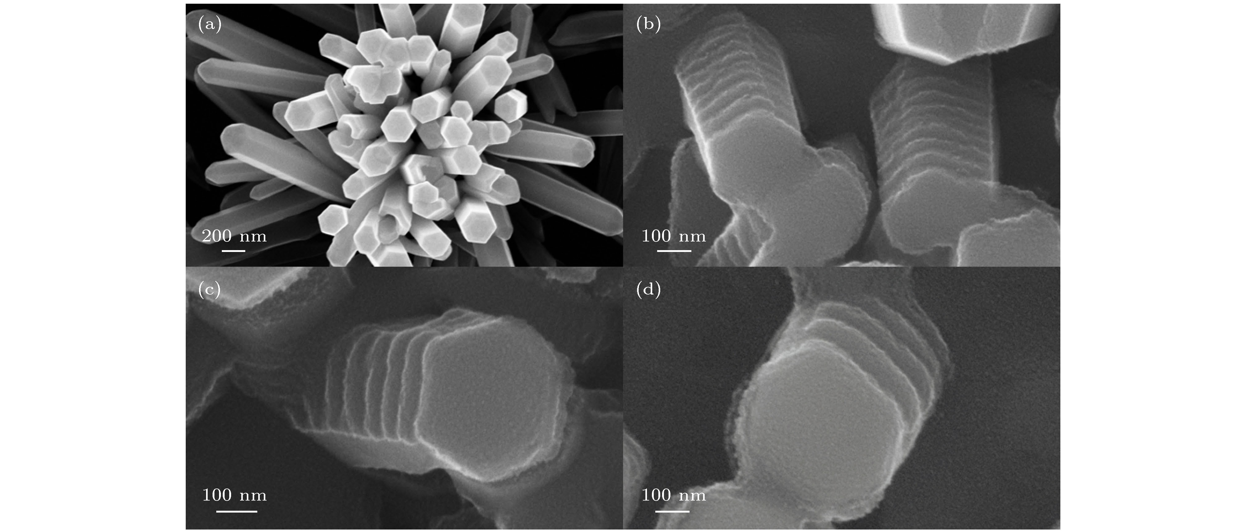
图 7 经不同团簇能量、剂量改性前后ZnO纳米棒的SEM图像 (a) 团簇辐照前; (b) 5 keV, 4 × 10¹⁶ ions/cm2; (c) 10 keV, 2 × 10¹⁶ ions/cm2; (d) 10 keV, 4 × 10¹⁶ ions/cm2
Figure 7. SEM images of ZnO nanorods before and after Ar GCIB irradiation at different cluster energy and dose: (a) Before cluster irradiation; (b) 5 keV, 4 × 10¹⁶ ions/cm2; (c) 10 keV, 2 × 10¹⁶ ions/cm2; (d) 10 keV, 4 × 10¹⁶ ions/cm2.
表 1 根据二维PSD计算的纳米波纹波长和数量
Table 1. Wavelength and number of nanowaves calculated from two-dimensional PSD.
入射角 30° 45° 60° y 轴 y 轴 x 轴 y 轴 最强频率/μm–1 2 6 2 4 波纹波长λ/μm 0.5 0.167 0.5 0.25 平均波纹数量N 10 30 10 20 -
[1] Yamada I, Matsuo J, Toyoda N, Kirkpatrick A 2001 Mater. Sci. Eng., R 34 231
 Google Scholar
[2] 郭尔夫, 韩纪锋, 李永青, 杨朝文, 周荣 2014 63 103601
 Google Scholar
Guo E F, Han J F, Li Y Q, Yang C W, Zhou R 2014 Acta Phys. Sin. 63 103601
 Google Scholar
[3] Becker E W, Bier K, Henkes W 1956 Z. Phys. 146 333
 Google Scholar
[4] Matsuo J, Abe H, Takaoka G H, Yamada I 1995 Nucl. Instrum. Meth. B 99 244
 Google Scholar
[5] Yamada I, Matsuo J, Insepov Z, Takeuchi D, Akizuki M, Toyoda N 1996 J. Vac. Sci. Technol., A 14 781
 Google Scholar
[6] Yamada I, Matsuo J 1998 Mater. Sci. Semicond. Process. 1 27
 Google Scholar
[7] Song J H, Kwon S N, Choi D K, Choi W K 2001 Nucl. Instrum. Meth. B 179 568
 Google Scholar
[8] Andreev A A, Chernysh V S, Ermakov Yu A, Ieshkin A E 2013 Vacuum 91 47
 Google Scholar
[9] Kirkpatrick A, Greer J 1999 AIP Conf. Proc. 475 405
[10] Qin W, Howson R P, Akizuki M, Matsuo J, Takaoka G, Yamada I 1998 Mater. Chem. Phys. 54 258
 Google Scholar
[11] Nose T, Inoue S, Toyoda N, Mochiji K, Mitamura T, Yamada I 2005 Nucl. Instrum. Meth. B 241 626
 Google Scholar
[12] Greer J A, Fenner D B, Hautala J, Allen L P, DiFilippo V, Toyoda N, Yamada I, Matsuo J, Minami E, Katsumata H 2000 Surf. Coat. Technol. 133 273
 Google Scholar
[13] Toyoda N, Yamada I 2008 Nucl. Instrum. Methods B 266 2529
 Google Scholar
[14] Lozano O, Chen Q Y, Tilakaratne B P, Seo H W, Wang X M, Wadekar P V, Chinta P V, Tu LW, Ho N J, Wijesundera D, Chu W K 2013 AIP Adv. 3 062107
 Google Scholar
[15] Saleem I, Tilakaratne B P, Li Y, Bao J, Wijesundera D N, Chu W K 2016 Nucl. Instrum. Methods B 380 20
 Google Scholar
[16] Bradley R M, Harper J M E 1988 J. Vac. Sci. Technol., A 6 2390
 Google Scholar
[17] Motta F C, Shipman P D, Bradley R M 2012 J. Phys. D: Appl. Phys. 45 122001
 Google Scholar
[18] Chao L C, Li Y K, Chang W C 2011 Mater. Lett. 65 1615
 Google Scholar
[19] Babonneau D, Camelio S, Simonot L, Pailloux F, Guerin P, Lamongie B, Lyon O 2011 EPL 93 26005
 Google Scholar
[20] Gkogkou D, Schreiber B, Shaykhutdinov T, Ly H K, Kuhlmann U, Gernert U, Facsko S, Hildebrandt P, Esser N, Hinrichs K, Weidinger I M, Oates T W H 2016 ACS Sens. 1 318
 Google Scholar
[21] Arranz M A, Colino J M, Palomares F J 2014 J. Appl. Phys. 115 183906
 Google Scholar
[22] Zhang K, Uhrmacher M, Hofsäss H, Krauser J 2008 J. Appl. Phys. 103 083507
 Google Scholar
[23] Zeng X M, Pelenovich V, Liu C S, Fu D J 2017 Chin. Phys. C 41 087003
 Google Scholar
[24] Zeng X M, Pelenovich V, Wang Z S, Zuo W B, Belykh S, Tolstogouzov A, Fu D J, Xiao X H 2019 Beilstein J. Nanotechnol. 10 135
 Google Scholar
[25] 瓦西里·帕里诺维奇, 曾晓梅, 付德君 2017 ZL 2017 1 0664910.3
Pelenovich V, Zeng X M, Fu D J 2017 ZL2017 1 0664910.3 (in Chinese)
[26] Pelenovich V, Zeng X M, Ieshkin A, Zuo W B, Chernysh V S, Tolstogouzov A B, Yang B, Fu D J 2019 Nucl. Instrum. Methods B 450 131
 Google Scholar
[27] Pelenovich V O, Zeng X M, Ieshkin A E, Chernysh V S, Tolstogouzov A B, Yang B, Fu D J 2019 J. Surf. Invest. 13 344
 Google Scholar
[28] Korobeishchikov N G, Nikolaev I V, Roenko M A 2019 Nucl. Instrum. Methods B 438 1
 Google Scholar
[29] Ieshkin A, Kireev D, Chernysh V, Molchanov A, Serebryakov A, Chirkin M 2019 Surf. Topogr. Metrol. Prop. 7 025016
 Google Scholar
[30] Insepov Z, Yamada I 1999 Nucl. Instrum. Methods B 148 121
 Google Scholar
[31] Toyoda N, Hagiwara N, Matsuo J, Yamada I 2000 Nucl. Instrum. Methods B 161 980
 Google Scholar
[32] Wang L, Kang Y, Liu X, Zhang S, Huang W, Wang S 2012 Sens. Actuators, B 162 237
 Google Scholar
[33] Shinde S D, Patil G E, Kajale D D, Gaikwad V B, Jain G H 2012 J. Alloys Compd. 528 109
 Google Scholar
[34] Inoue S, Toyoda N, Tsubakino H, Yamada I 2005 Nucl. Instrum. Methods B 237 449
 Google Scholar
[35] Sumie K, Toyoda N, Yamada I 2013 Nucl. Instrum. Methods B 307 290
 Google Scholar
[36] Insepov Z, Yamada I 1999 Nucl. Instrum. Methods B 153 199
 Google Scholar
[37] Moseler M, Rattunde O, Nordiek J, Haberland H 2000 Nucl. Instrum. Methods B 164 522
 Google Scholar
[38] Tilakaratne B P, Chen Q Y, Chu W K 2017 Materials 10 1056
 Google Scholar
[39] Pelenovich V, Zeng X M, Rakhimov R, Zuo W B, Tian C X, Fu D J, Yang B 2020 Mater. Lett. 264 127356
 Google Scholar
Catalog
Metrics
- Abstract views: 18318
- PDF Downloads: 234
- Cited By: 0


 DownLoad:


