


-
The violent inertial cavitation effect generated during high intensity focused ultrasound (HIFU) treatment may damage healthy tissues around the target area. Therefore, it is urgent to develop new technical approaches that can quantitatively monitor the acoustic cavitation motions in biological tissues with high precision in space and time, so as to ensure clinical safety and effectiveness. Compared with the traditional commercial ultrasonic gray value signal, the ultrasonic radio frequency (RF) signal can well retain more detailed information about the acoustic scattering signal. As a statistical parameter not based on mathematical function model, the information entropy can characterize the spatiotemporal evolution state of disorder of scatters inside tissues resulting from acoustic cavitation. Therefore, this paper proposes a real-time monitoring system for spatiotemporal evolution of acoustic cavitation based on the entropy analysis of ultrasonic RF signals. First, the original RF signal of scattered echoes generated by HIFU-induced cavitation bubbles inside the gel phantom is obtained by using a modified B-ultrasound system, and the two-dimensional mean filtering method is used to suppress the HIFU-induced strong interferences overlapping with cavitation monitoring imaging signals. Then, the dynamic variation range of the RF signal is expanded through data standardization processing, and the entropy image is reconstructed based on the sliding window information entropy analysis to demonstrate the spatiotemporal evolution status of the HIFU-induced cavitation behanviors. The experimental results indicate that the acoustic cavitation imaging algorithm based on RF signal entropy analysis should be more sensitive and accurate than the B-model gray scale imaging method for determining the onset time and spatial position of cavitation activities, which is helpful in ensuring the safety and efficacy of HIFU clinical treatment. Thepresent work will provide a useful tool for the spatiotemporal monitoring of the acoustic cavitation generated in tissues during HIFU treatment, and lays a solid theoretical and experimental foundation to establish an effective quantity-effect evaluation system for the cavitation related biological effect.
-
Keywords:
- acoustic cavitation /
- high intensity focused ultrasound /
- ultrasound radio frequency signal /
- information entropy
[1] Kennedy J E 2005 Nat. Rev. Cancer 5 321
 Google Scholar
[2] Izadifar Z, Izadifar Z, Chapman D, Babyn P 2020 J. Clin. Med. 9 460
Google Scholar
[3] 秦对, 邹青钦, 李章勇, 王伟, 万明习, 冯怡 2021 70 154701
Google Scholar
Qin D, Zou Q Q, Li Z Y, Wang W, Wan M X, Feng Y 2021 Acta Phys. Sin. 70 154701
Google Scholar
[4] Yang Y Y, Li Q, Guo X S, Tu J, Zhang D 2020 Ultrason. Sonochem. 67 105096
Google Scholar
[5] 于洁, 郭霞生, 屠娟, 章东 2015 64 094306
Google Scholar
Yu J, Guo X S, Tu J, Zhang D 2015 Acta Phys. Sin. 64 094306
Google Scholar
[6] Valle L F, Lehrer E J, Markovic D, Elashoff D, Levin-Epstein R, Karnes R J, Reiter R E, Rettig M, Calais J, Nickols N G, Dess R T, Spratt D E, Steinberg M L, Nguyen P L, Davis B J, Zaorsky N G, Kishan A U 2021 Eur. Urol. 80 280
Google Scholar
[7] Yoshizawa S, Ikeda T, Ito A, Ota R, Takagi S, Matsumoto Y 2009 Med. Biol. Eng. Comput. 47 851
Google Scholar
[8] Zderic V, Keshavarzi A, Noble M L, Paun M, Sharar S R, Crum L A, Martin R W, Vaezy S 2006 Ultrasonics 44 46
Google Scholar
[9] Ilyas A, Chen C J, Ding D, Romeo A, Buell T J, Wang T R, Kalani M Y S, Park M S 2018 Neurosurg Focus 44 E12
Google Scholar
[10] Bader K B, Vlaisavljevich E, Maxwell A D 2019 Ultrasound Med. Biol. 45 1056
Google Scholar
[11] 耿昊, 范庭波, 张喆, 屠娟, 郭霞生, 李发琪, 章东 2014 63 044301
Google Scholar
Geng H, Fan T B, Zhang Z, Tu J, Guo X S, Li F Q, Zhang D 2014 Acta Phys. Sin. 63 044301
Google Scholar
[12] Yang D X, Ni Z Y, Yang Y Y, Xu G Y, Tu J, Guo X S, Huang P T, Zhang D 2018 Ultrason. Sonochem. 49 111
Google Scholar
[13] Tu J, Swalwell J E, Giraud D, Cui W C, Chen W Z, Matula T J 2011 IEEE Trans. Ultrason. Ferroelectr. Freq. Control 58 955
Google Scholar
[14] Song R J, Zhang C B, Teng F M, Tu J, Guo X S, Fan Z, Zheng Y F, Zhang D 2021 Ultrason. Sonochem. 79 105790
[15] Fan P F, Yu J, Yang X, Tu J, Guo X S, Huang P T, Zhang D 2017 Chin. Phys. B 26 054301
Google Scholar
[16] 钱骏, 谢伟, 周小伟, 谭坚文, 王智彪, 杜永洪, 李雁浩 2022 71 037201
Google Scholar
Qian J, Xie W, Zhou X W, Tan J W, Wang Z B, Du Y H, Li Y H 2022 Acta Phys. Sin. 71 037201
Google Scholar
[17] Liu T, Yu D, Beitler J, Tridandapani S, Bruner D, Curran W, Yang X 2013 Med. Phys. 40 495
Google Scholar
[18] Zhang S, Li C, Zhou F Y, Wan M X, Wang S P 2014 J. Ultrasound Med. 33 959
Google Scholar
[19] Tsui P H, Chen C K, Kuo W H, Chang K J, Fang J, Ma H Y, Chou D 2017 Sci. Rep. 7 41004
Google Scholar
[20] Shankar P M 2000 IEEE Trans. Ultrason. Ferroelectr. Freq. Control 47 727
Google Scholar
[21] Tsui P H 2015 Entropy 17 6598
Google Scholar
[22] Hughes M S, Marsh J N, Wallace K D, Donahue T A, Connolly A M, Lanza G M, Wickline S A 2007 Ultrasound Med. Biol. 33 1236
Google Scholar
[23] Zhou Z H, Huang C C, Shung K K, Tsui P H, Fang J, Ma H Y, Wu S C, Lin C C 2014 PLoS One 9 e96195
Google Scholar
[24] Xu Z, Hall T L, Fowlkes J B, Cain C A 2007 J. Acoust. Soc. Am. 122 229
Google Scholar
[25] 张玫玫, 高凡, 屠娟, 吴意赟, 章东 2021 70 084302
Google Scholar
Zhang M M, Gao F, Tu J, Wu Y Y, Zhang D 2021 Acta Phys. Sin. 70 084302
Google Scholar
[26] Tuthill T A, Sperry R H, Parker K J 1988 Ultrasonic Imaging 10 81
Google Scholar
[27] Fan T B, Tu J, Luo L J, Guo X S, Huang P T, Zhang D 2016 Chin. Phys. Lett. 33 084302
Google Scholar
[28] 陈楚怡, 于洁, 陈功, 马勇, 郭霞生, 屠娟, 章东 2015 声学学报 40 563
Google Scholar
Chen C Y, Yu J, Chen G, Ma Y, Guo X S, Tu J, Zhang D 2015 Acta Acustica 40 563
Google Scholar
[29] Yu J, Chen C Y, Chen G, Guo X S, Ma Y, Tu J, Zhang D 2014 Chin. Phys. Lett. 31 034302
Google Scholar
[30] Lee J S 1980 IEEE Trans. Pattern Anal. Mach. Intell. 2 165
Google Scholar
[31] Liao P S, Chew T S, Chung P C 2001 J. Inf. Sci. Eng. 17 713
[32] 吴俊, 汪源源, 陈悦, 余锦华, 庞芸 2014 光学精密工程 22 1312
Google Scholar
Wu J J, Wang Y Y, Chen Y, Yu J H, Pang Y 2014 Opt. and Precision Eng. 22 1312
Google Scholar
[33] Tsui P H, Chang C C 2007 Ultrasound Med. Biol. 33 608
Google Scholar
[34] Tsui P H, Wan Y L 2016 Entropy 18 341
Google Scholar
[35] Bailey M R, Khokhlova V A, Sapozhnikov O A, Kargl S G, Crum L A 2003 Acoust. Phys. 49 369
Google Scholar
[36] Vaezy S, Shi X G, Martin R W, Chi E, Nelson P I, Bailey M R, Crum L A 2001 Ultrasound Med. Biol. 27 33
Google Scholar
-
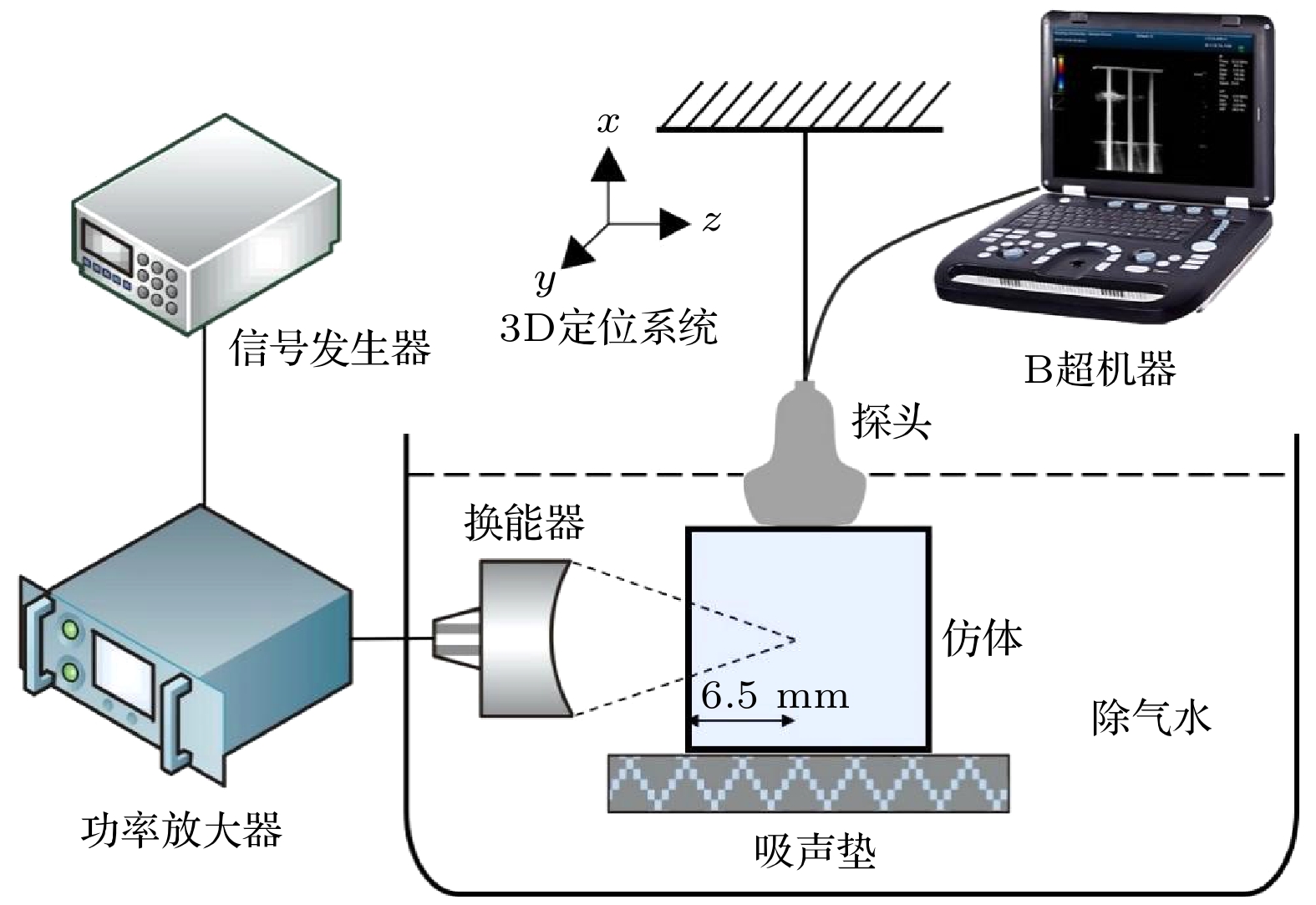
图 1 实验原理图
Figure 1. Diagram of experimental schematic.
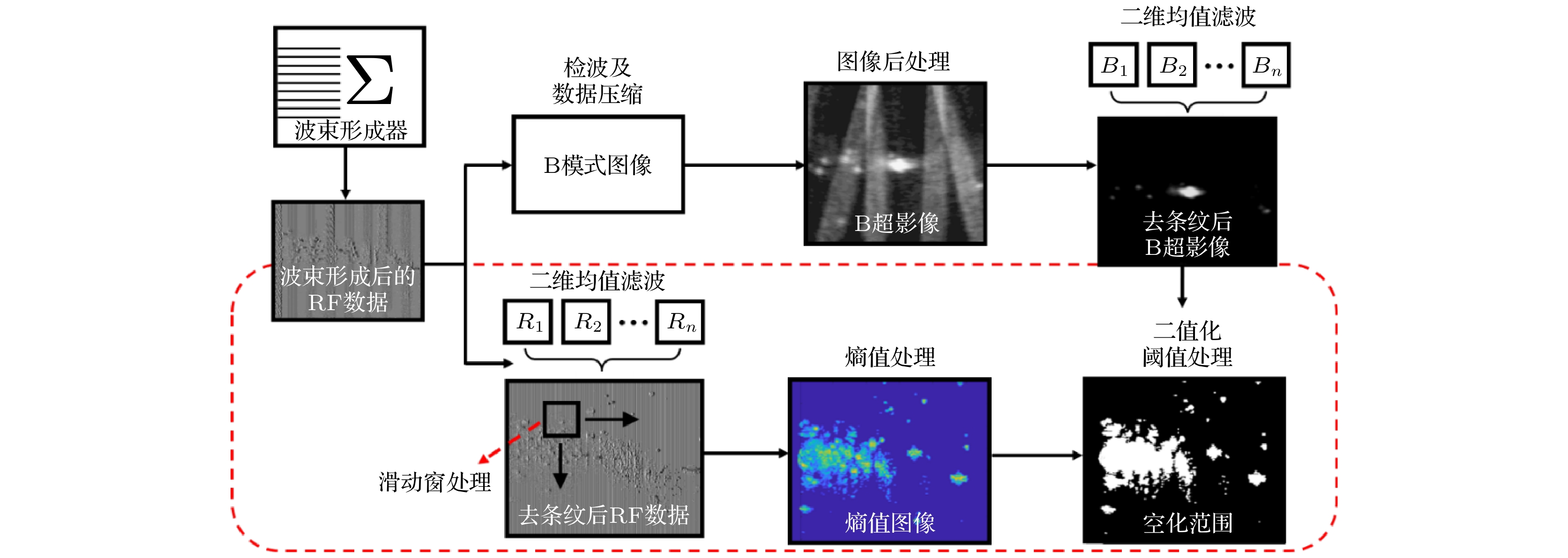
图 2 基于B超灰度图像与RF信号熵分析监测HIFU空化区域算法示意图
Figure 2. The algorithmic scheme designed for cavitation monitoring imaging based on B-mode and RF entropy images.
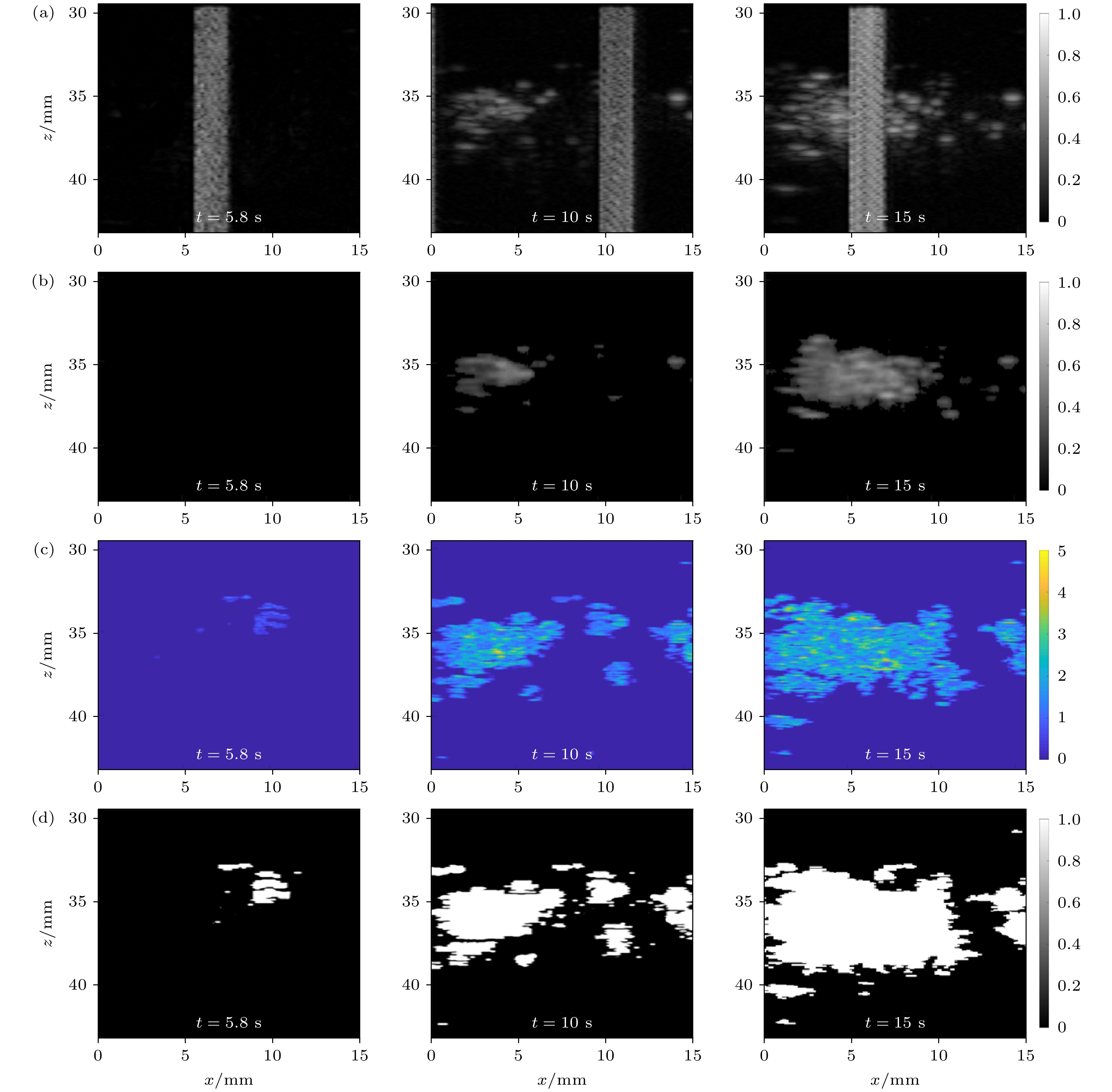
图 3 不同时刻治疗区域的图像 (a) 初步B模式图像; (b) 去干扰后初步B模式图像; (c) 熵值图像; (d) 经二值化处理后的熵值图像
Figure 3. Images at different treatment moments: (a) Preliminary B-mode images; (b) preliminary B-mode images after de-interference; (c) entropy images; (d) binary images at different treatment moments.
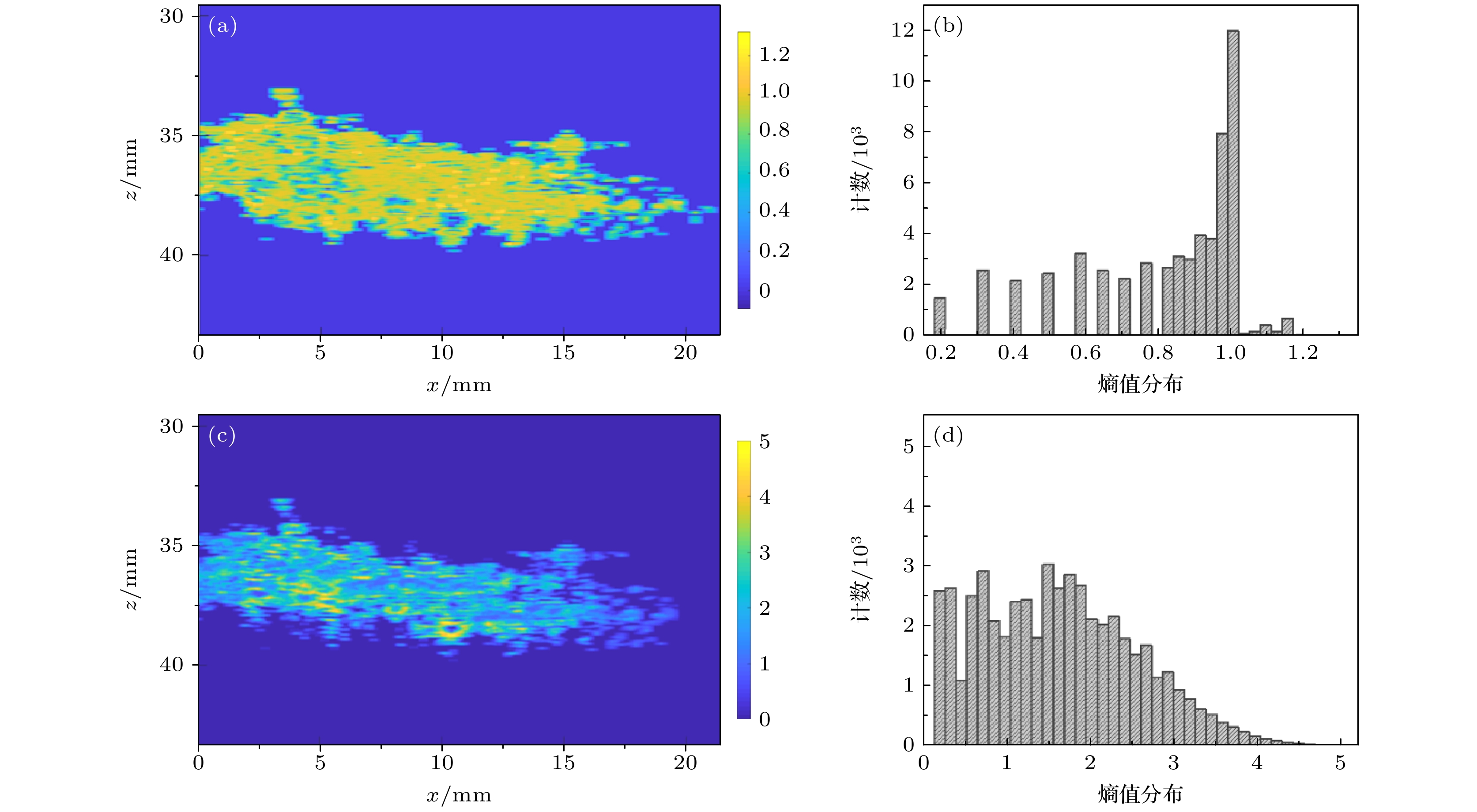
图 4 标准化操作前后熵值图像及幅值分布图 (a) 未经标准化处理得到的熵值图像; (b) 未经标准化得到的熵值分布直方图; (c) 经过标准化处理得到的熵值图像; (d) 经过标准化得到的熵值分布直方图
Figure 4. Entropy image and amplitude distribution before and after standardization: (a) Entropy image obtained without normalization; (b) histogram of entropy distribution obtained without normalization; (c) entropy image obtained with normalization; (d) histogram of entropy distribution obtained with normalization

图 5 不同声压下分别基于灰度值法与熵值法得到的实时空化范围趋势图
Figure 5. Real-time cavitation range trend based on gray value method and entropy value method respectively under different sound pressure.
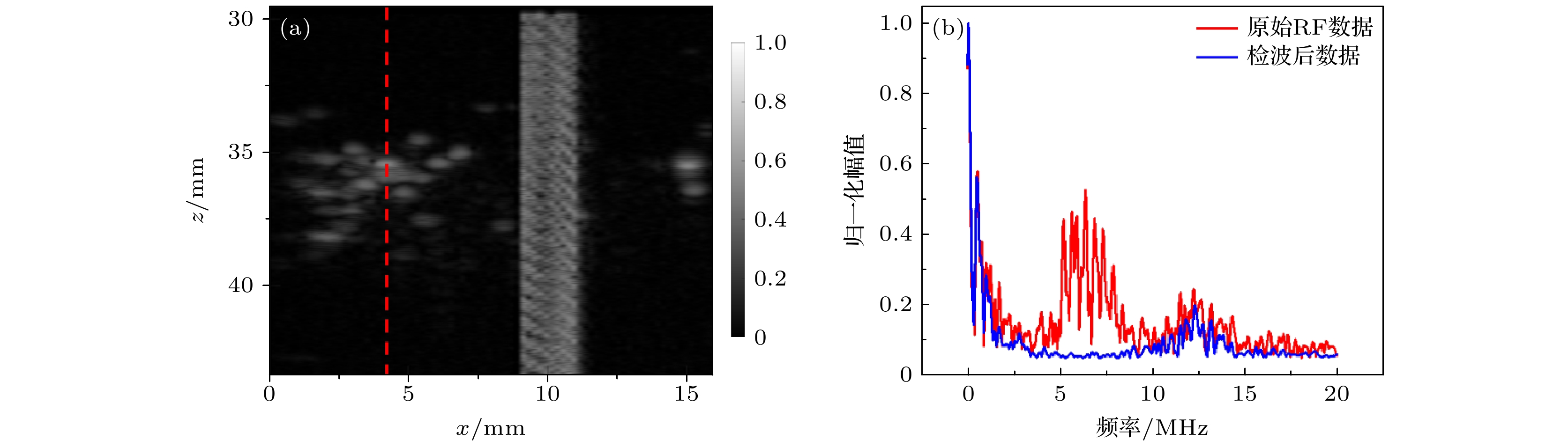
图 6 原始BFRF数据与检波后BFRF数据的频谱对比 (a) 初步B模式图像; (b) 检波前后数据频谱
Figure 6. Comparison of frequency spectrum of the raw BFRF data and the BFRF data after demodulation: (a) Preliminary B-mode image; (b) data frequency spectrum pre- and post- demodulation
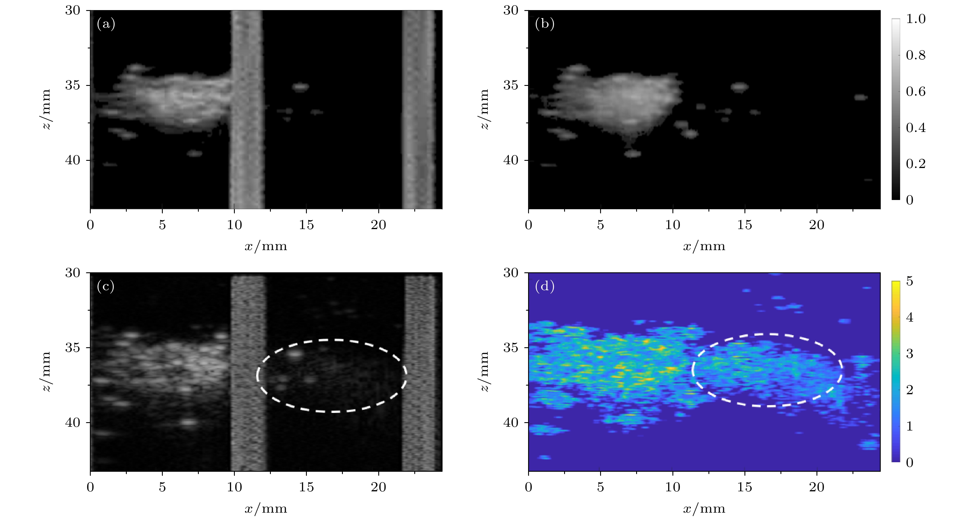
图 7 一帧B超影像与熵值图像反映空化范围的典型例子 (a) 一帧B超影像; (b) 去干扰后B超影像; (c) 同一帧基于BFRF的初步B模式图像; (d) 同一帧熵值图像
Figure 7. A typical example of a frame of ultrasound image and entropy image reflecting the range of cavitation: (a) A frame of ultrasound image; (b) the de-interfered ultrasound image; (c) the same frame of BFRF-based preliminary B-mode image; (d) the same frame of entropy image.
-
[1] Kennedy J E 2005 Nat. Rev. Cancer 5 321
Google Scholar
[2] Izadifar Z, Izadifar Z, Chapman D, Babyn P 2020 J. Clin. Med. 9 460
Google Scholar
[3] 秦对, 邹青钦, 李章勇, 王伟, 万明习, 冯怡 2021 70 154701
Google Scholar
Qin D, Zou Q Q, Li Z Y, Wang W, Wan M X, Feng Y 2021 Acta Phys. Sin. 70 154701
Google Scholar
[4] Yang Y Y, Li Q, Guo X S, Tu J, Zhang D 2020 Ultrason. Sonochem. 67 105096
Google Scholar
[5] 于洁, 郭霞生, 屠娟, 章东 2015 64 094306
Google Scholar
Yu J, Guo X S, Tu J, Zhang D 2015 Acta Phys. Sin. 64 094306
Google Scholar
[6] Valle L F, Lehrer E J, Markovic D, Elashoff D, Levin-Epstein R, Karnes R J, Reiter R E, Rettig M, Calais J, Nickols N G, Dess R T, Spratt D E, Steinberg M L, Nguyen P L, Davis B J, Zaorsky N G, Kishan A U 2021 Eur. Urol. 80 280
Google Scholar
[7] Yoshizawa S, Ikeda T, Ito A, Ota R, Takagi S, Matsumoto Y 2009 Med. Biol. Eng. Comput. 47 851
Google Scholar
[8] Zderic V, Keshavarzi A, Noble M L, Paun M, Sharar S R, Crum L A, Martin R W, Vaezy S 2006 Ultrasonics 44 46
Google Scholar
[9] Ilyas A, Chen C J, Ding D, Romeo A, Buell T J, Wang T R, Kalani M Y S, Park M S 2018 Neurosurg Focus 44 E12
Google Scholar
[10] Bader K B, Vlaisavljevich E, Maxwell A D 2019 Ultrasound Med. Biol. 45 1056
Google Scholar
[11] 耿昊, 范庭波, 张喆, 屠娟, 郭霞生, 李发琪, 章东 2014 63 044301
Google Scholar
Geng H, Fan T B, Zhang Z, Tu J, Guo X S, Li F Q, Zhang D 2014 Acta Phys. Sin. 63 044301
Google Scholar
[12] Yang D X, Ni Z Y, Yang Y Y, Xu G Y, Tu J, Guo X S, Huang P T, Zhang D 2018 Ultrason. Sonochem. 49 111
Google Scholar
[13] Tu J, Swalwell J E, Giraud D, Cui W C, Chen W Z, Matula T J 2011 IEEE Trans. Ultrason. Ferroelectr. Freq. Control 58 955
Google Scholar
[14] Song R J, Zhang C B, Teng F M, Tu J, Guo X S, Fan Z, Zheng Y F, Zhang D 2021 Ultrason. Sonochem. 79 105790
[15] Fan P F, Yu J, Yang X, Tu J, Guo X S, Huang P T, Zhang D 2017 Chin. Phys. B 26 054301
Google Scholar
[16] 钱骏, 谢伟, 周小伟, 谭坚文, 王智彪, 杜永洪, 李雁浩 2022 71 037201
Google Scholar
Qian J, Xie W, Zhou X W, Tan J W, Wang Z B, Du Y H, Li Y H 2022 Acta Phys. Sin. 71 037201
Google Scholar
[17] Liu T, Yu D, Beitler J, Tridandapani S, Bruner D, Curran W, Yang X 2013 Med. Phys. 40 495
Google Scholar
[18] Zhang S, Li C, Zhou F Y, Wan M X, Wang S P 2014 J. Ultrasound Med. 33 959
Google Scholar
[19] Tsui P H, Chen C K, Kuo W H, Chang K J, Fang J, Ma H Y, Chou D 2017 Sci. Rep. 7 41004
Google Scholar
[20] Shankar P M 2000 IEEE Trans. Ultrason. Ferroelectr. Freq. Control 47 727
Google Scholar
[21] Tsui P H 2015 Entropy 17 6598
Google Scholar
[22] Hughes M S, Marsh J N, Wallace K D, Donahue T A, Connolly A M, Lanza G M, Wickline S A 2007 Ultrasound Med. Biol. 33 1236
Google Scholar
[23] Zhou Z H, Huang C C, Shung K K, Tsui P H, Fang J, Ma H Y, Wu S C, Lin C C 2014 PLoS One 9 e96195
Google Scholar
[24] Xu Z, Hall T L, Fowlkes J B, Cain C A 2007 J. Acoust. Soc. Am. 122 229
Google Scholar
[25] 张玫玫, 高凡, 屠娟, 吴意赟, 章东 2021 70 084302
Google Scholar
Zhang M M, Gao F, Tu J, Wu Y Y, Zhang D 2021 Acta Phys. Sin. 70 084302
Google Scholar
[26] Tuthill T A, Sperry R H, Parker K J 1988 Ultrasonic Imaging 10 81
Google Scholar
[27] Fan T B, Tu J, Luo L J, Guo X S, Huang P T, Zhang D 2016 Chin. Phys. Lett. 33 084302
Google Scholar
[28] 陈楚怡, 于洁, 陈功, 马勇, 郭霞生, 屠娟, 章东 2015 声学学报 40 563
Google Scholar
Chen C Y, Yu J, Chen G, Ma Y, Guo X S, Tu J, Zhang D 2015 Acta Acustica 40 563
Google Scholar
[29] Yu J, Chen C Y, Chen G, Guo X S, Ma Y, Tu J, Zhang D 2014 Chin. Phys. Lett. 31 034302
Google Scholar
[30] Lee J S 1980 IEEE Trans. Pattern Anal. Mach. Intell. 2 165
Google Scholar
[31] Liao P S, Chew T S, Chung P C 2001 J. Inf. Sci. Eng. 17 713
[32] 吴俊, 汪源源, 陈悦, 余锦华, 庞芸 2014 光学精密工程 22 1312
Google Scholar
Wu J J, Wang Y Y, Chen Y, Yu J H, Pang Y 2014 Opt. and Precision Eng. 22 1312
Google Scholar
[33] Tsui P H, Chang C C 2007 Ultrasound Med. Biol. 33 608
Google Scholar
[34] Tsui P H, Wan Y L 2016 Entropy 18 341
Google Scholar
[35] Bailey M R, Khokhlova V A, Sapozhnikov O A, Kargl S G, Crum L A 2003 Acoust. Phys. 49 369
Google Scholar
[36] Vaezy S, Shi X G, Martin R W, Chi E, Nelson P I, Bailey M R, Crum L A 2001 Ultrasound Med. Biol. 27 33
Google Scholar


 DownLoad:


Catalog
Metrics
- Abstract views: 8709
- PDF Downloads: 122
- Cited By: 0
