-
The laser-induced damage to potassium dihydrogen phosphate (KDP) crystal restricts the development of high power laser systems and attract the attention of researchers. The defects are essential for the understanding of the laser-induced damage to KDP crystals. The defects in KDP crystals are commonly related to
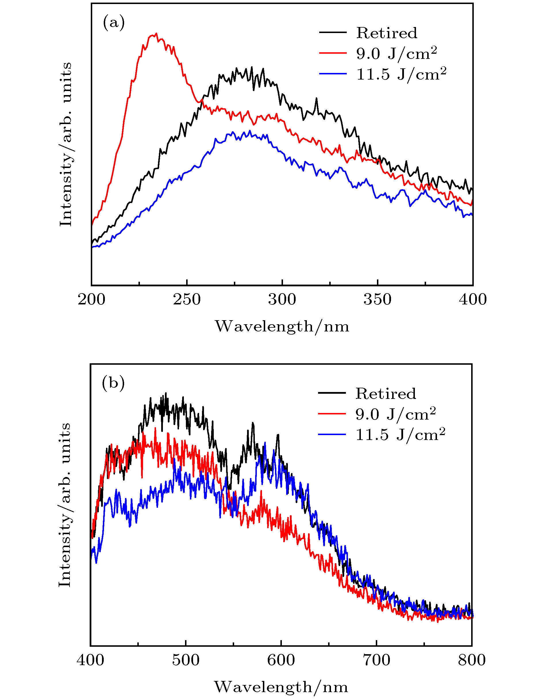
$ \rm H_2PO_4^{-} $ groups. The defects of KDP crystal have been studied extensively, however the changes of defects of KDP crystal with low fluence and high fluence have not been investigated sufficiently. The synchrotron radiation technology is a sensitive method of detecting the defects. The vacuum ultraviolet photoluminescence (PL) emission spectra can provide microscopic structural changes in KDP crystals. In this work, we investigate the defects of KDP crystals irradiated with different fluences by vacuum ultraviolet PL emission spectra. The vacuum ultraviolet spectra are obtained at the 4B8 beam line in Beijing synchrotron radiation facilities. Each KDP crystal spectrum is measured from 200 to 400 nm and 400 to 800 nm. The emission spectra of KDP crystal irradiated with different fluences are fitted for illustration. Each Gaussian curve represents a kind of defect. Comparing the retired components with KDP crystal irradiated by 11.5 J/cm2, the new band at 231.55 nm emerges in the spectra of KDP crystal irradiated by 9.0 J/cm2. The intrinsic luminescence band is assigned to the radiative annihilation of self-trapped excitons. According to our previous work, the short chain structures mainly exist in the crystal irradiated by 9.0 J/cm2, and the long chain structure is mainly in the crystal irradiated by 11.5 J/cm2. The retired components have the short, medium and long chain. The length of P—O bond in the short chain is shorter than that in the long chain structure. The overlap between phosphorus 3s orbitals and oxygen 2p increases, and the radiative annihilation of STEs becomes stronger. So the band at 231.55 nm emerges in the spectrum of KDP crystal irradiated by 9.0 J/cm2. It suggests that the structure of the retired component and the structure of KDP crystal irradiated by 9.0 J/cm2 are different. The results provide an insight into the defects in KDP crystals. It is meaningful to study the mechanism of laser-induced damage to KDP crystal.-
Keywords:
- potassium dihydrogen phosphate crystal /
- defect /
- photoluminescence
[1] Carr C W, Radousky H B, Demos S G 2003 Phys. Rev. Lett. 91 127402
 Google Scholar
[2] Boopathi K, Rajesh P, Ramasamy P, Manyum P 2013 Opt. Mater. 35 954
 Google Scholar
[3] De Yoreo J J, Burnham A K, Whitman P K 2002 Int. Mater. Rev. 47 113
 Google Scholar
[4] Schmid A, Kelly P, Bräunlich P 1977 Phys. Rev. B 16 4569
 Google Scholar
[5] Tien A C, Backus S, Kapteyn H, Murnane M, Mourou G 1999 Phys. Rev. Lett. 82 3883
 Google Scholar
[6] Swain J, Stokowski S, Milam D, Rainer F 1982 Appl. Phys. Lett. 40 350
 Google Scholar
[7] Yokotani A, Sasaki T, Yoshida K, Yamanaka T, Yamanaka C 1986 Appl. Phys. Lett. 48 1030
 Google Scholar
[8] Singleton M F, Cooper J F, Andresen B D, Milanovich F P 1988 Appl. Phys. Lett. 52 857
 Google Scholar
[9] Demos S G, Yan M, Staggs M, De Yoreo J J, Radousky H B 1998 Appl. Phys. Lett. 72 2367
 Google Scholar
[10] Jiang H, McNary J, Tom H W K, Yan M, Radousky H B, Demos S G 2002 Appl. Phys. Lett. 81 3149
 Google Scholar
[11] Demos S G, Staggs M, Radousky H B 2003 Phys. Rev. B 67 224102
 Google Scholar
[12] Davis J E, Hughes R S, Lee H W H 1993 Chem. Phys. Lett. 207 540
 Google Scholar
[13] Marshall C D 1994 J. Opt. Soc. Am. B: Opt. Phys. 11 774
 Google Scholar
[14] Chirila M M, Garces N Y, Halliburton L E, Demos S G, Land T A, Radousky H B 2003 J. Appl. Phys. 94 6456
 Google Scholar
[15] Pommiès M, Damiani D, Le Borgne X, Dujardin C, Surmin A, Birolleau J C, Pilon F, Bertussi B, Piombini H 2007 Opt. Commun. 275 372
 Google Scholar
[16] Paul De Mange, Christopher W. Carr, Raluca A. Negres, 2008 J. Appl. Phys. 104 103103
 Google Scholar
[17] Wang K, Fang C, Zhang J, Sun X, Wang S, Gu Q, Zhao X, Wang B 2006 J. Cryst. Growth 287 478
 Google Scholar
[18] Paul De Mange R A N, Christopher W C 2006 Opt. Express 14 5313
 Google Scholar
[19] Duchateau G, Geoffroy G, Dyan A, Piombini H, Guizard S 2011 Phys. Rev. B 83 075114
 Google Scholar
[20] Duchateau G, Geoffroy G, Belsky A, Fedorov N, Martin P, Guizard S 2013 J. Phys Condens Matter 25 435501
 Google Scholar
[21] Li X, Liu B A, Yan C, Liu C, Ju X 2018 Opt. Mater. Express 8 816
 Google Scholar
[22] Müller K A 1987 Ferroelectrics 72 273
 Google Scholar
[23] Setzler S D, Stevens K T, Halliburton L E 1998 Phys. Rev. B 57 2643
 Google Scholar
[24] Harris L B, Vella G J 1973 J. Chem. Phys. 58 4550
 Google Scholar
[25] Griscom D L, Friebele E J, Long K J, Fleming J W 1983 J. Appl. Phys. 54 3743
 Google Scholar
[26] Archidi M E, Haddad M, Nadiri A 1996 Nucl. Instrum. Methods Phys. Res. B 116 145
 Google Scholar
[27] Ehrt D, Ebeling P, Natura U 2000 J. Non-Cryst. Solids 263 240
 Google Scholar
[28] Chiodini N, Meinardi F, Morazzoni F, Paleari A, Scotti R, Di Martino D 2000 Appl. Phys. Lett. 76 3209
 Google Scholar
[29] Ogorodnikov I N, Pustovarov V A, Cheremnykh V S 2003 Opt. Spectrosc. 95 385
 Google Scholar
[30] Ogorodnikov I N, Shul’gin B V 2001 Opt. Spectrosc. 91 224
 Google Scholar
-
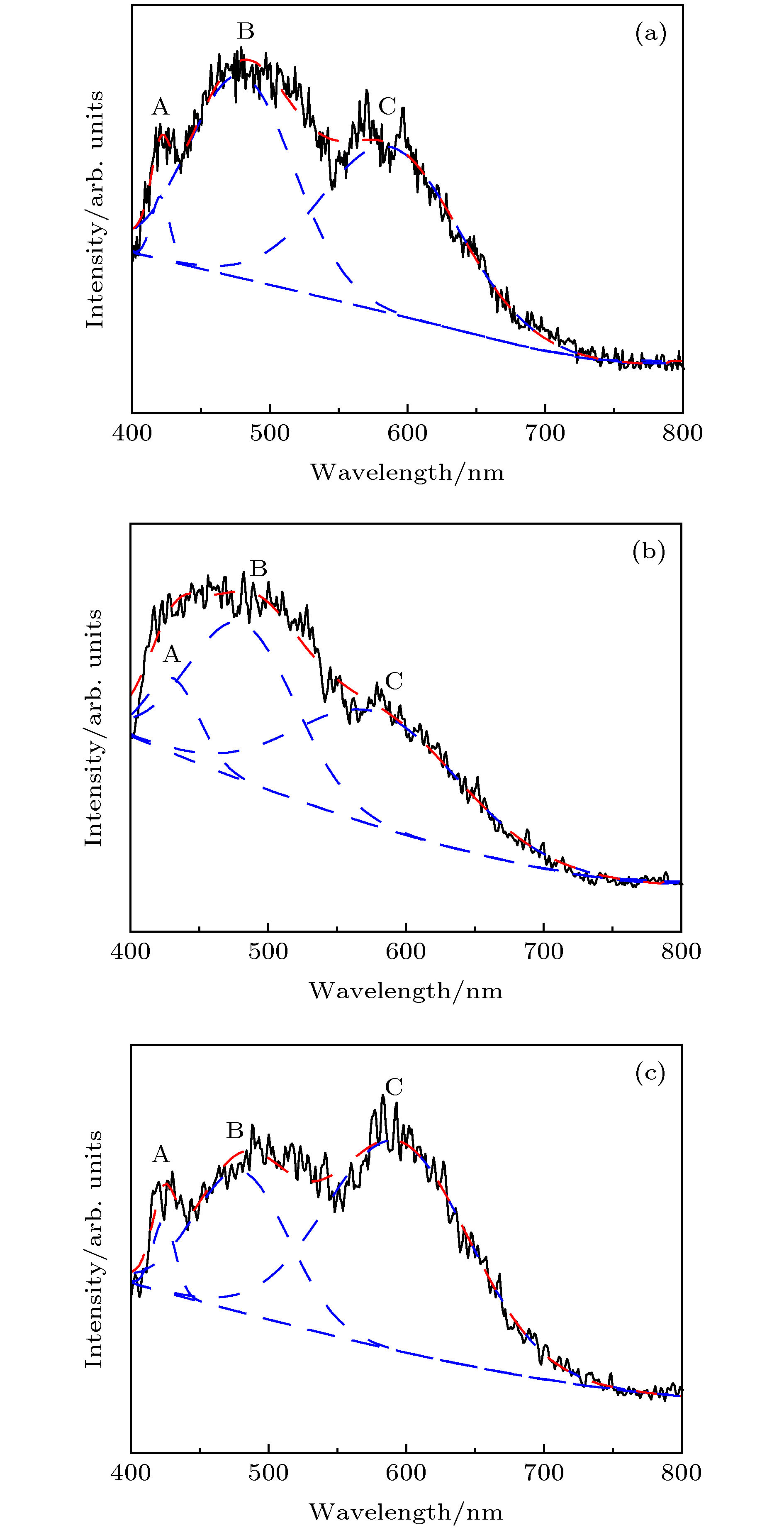
图 2 400—800 nm范围内不同通量辐照下KDP晶体的PL发射谱 (a) 退役元件; (b) 9.0 J/cm2; (c) 11.5 J/cm2. 黑色线是实验光谱, 红线是拟合叠加谱, 蓝线为高斯拟合曲线
Figure 2. PL emission spectra of KDP crystals with different flux irradiations measured from 400 to 800 nm: (a) Retired; (b) 9.0 J/cm2; (c) 11.5 J/cm2. The black solid lines represent the experiment spectra, the red dotted lines represent the simulated spectra, and the blue lines represent the Gaussian fitting curve.
图 3 200—400 nm范围内不同通量辐照下KDP晶体的PL发射谱 (a) 退役原件; (b) 9.0 J/m2; (c) 11.5 J/m2. 图中黑色线是实验光谱, 红线是拟合叠加谱, 蓝线为高斯拟合曲线
Figure 3. PL emission spectra of KDP crystals with different flux irradiations were measured from 200 to 400 nm: (a) Retired; (b) 9.0 J/m2; (c) 11.5 J/m2. The black solid lines represent the experiment spectra, the red dotted lines represent the simulated spectra and blue lines represent the Gaussian fitting curve.
表 1 400—800 nm范围内不同通量辐照下KDP晶体PL发射谱的高斯拟合参数
Table 1. Parameters of peaks with Gaussian fitting for samples irradiated by different flux irradiations measured from 400 to 800 nm.
Peak Position/nm Area/arb. units. FWHM/nm Retired 9.0 J/cm2 11.5 J/cm2 Retired 9.0 J/cm2 11.5 J/cm2 Retired 9.0 J/cm2 11.5 J/cm2 A 420.27 430.52 423.87 0.12 0.35 0.13 15.38 45.14 19.41 B 479.19 480.94 480.54 2.23 1.50 1.12 88.95 91.53 80.68 C 587.24 578.64 593.58 2.40 1.68 2.51 116.60 140.89 122.17 表 2 200—400 nm范围内不同通量辐照下KDP晶体PL发射谱的高斯拟合参数
Table 2. Parameters of peaks with Gaussian fitting for samples irradiated by different flux irradiations measured from 200 to 400 nm.
Peak Position/nm Area/arb. units FWHM/nm Retired 9.0 J/cm2 11.5 J/cm2 Retired 9.0 J/cm2 11.5 J/cm2 Retired 9.0 J/cm2 11.5 J/cm2 A 231.55 2.00 32.89 B 268.01 269.61 269.04 3.33 2.26 2.29 71.66 62.61 67.12 C 324.02 326.95 326.82 2.67 1.94 1.35 91.90 83.30 79.92 -
[1] Carr C W, Radousky H B, Demos S G 2003 Phys. Rev. Lett. 91 127402
 Google Scholar
[2] Boopathi K, Rajesh P, Ramasamy P, Manyum P 2013 Opt. Mater. 35 954
 Google Scholar
[3] De Yoreo J J, Burnham A K, Whitman P K 2002 Int. Mater. Rev. 47 113
 Google Scholar
[4] Schmid A, Kelly P, Bräunlich P 1977 Phys. Rev. B 16 4569
 Google Scholar
[5] Tien A C, Backus S, Kapteyn H, Murnane M, Mourou G 1999 Phys. Rev. Lett. 82 3883
 Google Scholar
[6] Swain J, Stokowski S, Milam D, Rainer F 1982 Appl. Phys. Lett. 40 350
 Google Scholar
[7] Yokotani A, Sasaki T, Yoshida K, Yamanaka T, Yamanaka C 1986 Appl. Phys. Lett. 48 1030
 Google Scholar
[8] Singleton M F, Cooper J F, Andresen B D, Milanovich F P 1988 Appl. Phys. Lett. 52 857
 Google Scholar
[9] Demos S G, Yan M, Staggs M, De Yoreo J J, Radousky H B 1998 Appl. Phys. Lett. 72 2367
 Google Scholar
[10] Jiang H, McNary J, Tom H W K, Yan M, Radousky H B, Demos S G 2002 Appl. Phys. Lett. 81 3149
 Google Scholar
[11] Demos S G, Staggs M, Radousky H B 2003 Phys. Rev. B 67 224102
 Google Scholar
[12] Davis J E, Hughes R S, Lee H W H 1993 Chem. Phys. Lett. 207 540
 Google Scholar
[13] Marshall C D 1994 J. Opt. Soc. Am. B: Opt. Phys. 11 774
 Google Scholar
[14] Chirila M M, Garces N Y, Halliburton L E, Demos S G, Land T A, Radousky H B 2003 J. Appl. Phys. 94 6456
 Google Scholar
[15] Pommiès M, Damiani D, Le Borgne X, Dujardin C, Surmin A, Birolleau J C, Pilon F, Bertussi B, Piombini H 2007 Opt. Commun. 275 372
 Google Scholar
[16] Paul De Mange, Christopher W. Carr, Raluca A. Negres, 2008 J. Appl. Phys. 104 103103
 Google Scholar
[17] Wang K, Fang C, Zhang J, Sun X, Wang S, Gu Q, Zhao X, Wang B 2006 J. Cryst. Growth 287 478
 Google Scholar
[18] Paul De Mange R A N, Christopher W C 2006 Opt. Express 14 5313
 Google Scholar
[19] Duchateau G, Geoffroy G, Dyan A, Piombini H, Guizard S 2011 Phys. Rev. B 83 075114
 Google Scholar
[20] Duchateau G, Geoffroy G, Belsky A, Fedorov N, Martin P, Guizard S 2013 J. Phys Condens Matter 25 435501
 Google Scholar
[21] Li X, Liu B A, Yan C, Liu C, Ju X 2018 Opt. Mater. Express 8 816
 Google Scholar
[22] Müller K A 1987 Ferroelectrics 72 273
 Google Scholar
[23] Setzler S D, Stevens K T, Halliburton L E 1998 Phys. Rev. B 57 2643
 Google Scholar
[24] Harris L B, Vella G J 1973 J. Chem. Phys. 58 4550
 Google Scholar
[25] Griscom D L, Friebele E J, Long K J, Fleming J W 1983 J. Appl. Phys. 54 3743
 Google Scholar
[26] Archidi M E, Haddad M, Nadiri A 1996 Nucl. Instrum. Methods Phys. Res. B 116 145
 Google Scholar
[27] Ehrt D, Ebeling P, Natura U 2000 J. Non-Cryst. Solids 263 240
 Google Scholar
[28] Chiodini N, Meinardi F, Morazzoni F, Paleari A, Scotti R, Di Martino D 2000 Appl. Phys. Lett. 76 3209
 Google Scholar
[29] Ogorodnikov I N, Pustovarov V A, Cheremnykh V S 2003 Opt. Spectrosc. 95 385
 Google Scholar
[30] Ogorodnikov I N, Shul’gin B V 2001 Opt. Spectrosc. 91 224
 Google Scholar
Catalog
Metrics
- Abstract views: 7249
- PDF Downloads: 91
- Cited By: 0


 DownLoad:


