


-
X-ray scintillation screens as the core component of X-ray imaging detectors have widespread applications in the medical imaging, security inspection, high energy physics, radiochemistry, and so on. For a long time, the development of X-ray scintillation screen mainly focuses on improving the light yield in order to enhance its detection efficiency. However, a novel tendency has recently emerged for ultrafast time performance of the X-ray imaging detector. The indium doping zinc oxide (ZnO:In) with high radiation hardness, higher light yield(>10000 photons/MeV) and subnanosecond decay time is a promising scintillation material for ultrafast detections. In order to satisfy the requirements of X-ray scintillation screens with ultrafast and high-spatial-resolution in the existing and upcoming high energy physics experiments, the ZnO:In nanorod arrays have been prepared on a 100-nm-thick ZnO-seeded substrate by hydrothermal reaction method and then treated by hydrogen plasma in present work. The results of SEM demonstrate the average diameter and length of the ZnO:In nanorods are about 0.5 and 12 μm, respectively. The XRD shows the ZnO:In nanorods are highly aligned perpendicular to the substrate along c-axis direction. The X-ray excited luminescence spectra show that two luminescence bands are observed, i.e. an ultraviolet emission peak located at about 395 nm and a visible emission band at 450–750 nm. It is particularly important to point out that hydrogen plasma treatment can enhance the ultraviolet emission of ZnO:In nanorod arrays and suppress its visible emission. The reason is attributed to the formation of shallow donors through hydrogen entering the ZnO and the combination of VO and Oi. In addition, the fluorescence decay times of the ultraviolet and visible emissions for the ZnO:In nanorod arrays are subnanosecond and nanosecond, respectively, satisfying the demand of the fast X-ray imaging. The spatial resolution of ZnO:In nanorod arrays has been characterized in X-ray imaging beamline at the Shanghai Synchrotron Radiation Facility. Under excitation of the X-ray beam with the energy of 20 keV, a system spatial resolution of 1.5 μm could be achieved by using an 12 μm thickness ZnO:In nanorod arrays as the scintillation screen, which is exceeded the highest level had ever been reported on ZnO:In nanorod arrays scintillation screen. In conclusion, this present work shows that it is a feasible solution for X-ray detection and imaging with high temporal and spatial resolution by using ZnO:In nanorod arrays as the X-ray scintillation screen.
-
Keywords:
- X-ray scintillation screen /
- ZnO:In nanorod arrays /
- ultrafast decay time /
- high spatial resolution
[1] Yanagida T 2018 Proc. Jpn. Acad., Ser. B 94 75
 Google Scholar
[2] Dujardin C, Auffray E, Bourret-Courchesne E, Dorenbos P, Lecoq P, Nikl M, Vasil'ev A N, Yoshikawa A, Zhu R Y 2018 IEEE Trans. Nucl. Sci. 65 1977
Google Scholar
[3] Nikl M 2006 Meas. Sci. Technol. 17 R37
Google Scholar
[4] Barnes, C W, Fernández, J C, Hartsfield, T M, Sandberg, R L, Sheffield, R L, Tapia, J P, Wang, Z 2018 AIP Conf. Proc. 1979 160003
Google Scholar
[5] Turk G, Reverdin C, Gontier D, Darbon S, Dujardin C, Ledoux G, Hamel M, Simic V, Normand S 2010 Rev. Sci. Instrum. 81 10E509
Google Scholar
[6] Atanov N, Baranov V, Budagov J, Cervelli F, Colao F, Cordelli M, Corradi G, Davydov Y I, Falco S D, Diociaiuti E, Donati S, Donghia R, Echenard B, Giovannella S, Glagolev V, Grancagnolo F, Happacher F, Hitlin D G, Martini M, Miscetti S, Miyashita T, Morescalchi L, Murat P, Pedreschi E, Pezzullo G, Porter F, Raffaelli F, Ricci M, Saputi A, Sarra I, Spinella F, Tassielli G, Tereshchenko V, Usubov Z, Zhu R Y 2018 J. Instrum. 13 C02037
Google Scholar
[7] Zhu R Y 2019 J. Phys. Conf. Ser. 1162 012022
Google Scholar
[8] Hu C, Zhang L, Zhu RY, Chen A, Wang Z, Ying L, Yu Z 2018 IEEE Trans. Nucl. Sci. 65 2097
Google Scholar
[9] Simpson P J, Tjossem R, Hunt A W, Lynn K G, Munné V 2003 Nucl. Instrum. Methods Phys. Res., Sect. A 505 82
Google Scholar
[10] Chen L, Ruan J, Xu M, He S, Hu J, Zhang Z, Liu J, Ouyang X 2019 Nucl. Instrum. Methods Phys. Res., Sect. A 933 71
Google Scholar
[11] Grigorjeva L, Grube J, Bite I, Zolotarjovs A, Smits K, Millers D, Rodnyi P, Chernenko K 2019 Radiat. Meas. 123 69
Google Scholar
[12] 邱志澈, 顾牡, 刘小林, 刘波, 黄世明, 倪晨 2016 光谱学与光谱分析 36 336
Google Scholar
Qiu Z C, Gu M, Liu X L, Liu B, Huang S M, Ni C 2016 Spectrosc. Spect. Anal. 36 336
Google Scholar
[13] Liu S, Gu M, Chen H, Sun Z, Liu X, Liu B, Huang S, Zhang J 2018 Nucl. Instrum. Methods Phys. Res., Sect. A 903 18
Google Scholar
[14] Li Q, Liu X, Gu M, Li F, Zhang J, Wu Q, Huang S, Liu S 2018 Appl. Surf. Sci. 433 815
Google Scholar
[15] Kobayashi M, Komori J, Shimidzu K, Izaki M, Uesugi K, Takeuchi A, Suzuki Y 2015 Appl. Phys. Lett. 106 081909
Google Scholar
[16] Izaki M, Kobayashi M, Shinagawa T, Koyama T, Uesugi K, Takeuchi A 2017 Phys. Status Solidi A 214 1700285
Google Scholar
[17] Li Q, Hao S, An R, Wang M, Sun Z, Wu Q, Gu M, Zhao J, Liu X, Zhang Z 2019 Appl. Surf. Sci. 493 1299
Google Scholar
[18] 倪晨, 顾牡, 王迪, 曹顿华, 刘小林, 黄世明 2009 光谱学与光谱分析 29 2291
Google Scholar
Ni C, Gu M, Wang D, Cao D H, Liu X L, Huang S M 2009 Spectrosc. Spect. Anal. 29 2291
Google Scholar
[19] Özgür Ü, Alivov Y I, Liu C, Teke A, Reshchikov M A, Doğan S, Avrutin V, Cho S J, Morkoç H 2005 J. Appl. Phys. 98 041301
Google Scholar
[20] Li Q, Liu X, Gu M, Huang S, Ni C, Liu B, Hu Y, Sun S, Zhang Z 2016 IEEE Trans. Nucl. Sci. 63 471
Google Scholar
[21] Li Q, Liu X, Gu M, Huang S, Zhang J, Ni C, Liu B, Hu Y, Wu Q, Zhao S 2016 Superlattices Microstruct. 98 351
Google Scholar
[22] Hofmann D M, Hofstaetter A, Leiter F, Zhou H, Henecker F, Meyer B K, Orlinskii S B, Schmidt J, Baranov P G 2002 Phys. Rev. Lett. 88 045504
Google Scholar
[23] Lavrov E V, Herklotz F, Weber J 2009 Phys. Rev. B 79 165210
Google Scholar
[24] Kano M, Wakamiya A, Yamanoi K, Sakai K, Takeda K, Cadatal-Raduban M, Nakazato T, Shimizu T, Sarukura N, Fukuda T 2012 IEEE Trans. Nucl. Sci. 59 2290
Google Scholar
[25] Yamanoi K, Sakai K, Cadatal-Raduban M, Nakazato T, Shimizu T, Sarukura N, Kano M, Wakamiya A, Fukuda T, Nagasono M, Togashi T, Matsubara S, Tono K, Higashiya A, Yabashi M, Kimura H, Ohashi H, Ishikawa T 2012 IEEE Trans. Nucl. Sci. 59 2298
Google Scholar
[26] 郭智敏, 倪培君 2010 兵器材料科学与工程 33 113
Google Scholar
Guo Z M, Ni P J, 2010 Ordnance Mater. Sci. Eng. 33 113
Google Scholar
[27] Chen H, Gu M, Sun Z, Liu X, Liu B, Zhang J, Huang S, Ni C 2019 Opt. Express 27 14871
Google Scholar
[28] Sowa K M, Last A, Korecki P 2017 Sci. Rep. 7 44944
Google Scholar
[29] Samei E, Flynn M J, Reimann D A 1998 Med. Phys. 25 102
Google Scholar
[30] Michail C, Valais I, Martini N, Koukou V, Kalyvas N, Bakas A, Kandarakis I, Fountos G 2016 Radiat. Meas. 94 8
Google Scholar
-
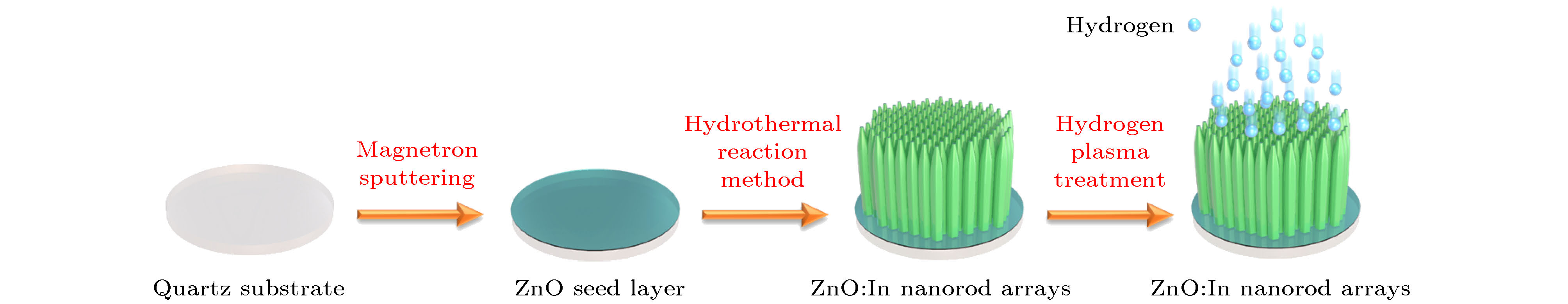
图 1 ZnO:In纳米棒阵列的制备流程示意图
Figure 1. The schematic illustration of the fabrication process of ZnO:In nanorod arrays.

图 2 ZnO:In纳米棒阵列的SEM (a)截面; (b)顶端; (c)表面; (d)斜视图
Figure 2. SEM images of ZnO:In nanorod arrays: (a) Cross-sectional; (b) top; (c) surface; (d) oblique views.
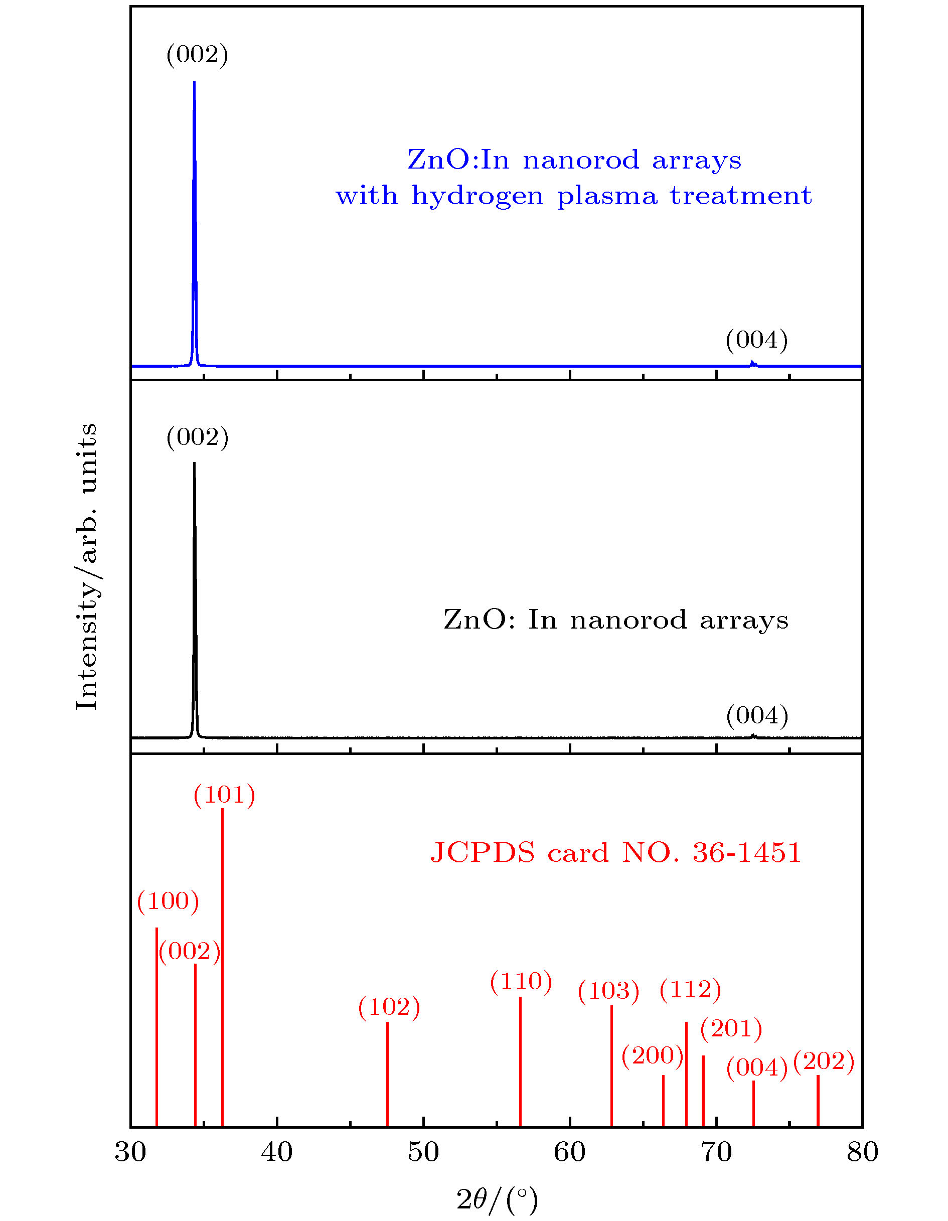
图 3 氢气氛等离子处理前后ZnO:In纳米棒阵列的XRD谱图
Figure 3. XRD patterns of the ZnO:In nanorod arrays before and after hydrogen plasma treatment.
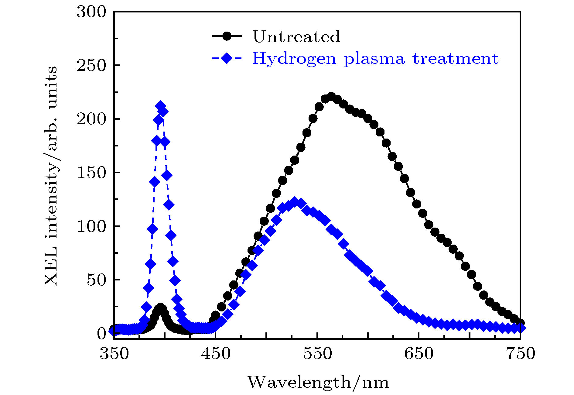
图 4 氢气氛等离子处理前后ZnO:In纳米棒阵列的XEL光谱
Figure 4. XEL spectra of the ZnO:In nanorod arrays before and after hydrogen plasma treatment.
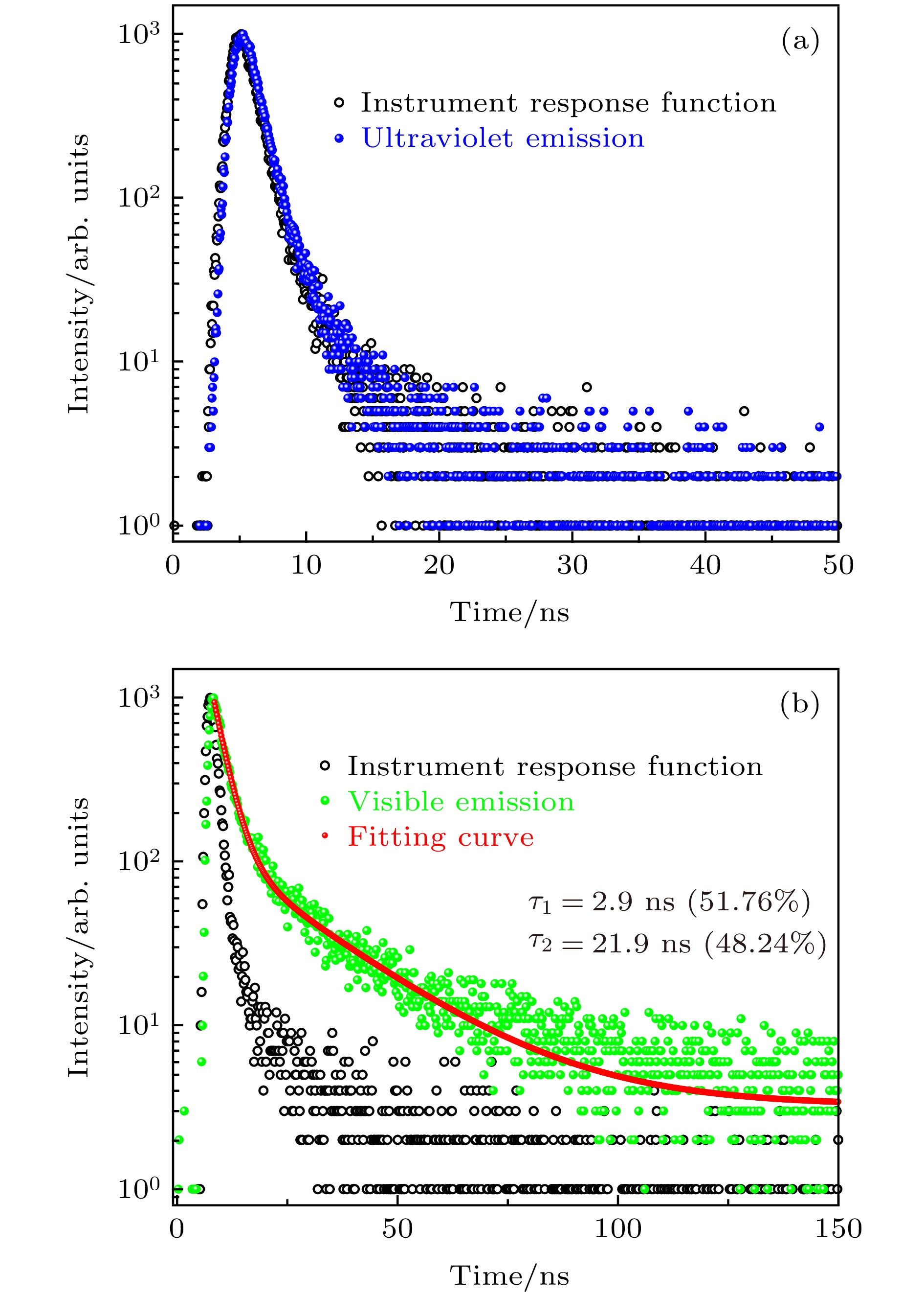
图 5 (a) ZnO:In纳米棒阵列的紫外发光衰减时间曲线(λex = 325 nm, λem = 395 nm); (b)可见发光衰减时间曲线(λex = 325 nm, λem = 530 nm)
Figure 5. The fluorescence decay curves of (a) ultravioletemission (λex = 325 nm, λem = 395 nm) and (b) visible emission (λex = 325 nm, λem = 530 nm) for the ZnO:In nanorod arrays.
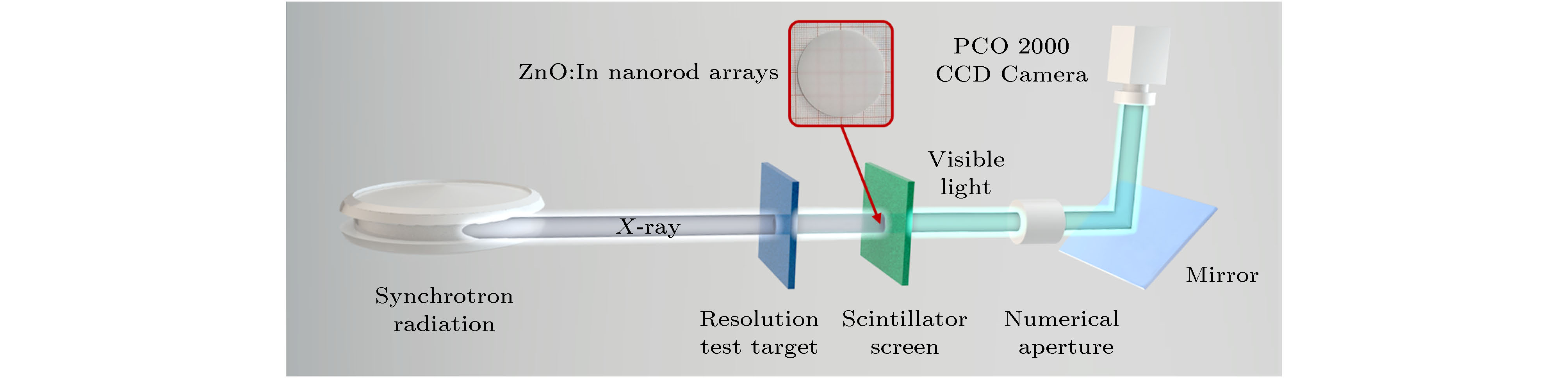
图 6 上海同步辐射光源BL13 W1线站的X射线成像测量设备示意图
Figure 6. Schematic diagram of the synchrotron radiation X-ray imaging measurement setup at BL13 W1, SSRF.
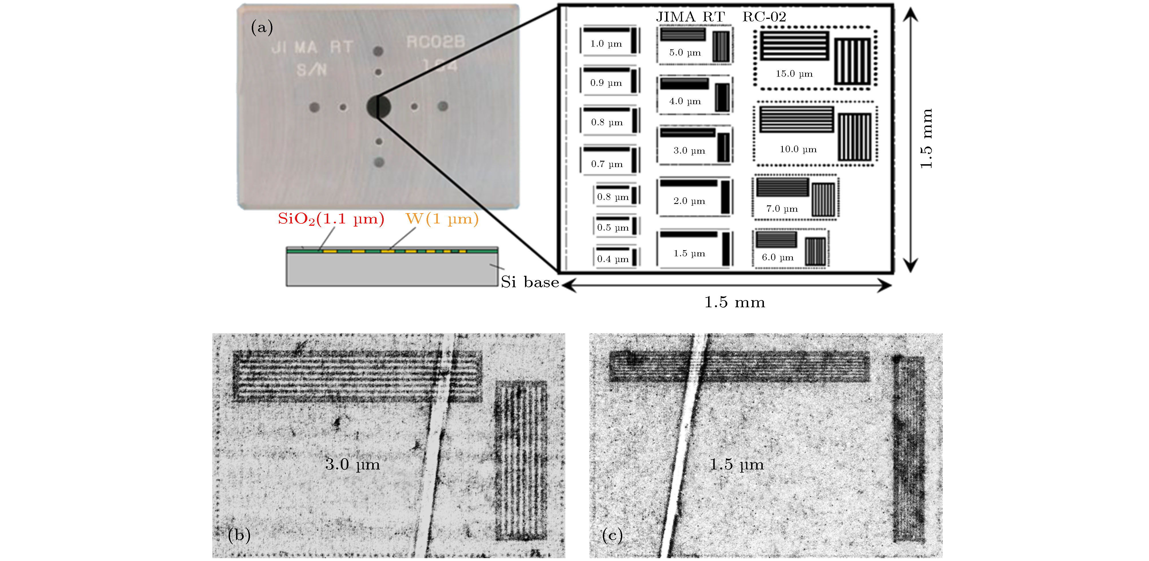
图 7 (a) JIMA RT RC-02型微米分辨率板实物图, 内部结构图示意图和微米分辨图案; 基于ZnO:In纳米棒阵列做闪烁转换屏的(b) 3 μm和(c) 1.5 μm的X射线成像图
Figure 7. (a) Physical, Schematic diagram of internal structure and Micron-resolved pattern of JIMA RT-02 micro-resolution plates; the X-ray images of (b) 3 μm and (c) 1.5 μm basedonZnO:In nanorod arrays as the scintillation screen.
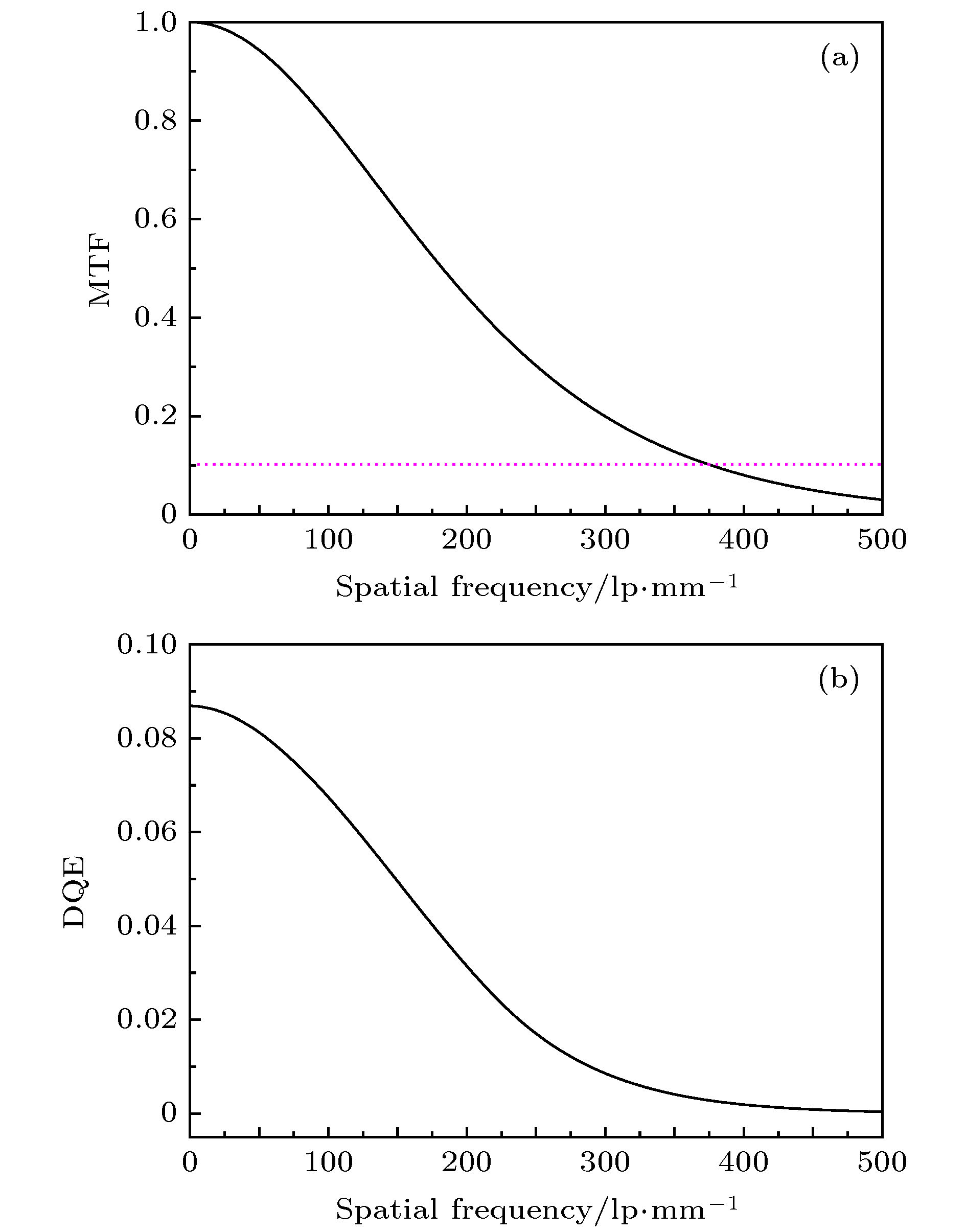
图 8 ZnO:In纳米棒阵列的X射线成像系统的(a) MTF曲线和(b) DQE曲线
Figure 8. (a) MTF and (b) DQE curves of the X-ray imaging system with ZnO:In nanorod arrays.
-
[1] Yanagida T 2018 Proc. Jpn. Acad., Ser. B 94 75
Google Scholar
[2] Dujardin C, Auffray E, Bourret-Courchesne E, Dorenbos P, Lecoq P, Nikl M, Vasil'ev A N, Yoshikawa A, Zhu R Y 2018 IEEE Trans. Nucl. Sci. 65 1977
Google Scholar
[3] Nikl M 2006 Meas. Sci. Technol. 17 R37
Google Scholar
[4] Barnes, C W, Fernández, J C, Hartsfield, T M, Sandberg, R L, Sheffield, R L, Tapia, J P, Wang, Z 2018 AIP Conf. Proc. 1979 160003
Google Scholar
[5] Turk G, Reverdin C, Gontier D, Darbon S, Dujardin C, Ledoux G, Hamel M, Simic V, Normand S 2010 Rev. Sci. Instrum. 81 10E509
Google Scholar
[6] Atanov N, Baranov V, Budagov J, Cervelli F, Colao F, Cordelli M, Corradi G, Davydov Y I, Falco S D, Diociaiuti E, Donati S, Donghia R, Echenard B, Giovannella S, Glagolev V, Grancagnolo F, Happacher F, Hitlin D G, Martini M, Miscetti S, Miyashita T, Morescalchi L, Murat P, Pedreschi E, Pezzullo G, Porter F, Raffaelli F, Ricci M, Saputi A, Sarra I, Spinella F, Tassielli G, Tereshchenko V, Usubov Z, Zhu R Y 2018 J. Instrum. 13 C02037
Google Scholar
[7] Zhu R Y 2019 J. Phys. Conf. Ser. 1162 012022
Google Scholar
[8] Hu C, Zhang L, Zhu RY, Chen A, Wang Z, Ying L, Yu Z 2018 IEEE Trans. Nucl. Sci. 65 2097
Google Scholar
[9] Simpson P J, Tjossem R, Hunt A W, Lynn K G, Munné V 2003 Nucl. Instrum. Methods Phys. Res., Sect. A 505 82
Google Scholar
[10] Chen L, Ruan J, Xu M, He S, Hu J, Zhang Z, Liu J, Ouyang X 2019 Nucl. Instrum. Methods Phys. Res., Sect. A 933 71
Google Scholar
[11] Grigorjeva L, Grube J, Bite I, Zolotarjovs A, Smits K, Millers D, Rodnyi P, Chernenko K 2019 Radiat. Meas. 123 69
Google Scholar
[12] 邱志澈, 顾牡, 刘小林, 刘波, 黄世明, 倪晨 2016 光谱学与光谱分析 36 336
Google Scholar
Qiu Z C, Gu M, Liu X L, Liu B, Huang S M, Ni C 2016 Spectrosc. Spect. Anal. 36 336
Google Scholar
[13] Liu S, Gu M, Chen H, Sun Z, Liu X, Liu B, Huang S, Zhang J 2018 Nucl. Instrum. Methods Phys. Res., Sect. A 903 18
Google Scholar
[14] Li Q, Liu X, Gu M, Li F, Zhang J, Wu Q, Huang S, Liu S 2018 Appl. Surf. Sci. 433 815
Google Scholar
[15] Kobayashi M, Komori J, Shimidzu K, Izaki M, Uesugi K, Takeuchi A, Suzuki Y 2015 Appl. Phys. Lett. 106 081909
Google Scholar
[16] Izaki M, Kobayashi M, Shinagawa T, Koyama T, Uesugi K, Takeuchi A 2017 Phys. Status Solidi A 214 1700285
Google Scholar
[17] Li Q, Hao S, An R, Wang M, Sun Z, Wu Q, Gu M, Zhao J, Liu X, Zhang Z 2019 Appl. Surf. Sci. 493 1299
Google Scholar
[18] 倪晨, 顾牡, 王迪, 曹顿华, 刘小林, 黄世明 2009 光谱学与光谱分析 29 2291
Google Scholar
Ni C, Gu M, Wang D, Cao D H, Liu X L, Huang S M 2009 Spectrosc. Spect. Anal. 29 2291
Google Scholar
[19] Özgür Ü, Alivov Y I, Liu C, Teke A, Reshchikov M A, Doğan S, Avrutin V, Cho S J, Morkoç H 2005 J. Appl. Phys. 98 041301
Google Scholar
[20] Li Q, Liu X, Gu M, Huang S, Ni C, Liu B, Hu Y, Sun S, Zhang Z 2016 IEEE Trans. Nucl. Sci. 63 471
Google Scholar
[21] Li Q, Liu X, Gu M, Huang S, Zhang J, Ni C, Liu B, Hu Y, Wu Q, Zhao S 2016 Superlattices Microstruct. 98 351
Google Scholar
[22] Hofmann D M, Hofstaetter A, Leiter F, Zhou H, Henecker F, Meyer B K, Orlinskii S B, Schmidt J, Baranov P G 2002 Phys. Rev. Lett. 88 045504
Google Scholar
[23] Lavrov E V, Herklotz F, Weber J 2009 Phys. Rev. B 79 165210
Google Scholar
[24] Kano M, Wakamiya A, Yamanoi K, Sakai K, Takeda K, Cadatal-Raduban M, Nakazato T, Shimizu T, Sarukura N, Fukuda T 2012 IEEE Trans. Nucl. Sci. 59 2290
Google Scholar
[25] Yamanoi K, Sakai K, Cadatal-Raduban M, Nakazato T, Shimizu T, Sarukura N, Kano M, Wakamiya A, Fukuda T, Nagasono M, Togashi T, Matsubara S, Tono K, Higashiya A, Yabashi M, Kimura H, Ohashi H, Ishikawa T 2012 IEEE Trans. Nucl. Sci. 59 2298
Google Scholar
[26] 郭智敏, 倪培君 2010 兵器材料科学与工程 33 113
Google Scholar
Guo Z M, Ni P J, 2010 Ordnance Mater. Sci. Eng. 33 113
Google Scholar
[27] Chen H, Gu M, Sun Z, Liu X, Liu B, Zhang J, Huang S, Ni C 2019 Opt. Express 27 14871
Google Scholar
[28] Sowa K M, Last A, Korecki P 2017 Sci. Rep. 7 44944
Google Scholar
[29] Samei E, Flynn M J, Reimann D A 1998 Med. Phys. 25 102
Google Scholar
[30] Michail C, Valais I, Martini N, Koukou V, Kalyvas N, Bakas A, Kandarakis I, Fountos G 2016 Radiat. Meas. 94 8
Google Scholar


 DownLoad:


Catalog
Metrics
- Abstract views: 11520
- PDF Downloads: 283
- Cited By: 0
