-
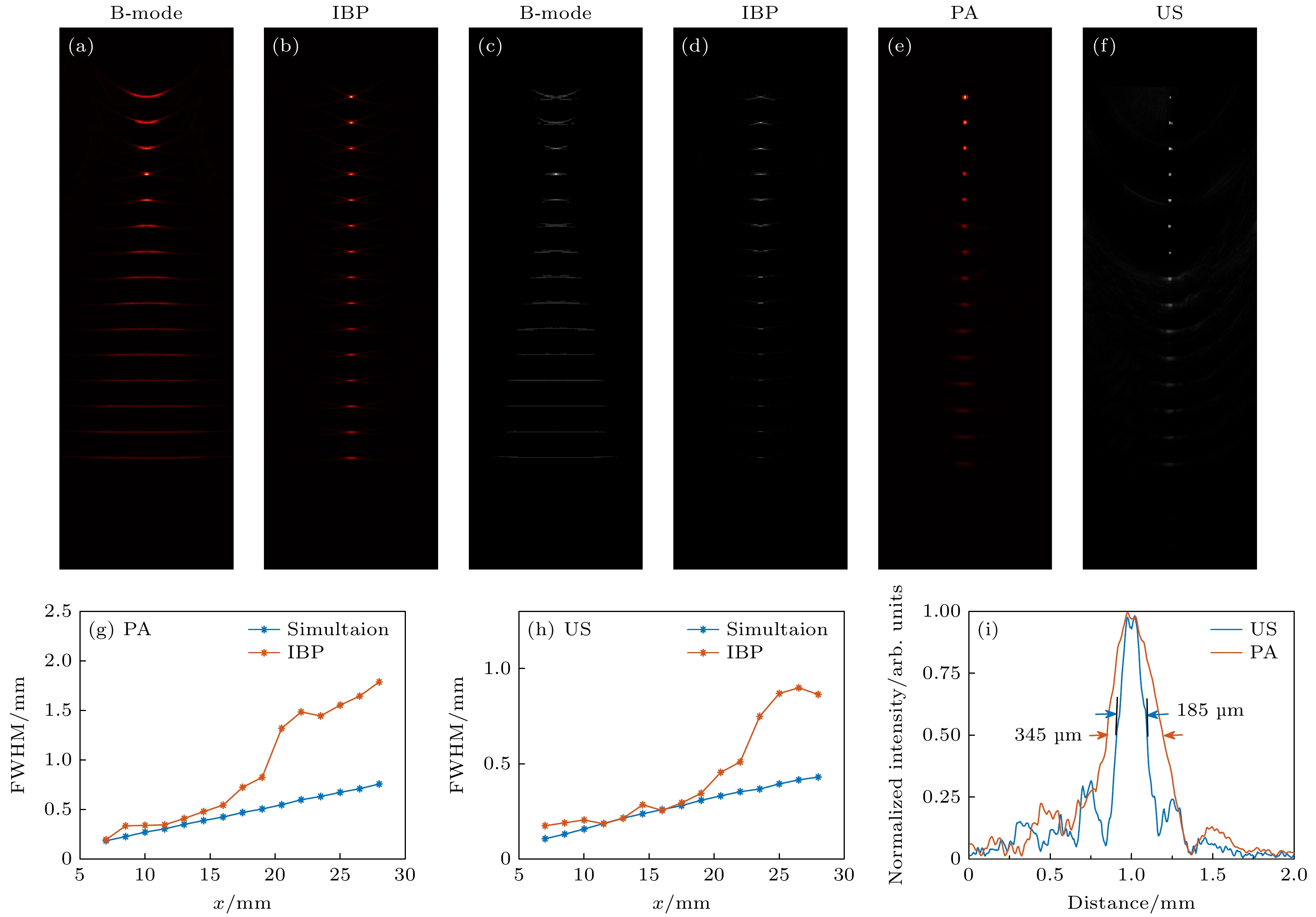
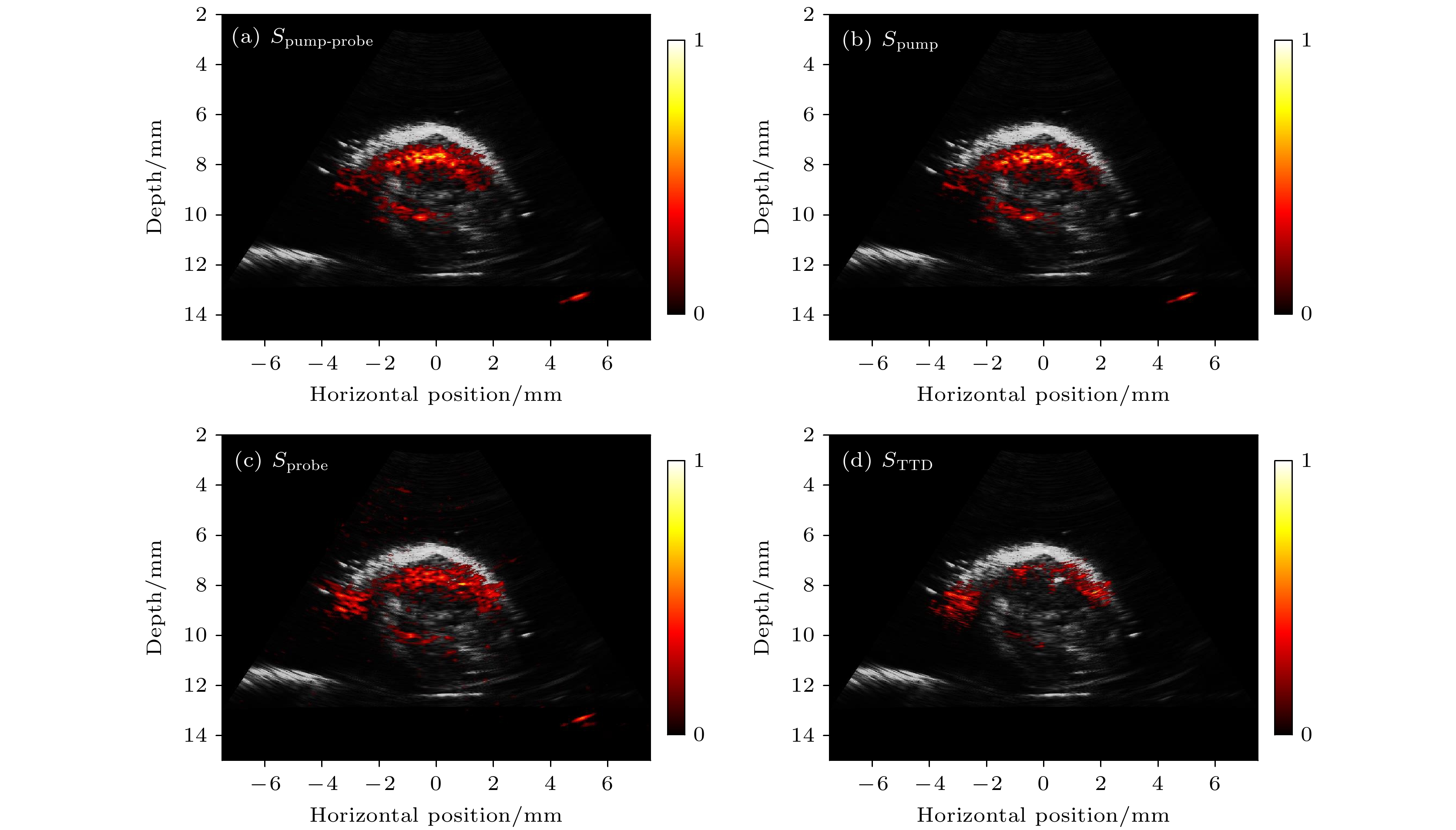
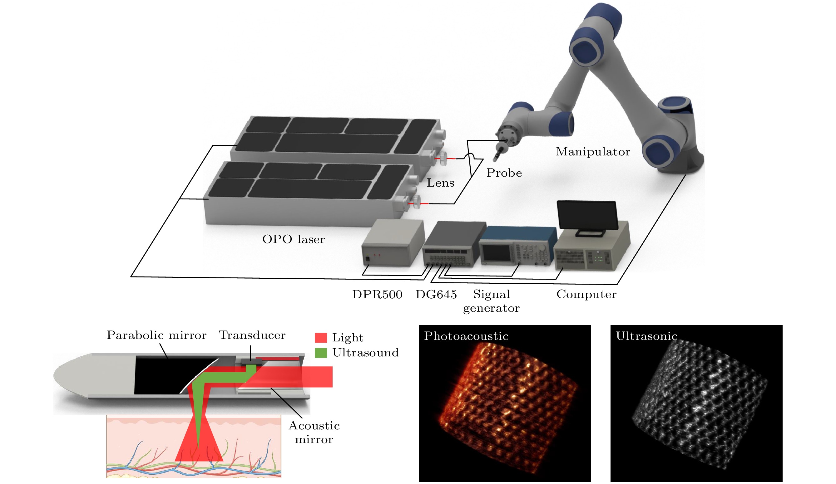
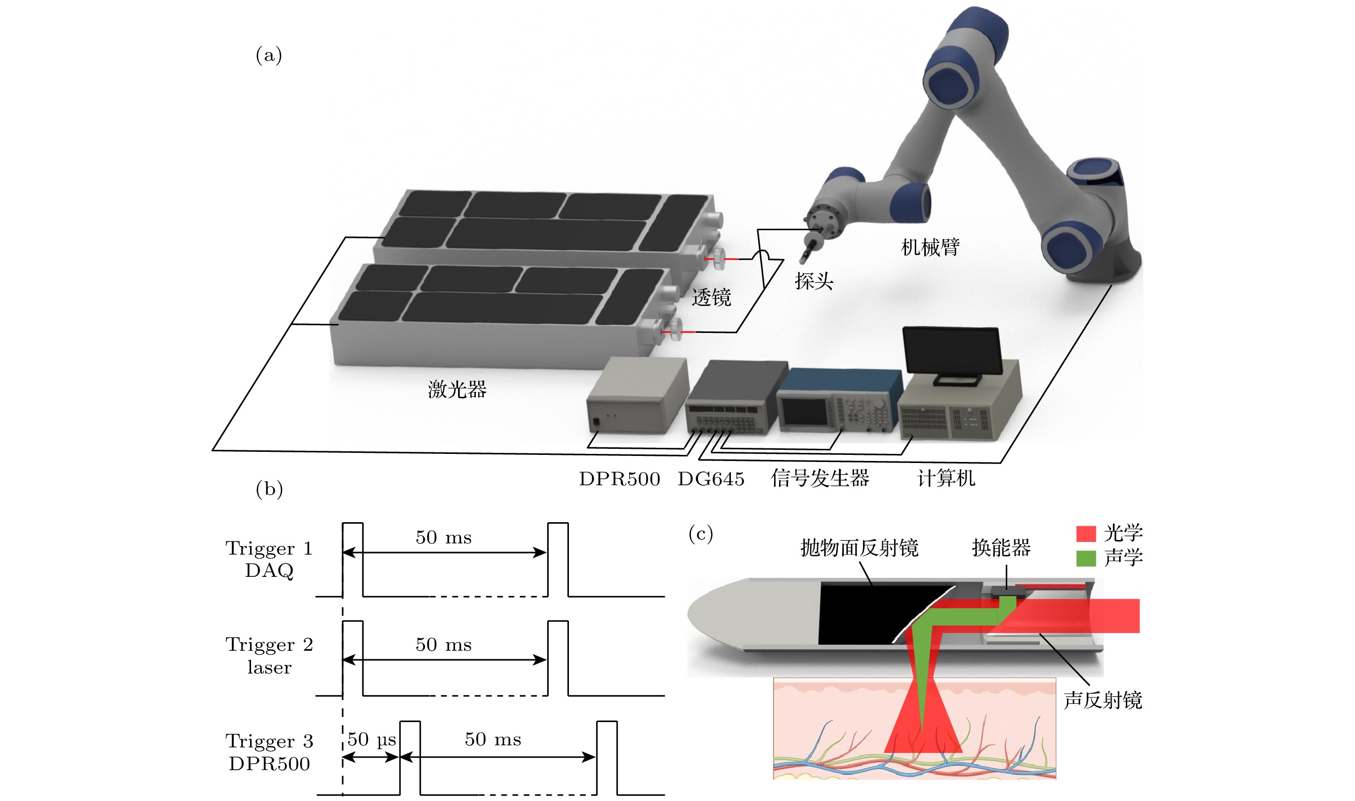
结直肠癌是全球癌症死亡的主要原因之一, 常用的光学或超声消化道内镜仍然存在穿透深度低、对比度差、功能/分子成像能力不足等问题. 本文介绍了一种小型化的手持式光声/超声双模态内窥探头, 旨在克服现有技术在穿透深度和分子成像能力方面的限制. 实验结果表明, 该探头在组织12 mm深度下分别达到了345 μm的光声横向分辨率和185 μm的超声横向分辨率, 并具有良好的对复杂结构目标成像的能力. 本文还利用泵浦探测技术排除了血液背景的干扰, 实现了肿瘤深层组织内亚甲基蓝分子的高特异性成像. 这种小型化手持式光声/超声双模态内窥探头兼具大成像深度、高空间分辨和高特异性分子成像的特点, 有望成为结直肠癌等消化道肿瘤诊断的重要工具, 为早期诊断和治疗监测提供强有力的支持.Colorectal cancer is one of the leading causes of cancer-related deaths worldwide. Traditional gastrointestinal endoscopes for colorectal cancer mainly rely on optical endoscope and ultrasound endoscope. Owing to significant light scattering in tissues the optical endoscope is limited to superficial tissue imaging, while the ultrasound endoscope, despite deeper penetration, provides limited molecular imaging capabilities. In this work, we build a miniaturized handheld photoacoustic/ultrasound dual-modality endoscopic probe to address these problems. It has a small size of 8 mm, and presents the dual advantages of high penetration depth and superior molecular imaging capability, marking a significant advancement over traditional methods. Results show that this probe achieves a high lateral resolution of 345 μm for photoacoustic imaging and 185 μm for ultrasound imaging at a depth of 12 mm within tissues. It also exhibits the ability to effectively image complex structural targets, as demonstrated by the imaging of a phantom with an embedded metal mesh. Furthermore, the probe adopts an innovative pump-probe method, which effectively mitigates interference from blood and other background tissues, thereby achieving high-specificity photoacoustic molecular imaging. This ability is first confirmed by imaging the distribution of methylene blue (MB) in a phantom, and then by observing the distribution of MB in the depth of tumor in mice. This handheld photoacoustic/ultrasound endoscopic probe has the advantages of small size, high penetration depth, high spatial resolution, and superior molecular imaging ability, and is expected to become an important diagnostic tool for colorectal cancer and other gastrointestinal cancer. This study can provide strong support for early diagnosis and treatment monitoring, potentially revolutionizing the detection and management of these diseases.
-
Keywords:
- photoacoustic endoscopy /
- multimode imaging /
- pump-probe /
- molecular imaging
[1] Bray F, Laversanne M, Sung H, Ferlay J, Siegel R L, Soerjomataram I, Jemal A 2024 CA-Cancer J. Clin. 74 229263
 Google Scholar
[2] Gora M J, Suter M J, Tearney G J, Li X 2017 Biomed. Opt. Express 8 2405
 Google Scholar
[3] Rex D K, Boland C R, Dominitz J A, Giardiello F M, Johnson D A, Kaltenbach T, Levin T R, Lieberman D, Robertson D J 2017 Am. J. Gastroenterol. 112 10161030
 Google Scholar
[4] Gora M J, Simmons L H, Quénéhervé L, Grant C N, Carruth R W, Lu W, Tiernan A, Dong J, Walker-Corkery B, Soomro A, Rosenberg M, Metlay J P, Tearney G J 2016 J. Biomed. Opt. 21 104001
 Google Scholar
[5] Pahlevaninezhad H, Khorasaninejad M, Huang Y W, Shi Z, Hariri L P, Adams D C, Ding V, Zhu A, Qiu C W, Capasso F, Suter M J 2018 Nat. Photonics 12 540547
 Google Scholar
[6] Krill T, Baliss M, Roark R, Sydor M, Samuel R, Zaibaq J, Guturu P, Parupudi S 2019 J. Thorac. Dis. 11 16021609
 Google Scholar
[7] Wang X, Seetohul V, Chen R, Zhang Z, Qian M, Shi Z, Yang G, Mu P, Wang C, Huang Z, Zhou Q, Zheng H, Cochran S, Qiu W 2017 IEEE Trans. Med. Imaging 36 19221929
 Google Scholar
[8] Lin L, Wang L V 2022 Nat. Rev. Clin. Oncol. 19 365384
 Google Scholar
[9] Attia A B E, Balasundaram G, Moothanchery M, Dinish U S, Bi R, Ntziachristos V, Olivo M 2019 Photoacoustics 16 100144
 Google Scholar
[10] Li Y, Lu G X, Zhou Q F, Chen Z P 2021 Photonics 8 281
 Google Scholar
[11] Wang L V, Hu S 2012 Science 335 14581462
 Google Scholar
[12] Guo H, Li Y, Qi W, Xi L 2020 J. Biophotonics 13 e202000217
 Google Scholar
[13] Xia J, Yao J, Wang L V 2014 Progr. Electromagnet. Res. 147 122
 Google Scholar
[14] Yang J M, Favazza C, Chen R, Yao J, Cai X, Maslov K, Zhou Q, Shung K K, Wang L V 2012 Nat. Med. 18 12971302
 Google Scholar
[15] Leng X, Chapman W, Rao B, Nandy S, Chen R, Rais R, Gonzalez I, Zhou Q, Chatterjee D, Mutch M, Zhu Q 2018 Biomed. Opt. Express 9 51595172
 Google Scholar
[16] Vu T, Razansky D, Yao J 2019 J. Opt. 21 103001
 Google Scholar
[17] Wang L V 2009 Nat. Photonics 3 503509
 Google Scholar
[18] He H, Wissmeyer G, Ovsepian S V., Buehler A, Ntziachristos V 2016 Opt. Lett. 41 27082710
 Google Scholar
[19] Xiao J, Jiang J, Zhang J, Wang Y, Wang B 2022 Opt. Express 30 35014
 Google Scholar
[20] Tan J W Y, Lee C H, Kopelman R, Wang X 2018 Sci. Rep. 8 9290
 Google Scholar
-
图 2 光声/超声分辨率测试实验结果 (a)—(d) 传统B-mode重建算法和IBP重建算法仿真结果对比; (e)—(f) 不同深度下点目标重建的光声/超声图像; (g) 从图(b), (e)中提取的光声模态下不同深度的FWHM; (h) 从图(d), (f)中提取的超声模态下不同深度的FWHM; (i) 在12 mm处获得的光声/超声横向轮廓及其横向分辨率
Fig. 2. Field test experimental result: (a)–(d) Simulation comparison of traditional B-mode and IBP reconstruction algorithms; (e)–(f) the photoacoustic/ultrasound images of different depth of the point target; (g) FWHM in photoacoustic at different depth obtained from panel (b), (e); (h) FWHM in ultrasound at different depth obtained from panel (d), (f); (i) photoacoustic/ultrasound lateral profiles of the point target obtained at 12 mm.
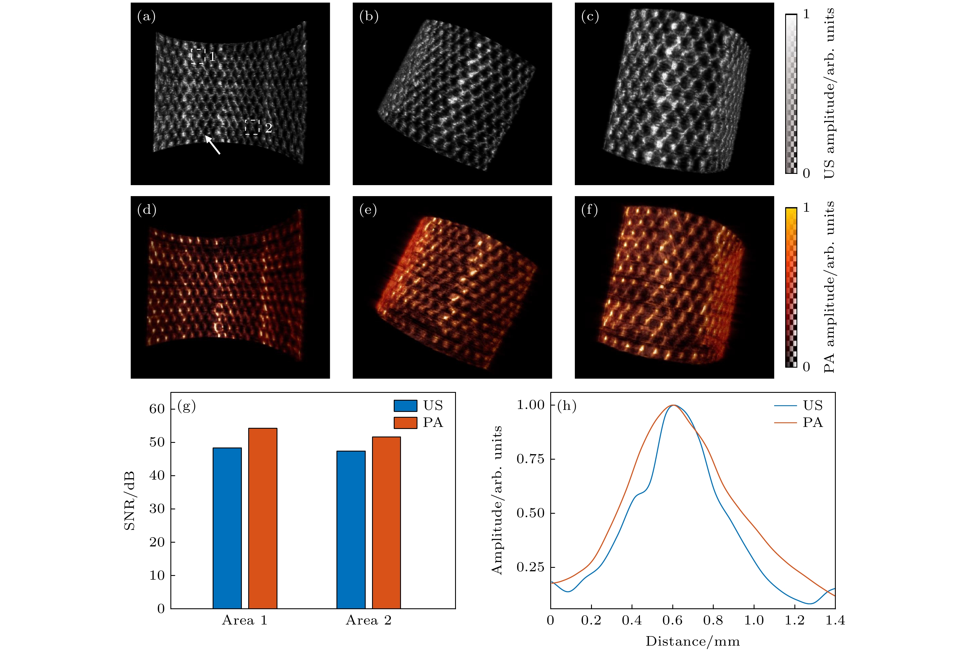
图 3 金属网格三维光声/超声成像实验结果 (a)—(c) 不同视角下金属网格的光声图像; (d)—(f) 相应的超声图像; (g) 不同区域的光声/超声图像的PSNR; (h) 在光声/超声图像中提取的金属丝轮廓信号的横向分布
Fig. 3. Experimental results of 3D photoacoustic/ultrasound imaging of a metal grid: (a)–(c) Photoacoustic images of metal grid from different perspectives; (d)–(f) corresponding ultrasound images of the same areas as in panel (a)–(c); (g) PSNR comparison of photoacoustic and ultrasound images in different regions; (h) contours of metal grid extracted from the photoacoustic/ultrasound images.
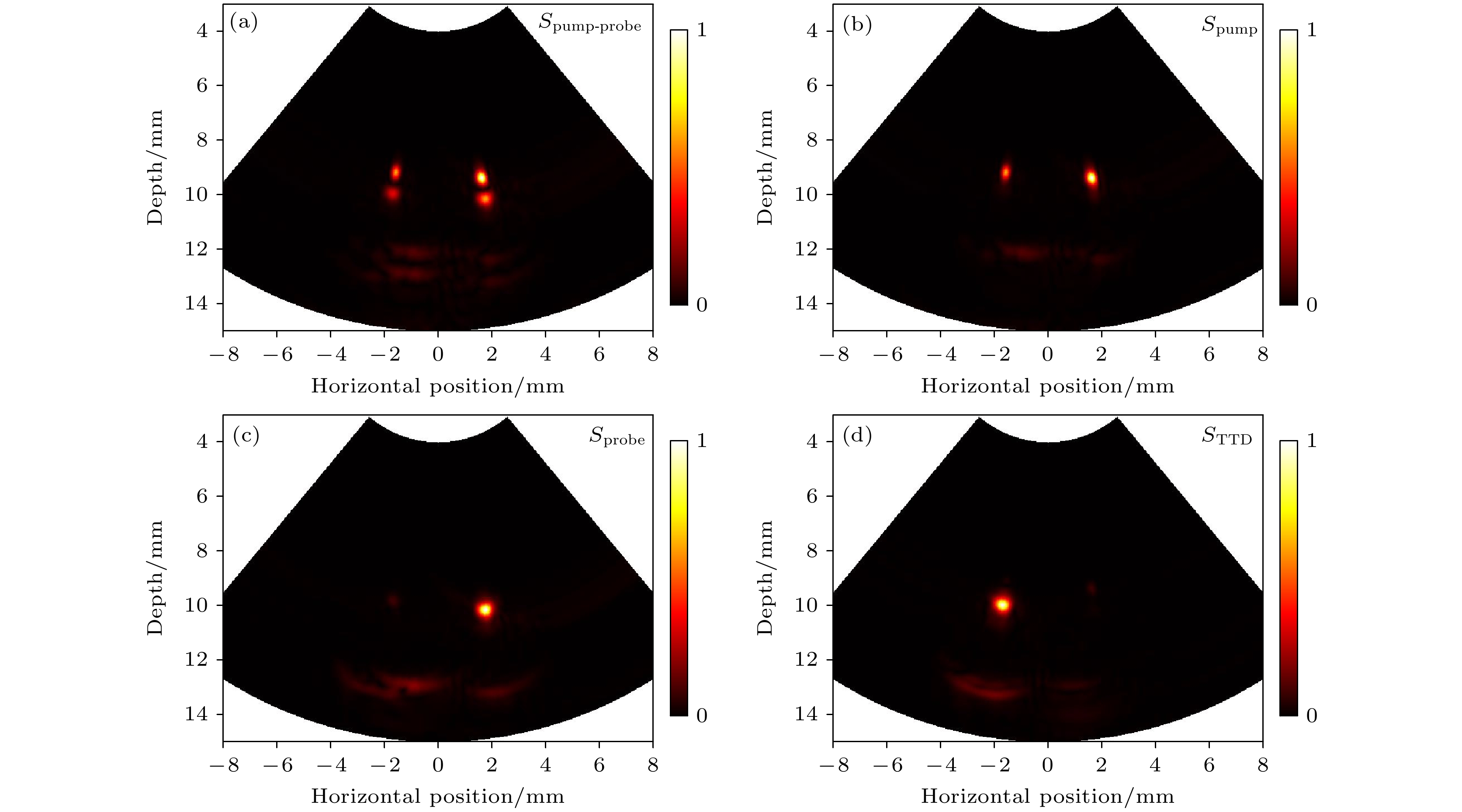
图 4 光声泵浦仿体成像实验结果 (a)—(d) 分别为Spump-probe, Spump, Sprobe, STTD重建的光声图像, 其中左侧管内为MB溶液, 右侧为牛血红蛋白溶液
Fig. 4. Pump-probe photoacoustic imaging of phantom: (a)–(d) Reconstructed images of Spump-probe, Spump, Sprobe, and STTD. In the setup, the left tube contains methylene blue solution, while the right contains bovine hemoglobin solution.
-
[1] Bray F, Laversanne M, Sung H, Ferlay J, Siegel R L, Soerjomataram I, Jemal A 2024 CA-Cancer J. Clin. 74 229263
 Google Scholar
[2] Gora M J, Suter M J, Tearney G J, Li X 2017 Biomed. Opt. Express 8 2405
 Google Scholar
[3] Rex D K, Boland C R, Dominitz J A, Giardiello F M, Johnson D A, Kaltenbach T, Levin T R, Lieberman D, Robertson D J 2017 Am. J. Gastroenterol. 112 10161030
 Google Scholar
[4] Gora M J, Simmons L H, Quénéhervé L, Grant C N, Carruth R W, Lu W, Tiernan A, Dong J, Walker-Corkery B, Soomro A, Rosenberg M, Metlay J P, Tearney G J 2016 J. Biomed. Opt. 21 104001
 Google Scholar
[5] Pahlevaninezhad H, Khorasaninejad M, Huang Y W, Shi Z, Hariri L P, Adams D C, Ding V, Zhu A, Qiu C W, Capasso F, Suter M J 2018 Nat. Photonics 12 540547
 Google Scholar
[6] Krill T, Baliss M, Roark R, Sydor M, Samuel R, Zaibaq J, Guturu P, Parupudi S 2019 J. Thorac. Dis. 11 16021609
 Google Scholar
[7] Wang X, Seetohul V, Chen R, Zhang Z, Qian M, Shi Z, Yang G, Mu P, Wang C, Huang Z, Zhou Q, Zheng H, Cochran S, Qiu W 2017 IEEE Trans. Med. Imaging 36 19221929
 Google Scholar
[8] Lin L, Wang L V 2022 Nat. Rev. Clin. Oncol. 19 365384
 Google Scholar
[9] Attia A B E, Balasundaram G, Moothanchery M, Dinish U S, Bi R, Ntziachristos V, Olivo M 2019 Photoacoustics 16 100144
 Google Scholar
[10] Li Y, Lu G X, Zhou Q F, Chen Z P 2021 Photonics 8 281
 Google Scholar
[11] Wang L V, Hu S 2012 Science 335 14581462
 Google Scholar
[12] Guo H, Li Y, Qi W, Xi L 2020 J. Biophotonics 13 e202000217
 Google Scholar
[13] Xia J, Yao J, Wang L V 2014 Progr. Electromagnet. Res. 147 122
 Google Scholar
[14] Yang J M, Favazza C, Chen R, Yao J, Cai X, Maslov K, Zhou Q, Shung K K, Wang L V 2012 Nat. Med. 18 12971302
 Google Scholar
[15] Leng X, Chapman W, Rao B, Nandy S, Chen R, Rais R, Gonzalez I, Zhou Q, Chatterjee D, Mutch M, Zhu Q 2018 Biomed. Opt. Express 9 51595172
 Google Scholar
[16] Vu T, Razansky D, Yao J 2019 J. Opt. 21 103001
 Google Scholar
[17] Wang L V 2009 Nat. Photonics 3 503509
 Google Scholar
[18] He H, Wissmeyer G, Ovsepian S V., Buehler A, Ntziachristos V 2016 Opt. Lett. 41 27082710
 Google Scholar
[19] Xiao J, Jiang J, Zhang J, Wang Y, Wang B 2022 Opt. Express 30 35014
 Google Scholar
[20] Tan J W Y, Lee C H, Kopelman R, Wang X 2018 Sci. Rep. 8 9290
 Google Scholar
计量
- 文章访问数: 4984
- PDF下载量: 102
- 被引次数: 0


 下载:
下载:
