-
21世纪以来, 扫描探针显微镜(scanning probe microscope, SPM)在微纳尺度形貌表征、物性测量及微纳加工等领域发挥着越来越重要的作用. 为了使扫描探针显微镜获得更稳定的运行环境、更高的能量分辨率, 人们研发了具备超高真空(ultra high vacuum, UHV)和低温(low temperature, LT)环境的SPM系统(UHV-LT-SPM). 目前, 大多数的UHV-LT-SPM系统通过向连续流式低温恒温器或低温杜瓦中输送液态氦-4(4He), 使SPM的温度达到约4.2 K. 然而由于4He元素在自然界中含量低且因需求日益增长, 导致液氦价格急剧飙升, 严重影响到了4He相关低温设备的正常运行. 为应对上述问题, 干式制冷技术成为新一代低温技术的发展方向. 在此背景下, 将干式制冷技术与扫描探针显微镜相结合, 搭建干式低温扫描探针显微镜, 成为了目前扫描探针仪器领域的研究重点之一. 本文主要从扫描探针显微镜系统设计、降温设计、减振方法以及其设备性能等方面, 介绍目前已经报道的几种干式LT-SPM系统. 最后总结了干式LT-SPM系统目前所遇见的问题和挑战, 探讨了该技术未来的发展方向.
Since the beginning of the 21st century, scanning probe microscopy (SPM) has played an increasingly important role in investigating the micro- and nanoscale surface characterization, physical property measurement, and micro/nano fabrication. To provide a more stable operating environment and higher energy resolution for SPM, researchers have developed low-temperature scanning probe microscopy (LT-SPM) systems that operate under the conditions of ultra-high vacuum and low temperature. Currently, most of LT-SPM systems have achieved temperatures around 4.2 K by supplying liquid helium-4 (4He) to continuous flow cryostats or low-temperature Dewars. However, due to the low natural abundance of 4He and its increasing demand, the significant increase in the price of liquid helium has seriously affected the normal operation of 4He-based low temperature equipment. To solve this problem, dry (cryogen-free) refrigeration technology has emerged as a promising alternative to the next-generation low-temperature systems. In this context, the integration of dry refrigeration technology with SPM to construct Dry-LT-SPM systems has become a key research focus in the field of scanning probe instruments. This paper mainly discusses several reported closed-cycle Dry-LT-SPM systems, focusing on aspects such as system design, refrigeration schemes, vibration reduction methods, and overall performance. Finally, this paper summarizes the current challenges and problems faced by Dry-LT-SPM systems and explores potential future developments in this field. -
Keywords:
- cryogen-free /
- low temperature /
- scanning probe microscopy /
- vibration
更正: 基于干式制冷的低温扫描探针显微镜研究进展 [ 2024, 73(22): 228701]
黄远志, 杨传浩, 何颂平, 马瑞松, 郇庆. 更正: 基于干式制冷的低温扫描探针显微镜研究进展. . doi: 10.7498/aps.73.249901
[1] Wu Z B, Gao Z Y, Chen X Y, et al. 2018 Rev. Sci. Instrum. 89 113705
 Google Scholar
[2] Bian K, Gerber C, Heinrich A J, Müller D J, Scheuring S, Jiang Y 2021 Nat. Rev. Method. Prime. 1 36
 Google Scholar
[3] Pettinger B, Schambach P, Villagómez C J, Scott N 2012 Annu. Rev. Phys. Chem. 63 379
 Google Scholar
[4] Watkins N J, Long J P, Kafafi Z H, Mäkinen A J 2007 Rev. Sci. Instrum. 78 053707
 Google Scholar
[5] Grafström S 2002 J. Appl. Phys. 91 1717
 Google Scholar
[6] Flores S M, Toca-Herrera J L 2009 Nanoscale 1 40
 Google Scholar
[7] Bharat B 2004 Handbook of Nanotechnology (Springer
[8] Baykara M Z, Morgenstern M, Schwarz A, Schwarz U D 2017 Handbook of Nanotechnology (Berlin: Springer) pp769–808
[9] Behler S, Rose M K, Dunphy J C, Ogletree D F, Salmeron M, Chapelier C 1997 Rev. Sci. Instrum. 68 2479
 Google Scholar
[10] Stipe B C, Rezaei M A, Ho W 1999 Rev. Sci. Instrum. 70 137
 Google Scholar
[11] Meyer G 1996 Rev. Sci. Instrum. 67 2960
 Google Scholar
[12] Elrod S A, Lozanne A L D, Quate C F 1984 Applied Physics Letters 45 1240
 Google Scholar
[13] He G, Wei Z X, Feng Z P, Yu X D, Zhu B Y, Liu L, Jin K, Yuan J, Huan Q 2020 Rev. Sci. Instrum. 91 013904
 Google Scholar
[14] Chaudhary S, Panda J J, Mundlia S, Mathimalar S, Ahmedof A, Raman K V 2021 Rev. Sci. Instrum. 92 023906
 Google Scholar
[15] Zhao Z, Wang C 2019 Engineering and Technologies: Principles and Applications of Cryogen-Free Systems (CRC Press
[16] Wong D, Jeon S, Nuckolls K P, Oh M, Kingsley S C J, Yazdani A 2020 Rev. Sci. Instrum. 91 023703
 Google Scholar
[17] Hackley J D, Kislitsyn D A, Beaman D K, Ulrich S, Nazin G V 2014 Rev. Sci. Instrum. 85 103704
 Google Scholar
[18] Zhang S, Huang D, Wu S W 2016 Rev. Sci. Instrum. 87 063701
 Google Scholar
[19] Kasai J, Koyama T, Yokota M, Iwaya K 2022 Rev. Sci. Instrum. 93 043711
 Google Scholar
[20] Huang H M, Shuai M M, Yang Y L, Song R, Liao Y H, Yin L M, Shen J 2022 Rev. Sci. Instrum. 93 073703
 Google Scholar
[21] Meng W J, Wang J H, Hou Y B, et al. 2019 Ultramicroscopy 205 20
 Google Scholar
[22] Coe A M, Li G H, Andrei E Y 2024 Rev. Sci. Instrum. 95 083702
 Google Scholar
[23] Ma R S, Li H, Shi C S, et al. 2023 Rev. Sci. Instrum. 94 093701
 Google Scholar
-
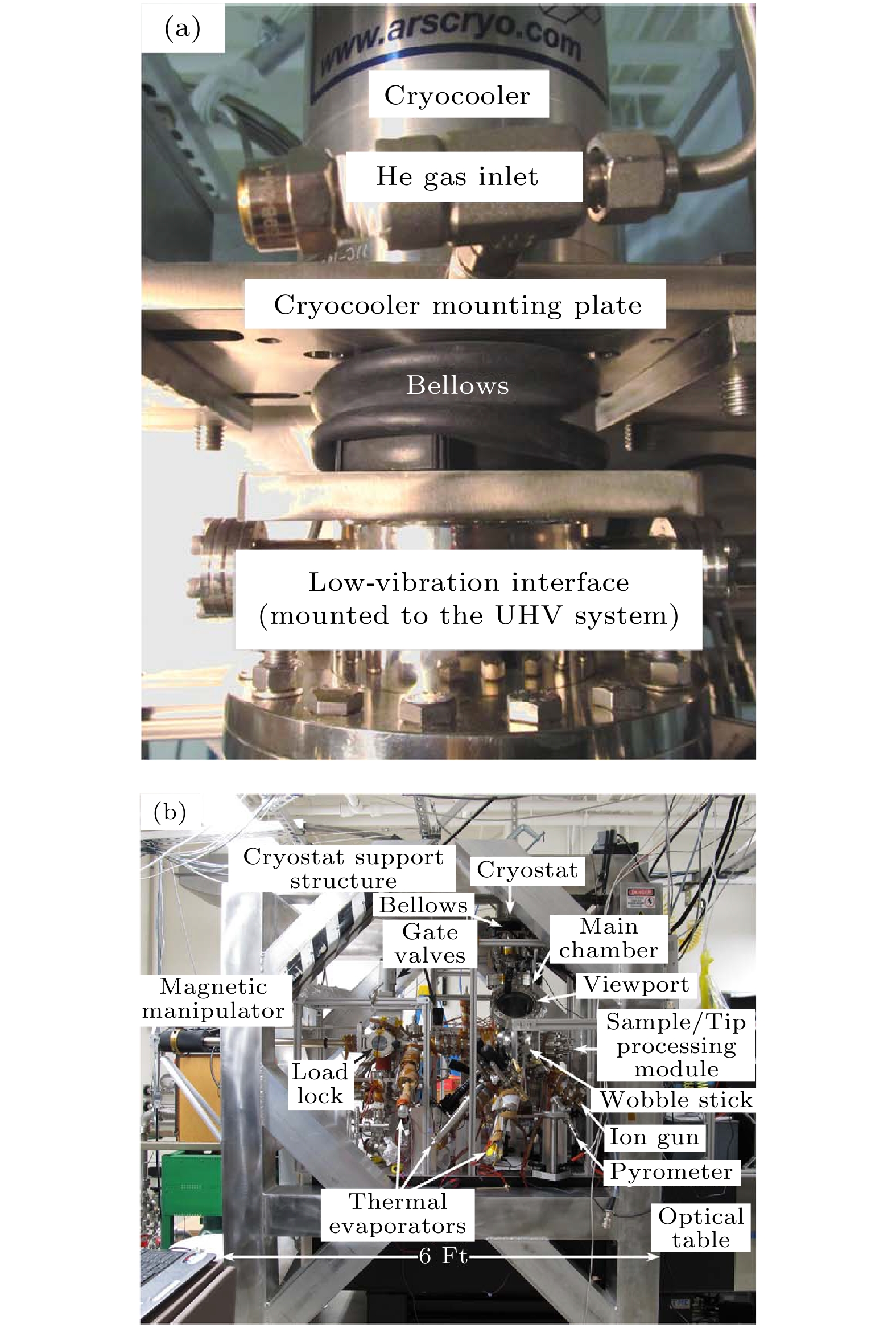
图 2 干式制冷部分实物图[17] (a) GM机与SPM部分的连接部分照片, 该方案采用橡胶波纹管连接制冷机和STM上方的二级热交换器; (b) LT-STM系统部分系统实物图, 低温恒温器安装在LT-STM系统上方的刚性支架上, 刚性支架与LT-STM系统没有刚性接触
Fig. 2. Photograph of Dry refrigeration[17]: (a) Photo of the connection part between the GM cryocooler cold head and the SPM part, this solution uses rubber bellows to connect the refrigerator and cold finger above the STM; (b) photo of the LT-STM system, the cryostat is mounted on a rigid support above the LT-STM system, the rigid support has no rigid contact with the LT-STM system.
图 5 (a) Dry-LT-STM系统示意图[18], 制冷机(蓝色)安装在刚性支架上, LT-STM系统放在含有气腿(橙色)的实验台上, 两者通过橡胶波纹管与LT-STM系统连接; (b)闭循环制冷部分示意图, 氦气在制冷机(紫色)和热交换器界面(青色)之间, 氦气由两级橡胶管(黑色)密封; (c) STM扫描探头示意图, 激光通过两种透镜聚焦在STM上, 并通过雪崩光电二极管或光谱仪从STM收集光信号
Fig. 5. (a) Schematic diagram of the Dry-LT-STM system[18], the cryocooler (blue) is mounted on a rigid frame and the LT-STM system is placed on the vibration isolation table containing the gas legs (orange), both of which are connected to the LT-STM system via rubber bellows; (b) schematic diagram of the closed-cycle refrigeration section, helium gas is filled between the cryocooler (purple) and stage interfaces (cyan), the helium is sealed by two-stage rubber bellow (black); (c) schematic of the STM scanning head. The laser is focused on the STM by two lenses and the optical signal is collected from the STM by means of a APD or a spectrometer.
图 7 (a)日本UNISOKU公司的LT-STM系统示意图; (b) PT制冷机和SPM扫描探头的示意图, PT制冷机两级冷台、PTFE波纹管和低温恒温器围成的区域充满氦气, 扫描腔则保持在超高真空状态[19]
Fig. 7. (a) Schematic diagram of the LT-STM system of Japanese UNISOKU company; (b) schematic of PT refrigerator (cryocooler) and SPM scanner, the area enclosed by the cooling stages, PTFE bellows and cryostat is filled with helium, while the SPM chamber is maintained in ultra-high vacuum condition[19].
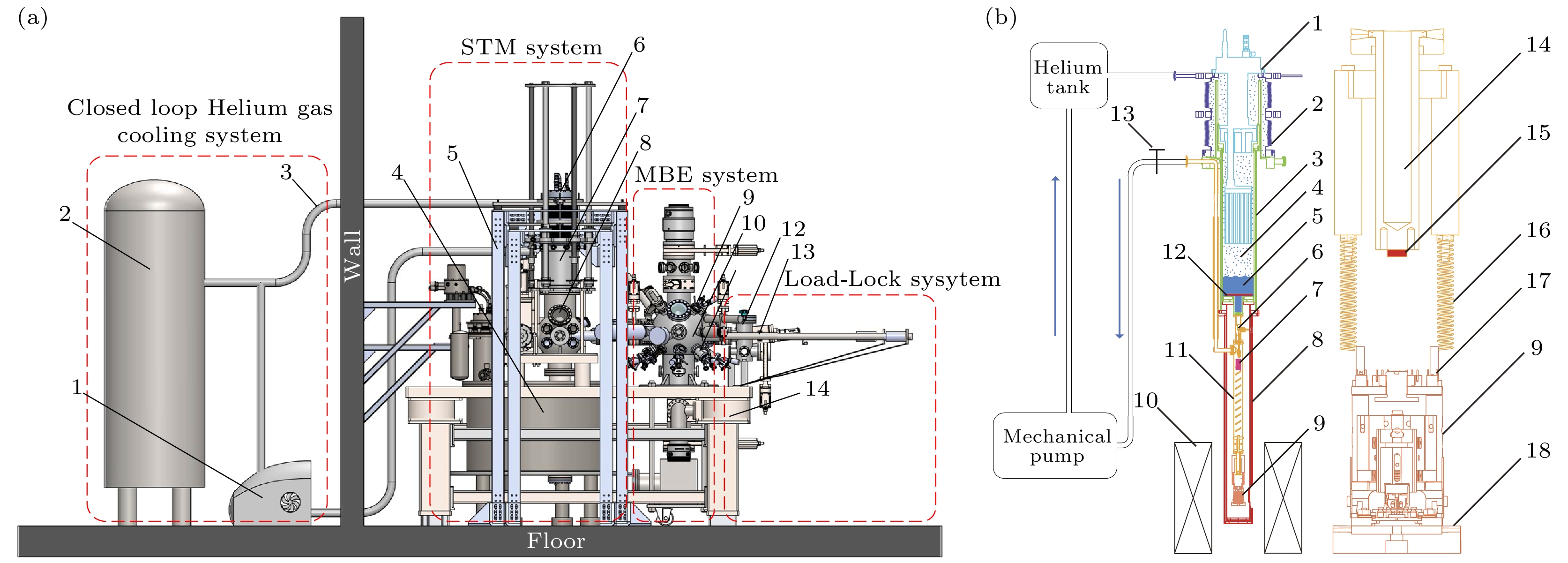
图 8 (a)搭建Dry-LT-SPM设备整体系统设计示意图[20]. 1-涡旋泵, 2-氦气罐, 3-氦气管道, 4-干式制冷超导磁体, 5-刚性支架, 6-PT制冷机, 7-低温恒温器, 8-STM腔体, 9-氩离子源, 10-MBE腔体, 11-MBE蒸发源, 12-快速进样腔, 13-传输杆, 14-主动减振平台. (b)制冷系统(左)和扫描探头(右)示意图, 1-PT制冷机, 2-金属焊接波纹管, 3-低温恒温器接口, 4-氦气, 5-液氦, 6-针阀, 7-AFM的前置放大器, 8-屏蔽罩, 9-扫描探头, 10-干式制冷超导磁体, 11-毛细管, 12-加热器–1, 13-排气阀, 14-1 K池, 15-加热器–2, 16-弹簧, 17-信号线插口, 18-磁阻尼铜板
Fig. 8. (a) Schematic diagram of the overall system design of the dry SPM equipment[20]. 1-scroll pump, 2-helium tank, 3-helium pipeline, 4-cryogen-free superconducting magnet, 5-supporting frame, 6-PT refrigerator, 7-cryostat, 8-STM chamber, 9-argon ion beam bombardment, 10-MBE chamber, 11-MBE evaporation sources, 12-load-lock chamber, 13-transfer rod, 14-active air damping. (b) Schematic diagram of the refrigeration system (left) and scanner (right), 1-PT cryocooler, 2-vibration-isolated bellows, 3-cryostat interface, 4-helium gas, 5-LHe, 6-needle valve, 7-the preamplifier of AFM, 8-thermal shield, 9-scanning head, 10-superconducting magnet, 11-pumping pipe, 12-heater-1, 13-exhaust valve, 14-1 K-pot, 15-heater-2, 16-spring, 17-socket, 18-copper plate for eddy current damping.
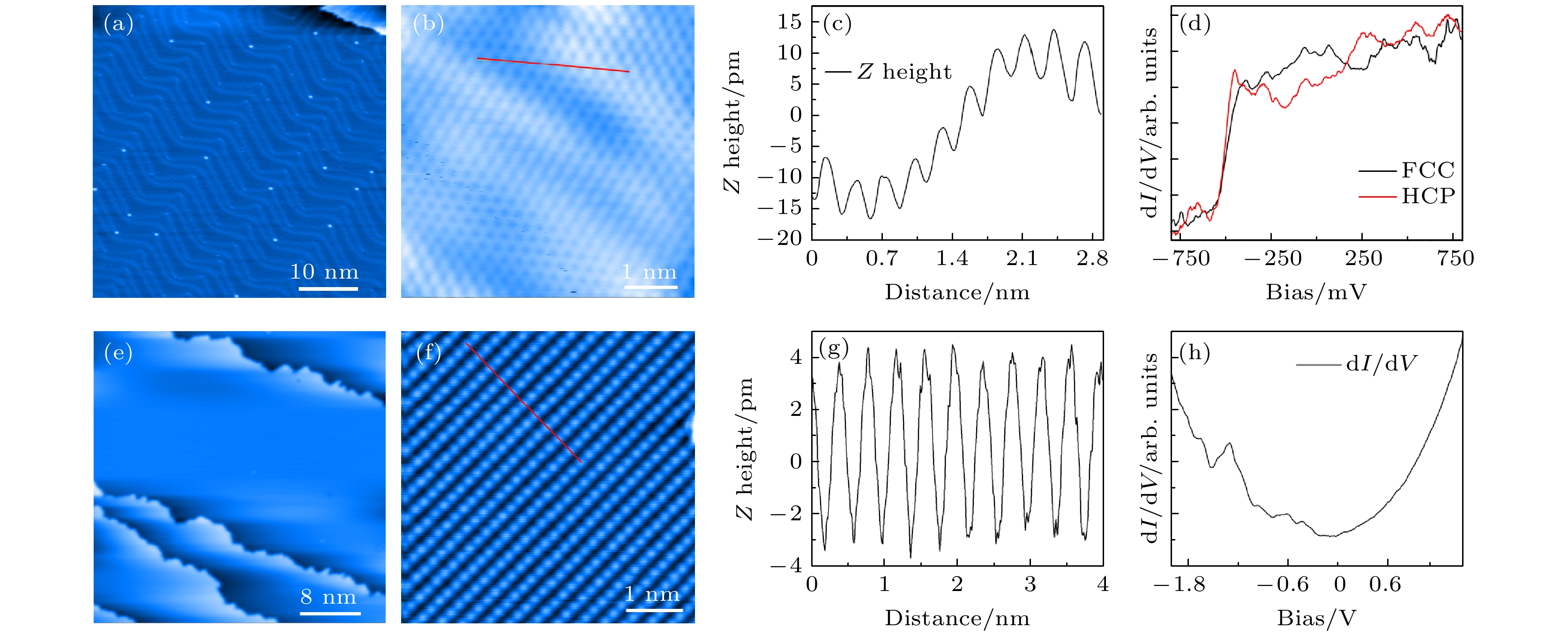
图 18 搭建Dry-LT-SPM的成像和隧道谱性能表征[23] (a) Au(111)表面的鱼骨形重构; (b) Au(111)表面的原子分辨; (c)金原子沿图(b)中红线的线剖面图; (d) Au(111)表面FCC和HCP位点的dI/dV谱; (e) Ag(110)表面的大范围STM 图像; (f) Ag(110)表面的原子分辨STM图像; (g) Ag原子沿图(f)中红线的线剖面图; (h) Ag(110)表面的dI/dV谱
Fig. 18. Imaging and tunneling spectrum performance characterization of the Dry-LT-SPM[23]: (a) Herringbone reconstruction of the Au(111) surface; (b) atomic resolution of the Au(111) surface; (c) line profile of gold atoms along the red line shown in panel (b); (d) dI/dV spectra of FCC and HCP sites on Au(111) surface; (e) large-scale STM image of the Ag(110) surface; (f) atomic-resolved STM image of the Ag(110) surface; (g) line profile of Ag atoms along the red line shown in panel (f); (h) dI/dV spectrum of Ag(110) surface.
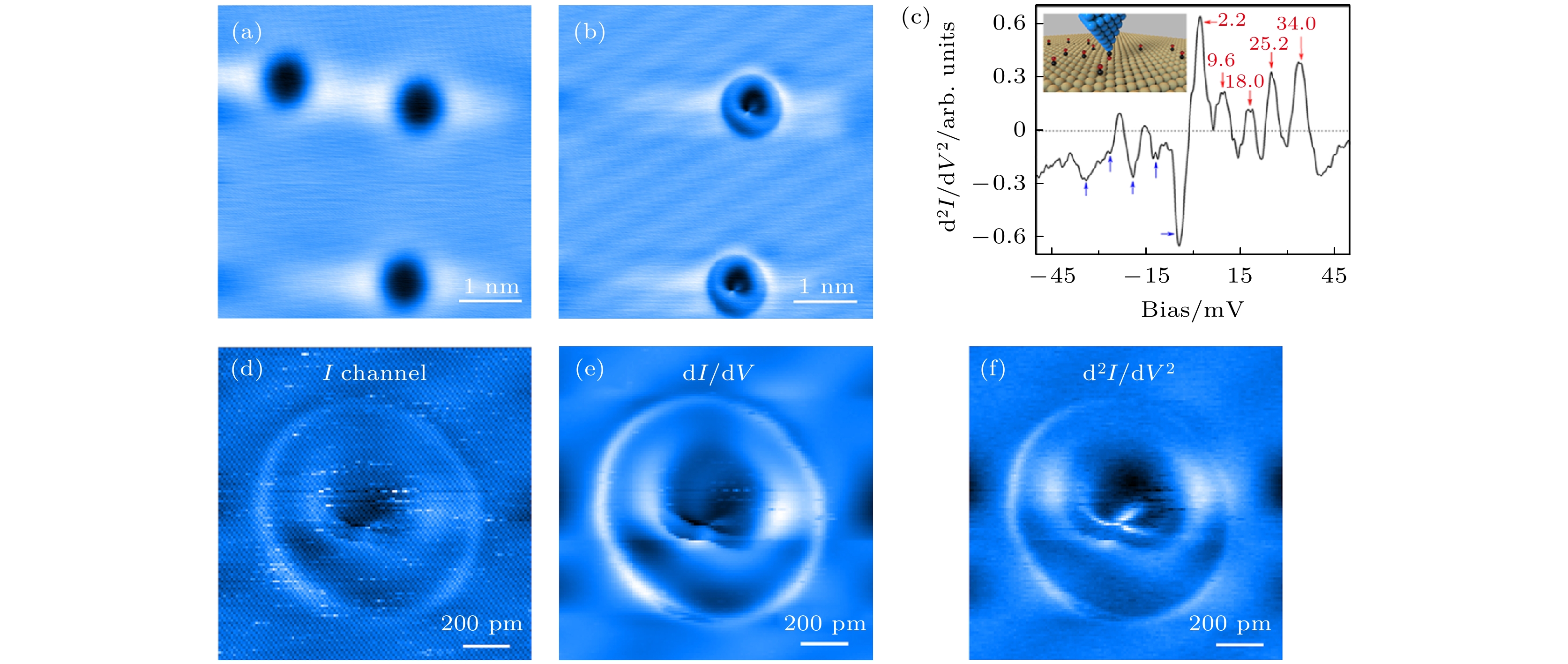
图 19 基于远端液化方案的SPM系统的谱学成像表征[23] (a) CO分子沉积在Ag(110)表面; (b)能够被探针捡起; (c)能获得高质量的IETS谱; (d)—(f)在CO分子上取得隧道电流谱, STS谱以及IETS谱学图像, 成像参数为110 pA, 9.6 mV
Fig. 19. Spectroscopic imaging performance of the SPM system based on the remote liquefaction scheme[23]: (a) CO molecules are deposited on the Ag(110) surface; (b) can be picked up by the probe; (c) high-quality IETS spectra can be obtained; (d)–(f) tunnelling current spectrum, STS spectrum and IETS spectroscopy images on CO molecules with setpoint of 110 pA and 9.6 mV.
-
[1] Wu Z B, Gao Z Y, Chen X Y, et al. 2018 Rev. Sci. Instrum. 89 113705
 Google Scholar
[2] Bian K, Gerber C, Heinrich A J, Müller D J, Scheuring S, Jiang Y 2021 Nat. Rev. Method. Prime. 1 36
 Google Scholar
[3] Pettinger B, Schambach P, Villagómez C J, Scott N 2012 Annu. Rev. Phys. Chem. 63 379
 Google Scholar
[4] Watkins N J, Long J P, Kafafi Z H, Mäkinen A J 2007 Rev. Sci. Instrum. 78 053707
 Google Scholar
[5] Grafström S 2002 J. Appl. Phys. 91 1717
 Google Scholar
[6] Flores S M, Toca-Herrera J L 2009 Nanoscale 1 40
 Google Scholar
[7] Bharat B 2004 Handbook of Nanotechnology (Springer
[8] Baykara M Z, Morgenstern M, Schwarz A, Schwarz U D 2017 Handbook of Nanotechnology (Berlin: Springer) pp769–808
[9] Behler S, Rose M K, Dunphy J C, Ogletree D F, Salmeron M, Chapelier C 1997 Rev. Sci. Instrum. 68 2479
 Google Scholar
[10] Stipe B C, Rezaei M A, Ho W 1999 Rev. Sci. Instrum. 70 137
 Google Scholar
[11] Meyer G 1996 Rev. Sci. Instrum. 67 2960
 Google Scholar
[12] Elrod S A, Lozanne A L D, Quate C F 1984 Applied Physics Letters 45 1240
 Google Scholar
[13] He G, Wei Z X, Feng Z P, Yu X D, Zhu B Y, Liu L, Jin K, Yuan J, Huan Q 2020 Rev. Sci. Instrum. 91 013904
 Google Scholar
[14] Chaudhary S, Panda J J, Mundlia S, Mathimalar S, Ahmedof A, Raman K V 2021 Rev. Sci. Instrum. 92 023906
 Google Scholar
[15] Zhao Z, Wang C 2019 Engineering and Technologies: Principles and Applications of Cryogen-Free Systems (CRC Press
[16] Wong D, Jeon S, Nuckolls K P, Oh M, Kingsley S C J, Yazdani A 2020 Rev. Sci. Instrum. 91 023703
 Google Scholar
[17] Hackley J D, Kislitsyn D A, Beaman D K, Ulrich S, Nazin G V 2014 Rev. Sci. Instrum. 85 103704
 Google Scholar
[18] Zhang S, Huang D, Wu S W 2016 Rev. Sci. Instrum. 87 063701
 Google Scholar
[19] Kasai J, Koyama T, Yokota M, Iwaya K 2022 Rev. Sci. Instrum. 93 043711
 Google Scholar
[20] Huang H M, Shuai M M, Yang Y L, Song R, Liao Y H, Yin L M, Shen J 2022 Rev. Sci. Instrum. 93 073703
 Google Scholar
[21] Meng W J, Wang J H, Hou Y B, et al. 2019 Ultramicroscopy 205 20
 Google Scholar
[22] Coe A M, Li G H, Andrei E Y 2024 Rev. Sci. Instrum. 95 083702
 Google Scholar
[23] Ma R S, Li H, Shi C S, et al. 2023 Rev. Sci. Instrum. 94 093701
 Google Scholar
计量
- 文章访问数: 3396
- PDF下载量: 130
- 被引次数: 0


 下载:
下载:
