-
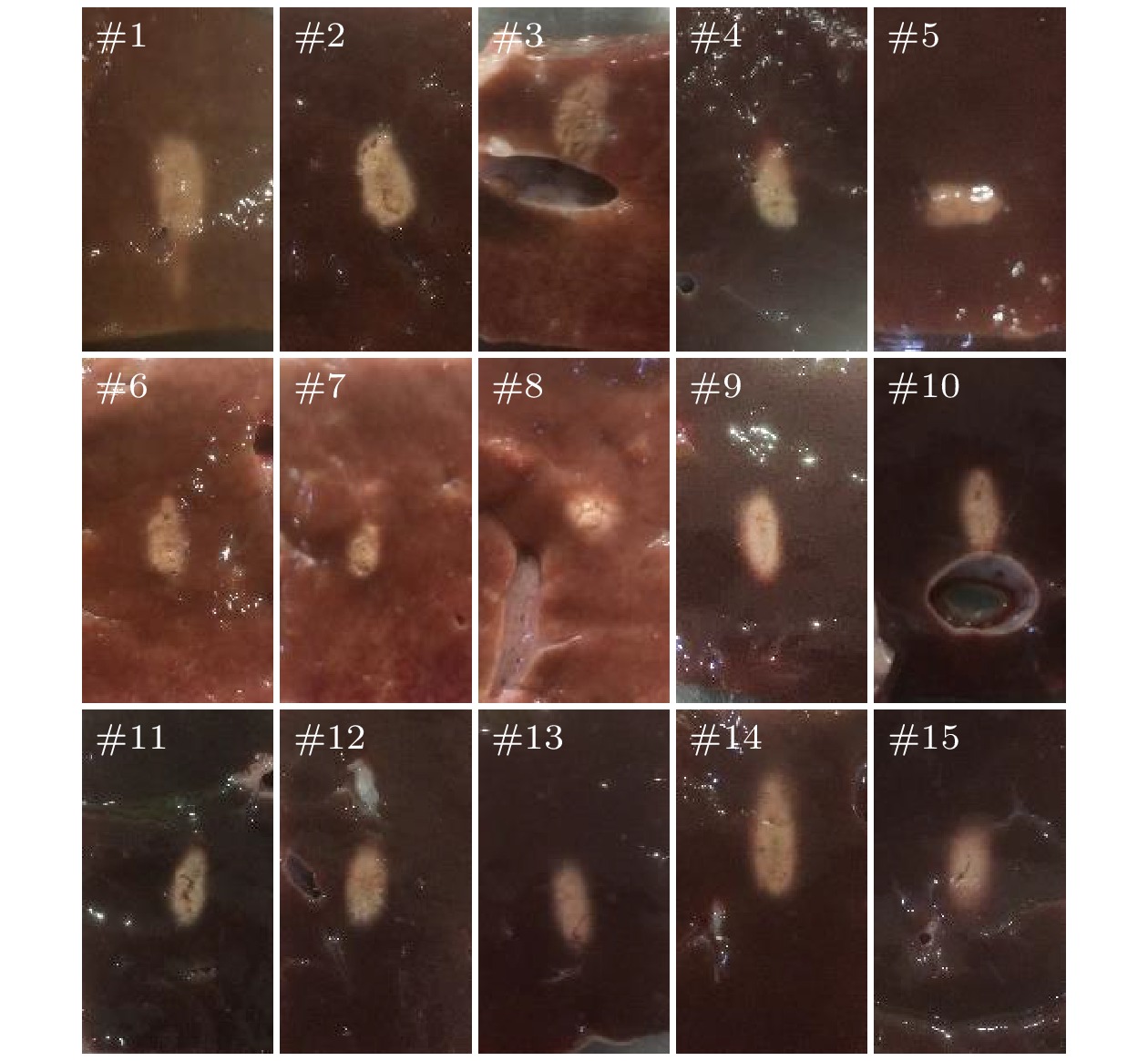
高强度聚焦超声(high intensity focused ultrasound, HIFU)焦域的实时监测是聚焦超声临床治疗面临的关键问题, 目前临床常采用B超图像强回声的变化实现焦域组织损伤的监测, 而B超图像出现的强回声大多与焦域处的空化及沸腾气泡相关, 无法准确、实时地监测治疗状态. HIFU治疗中焦域组织会伴随温度升高、空化、沸腾和组织特性等变化, 换能器表面的声学负载也在不断变化, 针对该问题, 本文构建了换能器电压电流实时检测平台, 通过测量换能器电学参数来感知焦域组织的状态变化. 以离体牛肝组织作为HIFU辐照对象, 并将相位差变化的结果与离体牛肝组织损伤的结果进行了对照, 实验结果表明, 在HIFU辐照过程中, 换能器电压与电流的相位会出现由相对平稳到大幅波动的过程, 此时停止辐照可见焦域出现明显损伤, 而此时B超图像灰度无明显变化; 此外, 当焦域出现空化时, 其波动幅度与范围将较之更大. 此方法可为HIFU焦域组织损伤监测提供一种新的研究方案和手段.
-
关键词:
- 高强度聚焦超声损伤监测 /
- 互相关 /
- 换能器电气特性
Real-time monitoring of high intensity focused ultrasound (HIFU) focal region is a key problem in clinical treatment of focused ultrasound. At present, the change of strong echo in B-ultrasound image is often used in clinical practice to monitor tissue damage in the focal area. However, the strong echo in B-ultrasound image is mostly related to cavitation and boiling bubbles in the focal area, which cannot monitor the treatment status accurately or in real time. In the HIFU treatment, the focal area tissue will be accompanied by changes in temperature, cavitation, boiling, and tissue characteristics. The acoustic load on the surface of the transducer is also constantly changing. To solve this problem, a real-time detection platform of transducer voltage and current is built in this work, which can sense the change of focal area tissue state by measuring the electrical parameters of the transducer. The experimental results show that the stability of the phase difference of the transducer driving signal will be different (the fluctuation amplitude will be different) when different media are placed on the surface of the transducer to change the acoustic load on the surface of the transducer. The fluctuation amplitude of the phase difference of the driving signal will be larger than that in the water when the iron plate is placed in the focal plane. However, the phase fluctuation amplitude will be much smaller than that in the water where the beef liver is placed. This shows that different acoustic loads can cause the electrical parameters of the transducer to change. The isolated bovine liver tissue is used as the HIFU irradiation object, and the results of the phase difference change are compared with the results of the isolated bovine liver tissue damage. The experimental results show that the phase of the transducer voltage and current will change from relatively stable to large fluctuations during the HIFU irradiation. At this time, obvious damage can be seen in the focal region when the irradiation is stopped, and the grayscale of B-ultrasound image has no significant change. In addition, when the cavitation occurs in the focal region, the fluctuation amplitude and range will turn larger. The damage area of the lower focal area under the monitoring method is smaller than that under B-ultrasonic monitoring, and the over input of radiation dose can be avoided. This method can provide a new research scheme and means for HIFU focal area tissue damage monitoring.-
Keywords:
- high intensity focused ultrasound damage monitoring /
- cross-correlation /
- electrical characteristics of transducer
[1] Fry W J, Fry F J 1960 IRE Trans. Med. Electron. 3 166
 Google Scholar
[2] Bailey M R, Khokhlova V A, Sapozhnikov O A, Kargl S G, Crum L A 2003 Acoust. Phys. 49 369
 Google Scholar
[3] Jenne J W, Preusser T, Günther M 2012 Z. Med. Phys. 22 311
 Google Scholar
[4] Jeng C J, Ou K Y, Long C Y, Chuang L, Ker C 2020 Taiwan. J. Obstet. Gyne. 59 865
 Google Scholar
[5] Peek M C L, Ahmed M, Napoli A, Ten Haken B, Mcwilliams S, Usiskin S I, Pinder S E, Van Hemelrijck M, Douek M 2015 Br. J. Surg. 102 873
 Google Scholar
[6] Schmid F A, Schindele D, Mortezavi A, Spitznagela T, Sulsera T, Schostakb M, Eberlia D 2020 Urol. Oncol. -Semin. Ori. 38 225
 Google Scholar
[7] Quadri S A, Waqas M, Khan I, Khan M A, Suriya S S, Farooqui M, Fiani B 2018 Neurosurg. Focus 44 1
 Google Scholar
[8] Charrel T, Aptel F, Birer A, Chavrier F, Romano F, Chapelon J Y, Denid P, Lafon C 2011 Ultrasound Med. Biol. 37 742
 Google Scholar
[9] Dogra V S, Zhang M, Bhatt S 2009 Ultrasound Clinics 4 307
[10] Rabkin B A, Zderic V, Crum L A, Vaezy S 2016 Ultrasound Med. Biol. 32 1721
[11] Adams C, McLaughlan J R, Carpenter T M, Freear S 2019 IEEE Trans. Ultrason. Ferroelectr. Freq. Control 67 239
 Google Scholar
[12] Thomas C R, Farny C H, Wu T, Holt G, Roy R A 2006 AIP Conference Proceedings 829 293
 Google Scholar
[13] Karaböce B, Gülmez Y, Bilgiç E, et al. 2014 IEEE International Symposium on Medical Measurements and Applications Lisboa, Portugal, June 11−12, 2014 p1
[14] Bentley J P 2005 Principles of Measurement Systems (4th Ed.) (London: Pearson Education) pp427−436
[15] 郭林伟, 林书玉, 许龙 2010 陕西师范大学学报(自然科学版) 38 39
 Google Scholar
Guo L W, Lin S Y, Xu L 2010 J. Shaanxi Normal Univ. (Nat. Sci. Ed.) 38 39
 Google Scholar
[16] 曹奕涛, 淳莉, 张宏军, 赵洪峰, 周小川 2019 空天防御 2 47
 Google Scholar
Cao Y T, Chun L, Zhang H J, Zhao X F, Zhou X C 2019 Air Space Defense 2 47
 Google Scholar
[17] 曾玲, 陈伟, 陶金 2017 电子测量技术 40 71
 Google Scholar
Ceng L, Chen W, Tao J 2017 Electron. Meas. Technol 40 71
 Google Scholar
[18] Liu J F, Liu J Y, Zhang T T, Li J C 2009 Seventh Annual Communication Networks and Services Research Conference Moncton, NB, Canada, May 11−13, 2009 p440
[19] Zhang L, Wu X 2006 Digital Signal Processing 16 682
 Google Scholar
[20] Lai X, Torp H 1999 IEEE Trans. Ultrason. Ferroelectr. Freq. Control 46 277
 Google Scholar
[21] Zhang S, Wan M, Zhong H, Xu C, Liao Z Z, Liu H Q, Wang S P 2009 Ultrasound Med. Biol. 35 1828
 Google Scholar
[22] Bornmann P, Hemsel T, Sextro W, Maeda T, Morita T 2012 IEEE International Ultrasonics Symposium Dresden, Germany, Oct. 7−10, 2012 p1141
[23] Tu J, Matula T J, Brayman A A, Crum L A 2006 Ultrasound Med. Biol. 32 281
 Google Scholar
[24] Xu H, He L B, Zhong B, Qiu J M, Tu J 2019 Ultrasonics Sonochemistry 56 77
 Google Scholar
[25] Saalbach K A, Twiefel J, Wallaschek J 2019 Ultrasonics 94 401
 Google Scholar
[26] Tan J W, Liao R J, Wang H, et al. 2011 International Symposium on Bioelectronics and Bioinformations Suzhou, China, November 3−5, 2011 p279
-
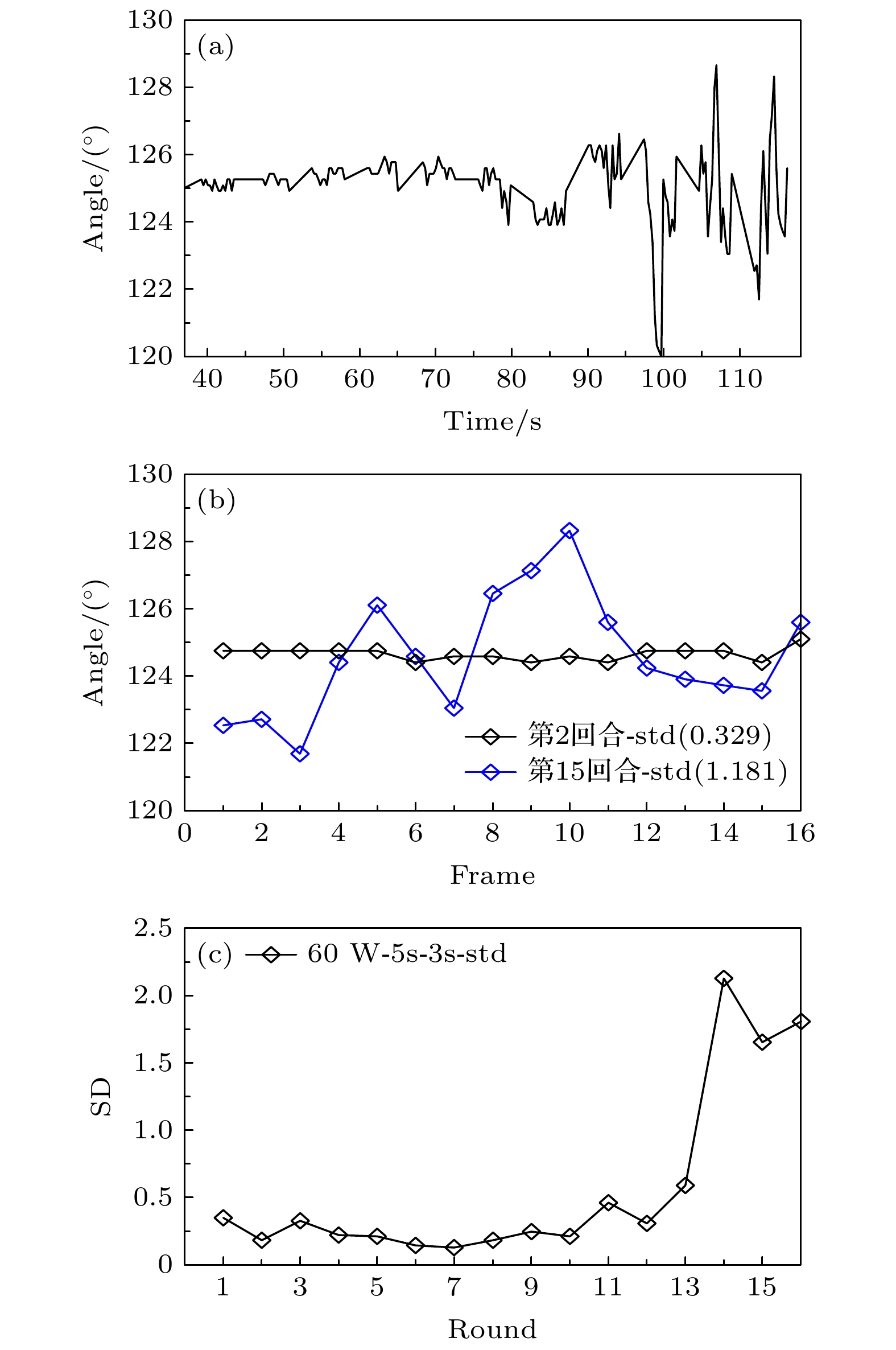
图 7 HIFU辐照中(无空化时)相位变化趋势 (a) HIFU辐照过程中相位变化过程; (b) 损伤前与损伤后相位波动对比; (c) 辐照期间每回合的相位标准差变化
Fig. 7. Phase change trend in HIFU irradiation (without cavitation): (a) Phase change process during HIFU irradiation; (b) comparison of phase fluctuation before and after damage; (c) phase standard deviation change per turn during irradiation.
图 9 HIFU辐照中出现空化时相位变化趋势 (a) HIFU辐照过程中相位变化过程; (b) 损伤前与损伤后相位波动对比; (c) 辐照期间每回合的相位标准差变化
Fig. 9. Phase change trend of cavitation appears in HIFU irradiation: (a) Phase change process during HIFU irradiation; (b) comparison of phase fluctuation before and after damage; (c) phase standard deviation change per turn during irradiation.
-
[1] Fry W J, Fry F J 1960 IRE Trans. Med. Electron. 3 166
 Google Scholar
[2] Bailey M R, Khokhlova V A, Sapozhnikov O A, Kargl S G, Crum L A 2003 Acoust. Phys. 49 369
 Google Scholar
[3] Jenne J W, Preusser T, Günther M 2012 Z. Med. Phys. 22 311
 Google Scholar
[4] Jeng C J, Ou K Y, Long C Y, Chuang L, Ker C 2020 Taiwan. J. Obstet. Gyne. 59 865
 Google Scholar
[5] Peek M C L, Ahmed M, Napoli A, Ten Haken B, Mcwilliams S, Usiskin S I, Pinder S E, Van Hemelrijck M, Douek M 2015 Br. J. Surg. 102 873
 Google Scholar
[6] Schmid F A, Schindele D, Mortezavi A, Spitznagela T, Sulsera T, Schostakb M, Eberlia D 2020 Urol. Oncol. -Semin. Ori. 38 225
 Google Scholar
[7] Quadri S A, Waqas M, Khan I, Khan M A, Suriya S S, Farooqui M, Fiani B 2018 Neurosurg. Focus 44 1
 Google Scholar
[8] Charrel T, Aptel F, Birer A, Chavrier F, Romano F, Chapelon J Y, Denid P, Lafon C 2011 Ultrasound Med. Biol. 37 742
 Google Scholar
[9] Dogra V S, Zhang M, Bhatt S 2009 Ultrasound Clinics 4 307
[10] Rabkin B A, Zderic V, Crum L A, Vaezy S 2016 Ultrasound Med. Biol. 32 1721
[11] Adams C, McLaughlan J R, Carpenter T M, Freear S 2019 IEEE Trans. Ultrason. Ferroelectr. Freq. Control 67 239
 Google Scholar
[12] Thomas C R, Farny C H, Wu T, Holt G, Roy R A 2006 AIP Conference Proceedings 829 293
 Google Scholar
[13] Karaböce B, Gülmez Y, Bilgiç E, et al. 2014 IEEE International Symposium on Medical Measurements and Applications Lisboa, Portugal, June 11−12, 2014 p1
[14] Bentley J P 2005 Principles of Measurement Systems (4th Ed.) (London: Pearson Education) pp427−436
[15] 郭林伟, 林书玉, 许龙 2010 陕西师范大学学报(自然科学版) 38 39
 Google Scholar
Guo L W, Lin S Y, Xu L 2010 J. Shaanxi Normal Univ. (Nat. Sci. Ed.) 38 39
 Google Scholar
[16] 曹奕涛, 淳莉, 张宏军, 赵洪峰, 周小川 2019 空天防御 2 47
 Google Scholar
Cao Y T, Chun L, Zhang H J, Zhao X F, Zhou X C 2019 Air Space Defense 2 47
 Google Scholar
[17] 曾玲, 陈伟, 陶金 2017 电子测量技术 40 71
 Google Scholar
Ceng L, Chen W, Tao J 2017 Electron. Meas. Technol 40 71
 Google Scholar
[18] Liu J F, Liu J Y, Zhang T T, Li J C 2009 Seventh Annual Communication Networks and Services Research Conference Moncton, NB, Canada, May 11−13, 2009 p440
[19] Zhang L, Wu X 2006 Digital Signal Processing 16 682
 Google Scholar
[20] Lai X, Torp H 1999 IEEE Trans. Ultrason. Ferroelectr. Freq. Control 46 277
 Google Scholar
[21] Zhang S, Wan M, Zhong H, Xu C, Liao Z Z, Liu H Q, Wang S P 2009 Ultrasound Med. Biol. 35 1828
 Google Scholar
[22] Bornmann P, Hemsel T, Sextro W, Maeda T, Morita T 2012 IEEE International Ultrasonics Symposium Dresden, Germany, Oct. 7−10, 2012 p1141
[23] Tu J, Matula T J, Brayman A A, Crum L A 2006 Ultrasound Med. Biol. 32 281
 Google Scholar
[24] Xu H, He L B, Zhong B, Qiu J M, Tu J 2019 Ultrasonics Sonochemistry 56 77
 Google Scholar
[25] Saalbach K A, Twiefel J, Wallaschek J 2019 Ultrasonics 94 401
 Google Scholar
[26] Tan J W, Liao R J, Wang H, et al. 2011 International Symposium on Bioelectronics and Bioinformations Suzhou, China, November 3−5, 2011 p279
计量
- 文章访问数: 6684
- PDF下载量: 134
- 被引次数: 0


 下载:
下载:

