-
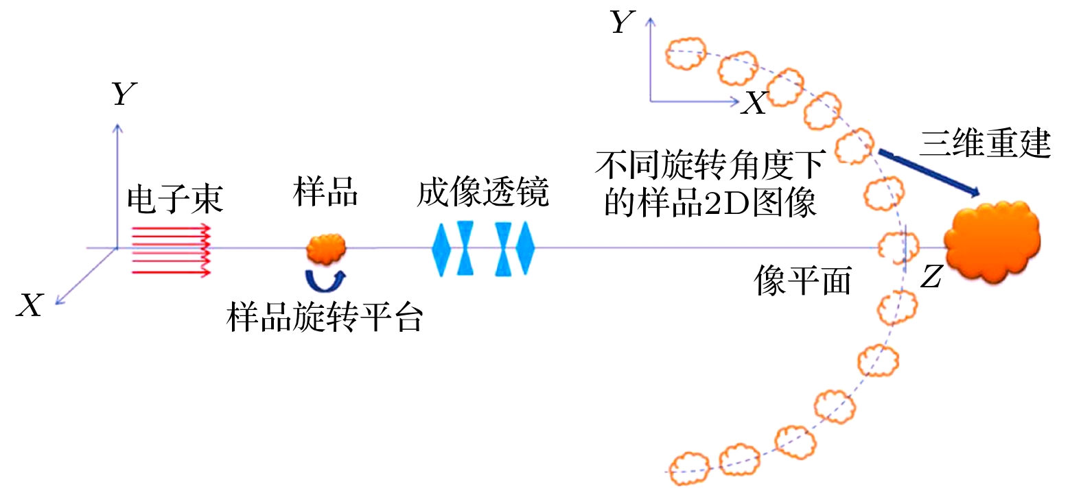
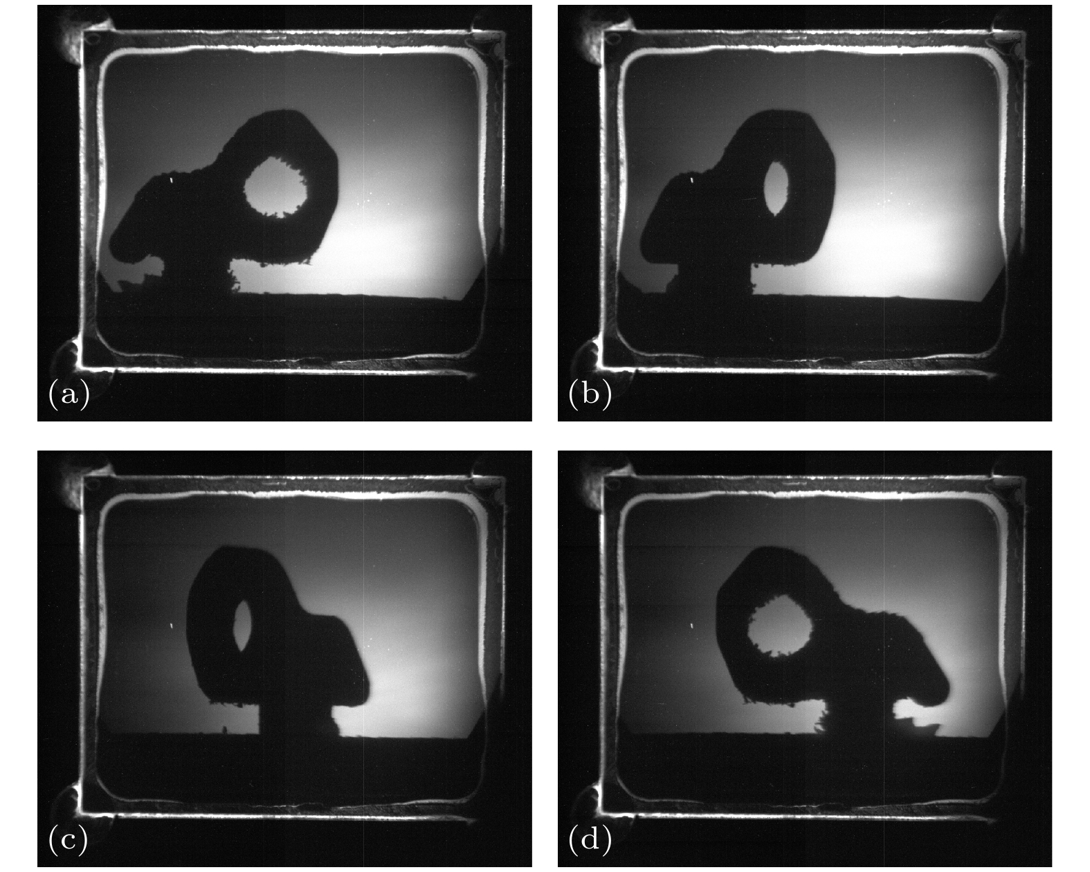
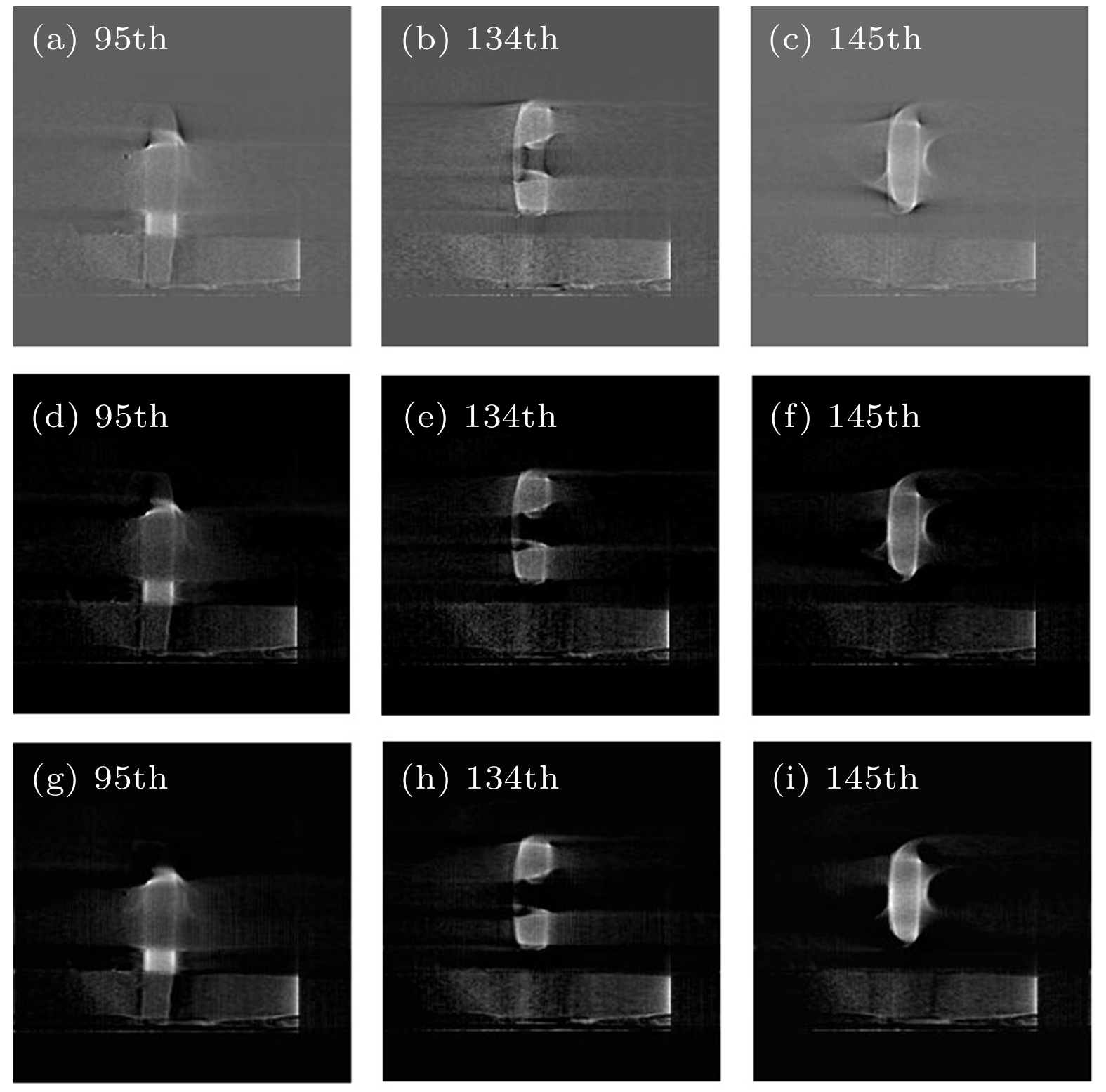
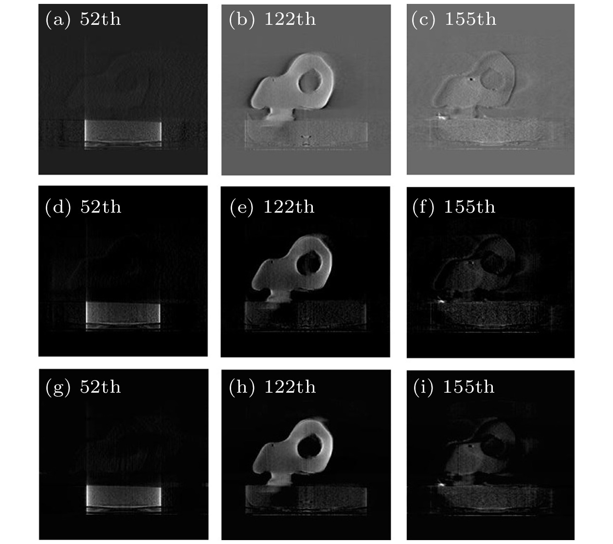
高能电子成像技术被首次提出作为温稠密物质和惯性约束聚变实验研究的高时空分辨诊断工具之一, 现已通过前期实验证明其对中尺度科学诊断的可行性. 为了进一步提高高能电子成像技术诊断样品的能力, 来获取样品内部信息, 将高能电子成像技术和三维重建算法结合, 提出了高能电子三维成像技术. 本文主要通过实验研究了高能电子三维成像技术的可行性. 不同三维重建算法重建样品的结果首次证实了高能电子三维成像技术的可行性, 使用的三维重建算法包括滤波反投影算法、迭代算法-代数重建技术和联立代数重建技术, 最终重建的x, y, z方向上的不同重建切片图像清楚地显示了样品的详细结构. 实验证实的高能电子三维成像技术将有利于拓展高能电子成像技术的应用领域, 尤其是在中尺度科学领域.High energy electron radiography (HEER) proposed first for real-time high spatial and temporal resolution diagnosis of warm dense matter (WDM) and inertial confinement fusion (ICF) has proved experimentally feasible for mesoscale sciences diagnosis. Until now, the spatial resolution of the images close to 1 μm has been reached experimentally which is better than that of X-rays and neutron radiography. However, traditional HEER obtains two-dimensional images which cannot accurately present the three-dimensional structure of the sample. To further improve the capability of HEER to diagnose and obtain the internal information of samples, three-dimensional high energy electron radiography (TDHEER) was put forward by combining HEER with three-dimensional (3D) reconstruction tomography technology. The validity and usage of the TDHEER method have been confirmed through simulation of the fully 3D diagnostic of static mesoscale sample. This paper focuses mainly on the experimental demonstration of the 3D high energy electron radiography. The feasibility of TDHEER is for the first time confirmed by the results achieved with different 3D reconstruction algorithms. The 3D reconstruction algorithms, analytical algorithm-filtered back projection (FBP), iterative algorithms-algebraic reconstruction technique (ART), and simultaneous algebraic reconstruction technique (SART) are used here. In this experiment, the less projected data are used, so it takes the less time to obtain two-dimensional (2D) HEER images and the reconstruction. In order to spend the time as little as possible and obtain the satisfactory quality of reconstruction result, there are three groups of projected image sets, 180, 36 and 18, acquired in our experiment. When all three algorithms are adopted in 180 projected images, the reconstructed images show that all three algorithms FBP, ART and SART are feasible for TDHEER. The different reconstructed slice images of the sample in X-, Y-, and Z- direction clearly show the detailed structure of the sample. The images reconstructed by ART and SART algorithm are equivalent. Comparing with ART and SART, the reconstruction results by FBP can show more details, but there are some artifacts. Because the 36 2D HEER images fail to satisfy the Nyquist sampling theory, the analytic algorithm FBP is not used. Taking the result of FBP reconstructed by 180 images as a standard reference to compare the result of ART with the results of SART, the images reconstructed by the SART algorithm are closer to the original images. Testing 18 images, the results of the ART and SART both have lots of artifacts but the SART algorithm spends less time in reconstruction. As fewer projected images are used, more artifacts are found in the reconstructed images. Therefore, it is advantageous to combine the SART algorithm with 36 HEER projected images, which obtains high-quality reconstruction images and spends less time. The feasibility of TDHEER is confirmed experimentally for the first time and all three dimensions of the sample structures are obtained. Of the three different 3D reconstruction algorithms, the SART algorithm is the most suitable for reconstructing the few-view images. The TDHEER technology will extend HEER’s application fields, especially for mesoscale sciences.
-
Keywords:
- high energy electron radiography /
- three-dimensional reconstruction algorithm /
- three-dimensional high energy electron radiography /
- mesoscale sciences
[1] Wei G, Qiu J, Jing C 2014 Pro. SPIE 9211 921104
[2] Zhao Y T, Zhang Z, Gai W, et al. 2016 Laser Part. Beams 34 338
 Google Scholar
[3] Merrill F, Harmon F, Hunt A, Mariam F, Morley K, Morris C, Saunders A, Schwartz C 2007 Nucl. Instrum. Methods Phys. Res., Sect. B 261 382
 Google Scholar
[4] Zhao Q T, Cao S C, Cheng R, Shen X K, Zhang Z M, Zhao Y T, Gai W, Du Y C 2014 Proceedings of the LINAC2014 Geneva, Switzerland, August 31–September 5, 2014 p76
[5] Zhao Q T, Cao S C, Liu M, et al. 2016 Nucl. Instrum. Methods Phys. Res., Sect. A 832 144
 Google Scholar
[6] Zhou Z, Du Y C, Cao S C, et al. 2018 Phys. Rev. Accel. Beams 21 074701
 Google Scholar
[7] Zhao Q T, Cao S. C, Cheng R, et al. 2018 Laser Part. Beams 36 313
 Google Scholar
[8] Zhao Q T, Cao S C, Shen X K, Wang Y R, Zong Y, Xiao J H, Zhu Y L, Zhou Y W, Liu M, Cheng R, Zhao Y T, Zhang Z M, Gai W 2017 Laser Part. Beams 35 579
 Google Scholar
[9] Zhou Z, Fang Y, Chen H, Wu Y P, Du Y C, Yan L X, Tang C X, Huang W H 2019 Phys. Rev. Appl. 11 034068
 Google Scholar
[10] Maddox B R, Park H S, Remington B A, et al. 2011 Phys. Plasmas 18 168
 Google Scholar
[11] Park H S, Maddox B R, Giraldez E, et al. 2008 Phys. Plasmas 15 3048
 Google Scholar
[12] Tian C, Yu M H, Shan L Q, Wu Y C, Zhang T K, Bi B, Zhang F, Zhang Q Q, Liu D X, Wang W W, Yuan Z Q, Yang S Q, Yang L, Zhou W M, Gu Y Q, Zhang B H 2019 Nucl. Fusion 59 046012
 Google Scholar
[13] Higginson D P, Vassura L, Gugiu M, et al. 2015 Phys. Rev. Lett. 115 054802
 Google Scholar
[14] Strobl M, Manke I, Kardjilov N, Hilger A, Dawson M, Banhart J 2009 J. Phys. D: Appl. Phys. 42 243001
 Google Scholar
[15] Merrill F E, Bower D, Buckles R, et al. 2012 Rev. Sci. Instrum. 83 051003
 Google Scholar
[16] King N S P, Ables E, Adams K, et al. 1999 Nucl. Instrum. Methods Phys. Res., Sect. A 424 84
 Google Scholar
[17] Zhao Q T, Ma Y Y, Xiao J H, Cao S C, Zhang Z M 2019 Appl. Sci. 9 3764
 Google Scholar
[18] 谢一冈, 陈昌, 王曼, 吕军光, 孟祥承, 王锋, 顾树棣, 过雅南 2003 粒子探测器与数据获取 (北京: 科学出版社) 第5−16页
Xie Y G, Cheng C, Wang M, Lv J G, Meng X G, Wang F, Gu S D, Guo Y N 2003 Particle Detector and Data Acquisition (Beijing: Science Press) pp5−16 (in Chinese)
[19] Padole A, Khawaja Rd Ali, Kalra M K, Singh S 2015 Am. J. Roentgenol. 204 384
 Google Scholar
[20] Chen B X, Yang M, Zhang Z, Bian J G, Han X, Sidky E, Pan X C 2014 Biochim. Biophys. Acta 1581 1856
 Google Scholar
[21] Schofield R, King L, Tayal U, Castellano I, Stirrup J, Pontana F, Earls J, Nicol E 2020 J. Cardiovasc Comput Tomogr. 14 219
 Google Scholar
[22] Gordon R, Bender R, Herman G T 1970 J. Theor. Biol. 29 471
 Google Scholar
[23] Trampert J, Leveque J J 1990 J. Geophys. Res. B:Solid Earth 95 12553
 Google Scholar
[24] Zhu Y L, Yuan P, Cao S C, et al. 2018 Nucl. Instrum. Methods Phys. Res., Sect. A 911 74
 Google Scholar
[25] Blackledget J M 2006 Digital Signal Processing (2nd Ed.) (Cambridge: Woodhead Press) pp522−540
[26] 拉斐尔C, 理查E, 史蒂文L 著 (阮秋琦 译) 2014 数字图像处理(MATLAB版) (第二版) (北京: 电子工业出版社) 第45−48页
Rafael C, Richard E, Steven L (translated by Ruan Q Q) 2014 Digital Image Processing Using MATLAB (2nd Ed.) (Beijing: Electronics Industry Press) pp45−48 (in Chinese)
[27] Vetterli M, Herley C 1992 IEEE Trans. Acoust., Speech, Signal Process 40 2207
 Google Scholar
[28] Zhang Y, Zhang W H, Lei Y J, Zhou J L 2014 J. Opt. Soc. Am. A 31 981
 Google Scholar
[29] Soleimani M., Pengpen T 2015 Philos Trans. R. Dov. London, Ser. A 373 20140399
 Google Scholar
[30] Sidky E Y, Kao C M, Pan X C 2009 J. X-Ray Sci. Technol. 14 119
 Google Scholar
[31] Liu Y, Ma J H, Fan Y, Liang Z R 2012 Phys. Med. Biol. 57 7923
 Google Scholar
[32] Deng L Z, Mi D L, He P, Feng P, Yu P W, Chen M Y, Li Z C, Wang J, Wei B 2015 Bio-med. Mater. Eng. 26 1685
 Google Scholar
-
表 1 ART, SART重建切片的d和r值
Table 1. Value of d and r of the image reconstructed by ART and SART.
切片位置 重建算法 d r Y 100th ART 11.0736 0.8820 SART 10.9060 0.8672 Y 127th ART 9.0947 0.9076 SART 8.9520 0.8933 -
[1] Wei G, Qiu J, Jing C 2014 Pro. SPIE 9211 921104
[2] Zhao Y T, Zhang Z, Gai W, et al. 2016 Laser Part. Beams 34 338
 Google Scholar
[3] Merrill F, Harmon F, Hunt A, Mariam F, Morley K, Morris C, Saunders A, Schwartz C 2007 Nucl. Instrum. Methods Phys. Res., Sect. B 261 382
 Google Scholar
[4] Zhao Q T, Cao S C, Cheng R, Shen X K, Zhang Z M, Zhao Y T, Gai W, Du Y C 2014 Proceedings of the LINAC2014 Geneva, Switzerland, August 31–September 5, 2014 p76
[5] Zhao Q T, Cao S C, Liu M, et al. 2016 Nucl. Instrum. Methods Phys. Res., Sect. A 832 144
 Google Scholar
[6] Zhou Z, Du Y C, Cao S C, et al. 2018 Phys. Rev. Accel. Beams 21 074701
 Google Scholar
[7] Zhao Q T, Cao S. C, Cheng R, et al. 2018 Laser Part. Beams 36 313
 Google Scholar
[8] Zhao Q T, Cao S C, Shen X K, Wang Y R, Zong Y, Xiao J H, Zhu Y L, Zhou Y W, Liu M, Cheng R, Zhao Y T, Zhang Z M, Gai W 2017 Laser Part. Beams 35 579
 Google Scholar
[9] Zhou Z, Fang Y, Chen H, Wu Y P, Du Y C, Yan L X, Tang C X, Huang W H 2019 Phys. Rev. Appl. 11 034068
 Google Scholar
[10] Maddox B R, Park H S, Remington B A, et al. 2011 Phys. Plasmas 18 168
 Google Scholar
[11] Park H S, Maddox B R, Giraldez E, et al. 2008 Phys. Plasmas 15 3048
 Google Scholar
[12] Tian C, Yu M H, Shan L Q, Wu Y C, Zhang T K, Bi B, Zhang F, Zhang Q Q, Liu D X, Wang W W, Yuan Z Q, Yang S Q, Yang L, Zhou W M, Gu Y Q, Zhang B H 2019 Nucl. Fusion 59 046012
 Google Scholar
[13] Higginson D P, Vassura L, Gugiu M, et al. 2015 Phys. Rev. Lett. 115 054802
 Google Scholar
[14] Strobl M, Manke I, Kardjilov N, Hilger A, Dawson M, Banhart J 2009 J. Phys. D: Appl. Phys. 42 243001
 Google Scholar
[15] Merrill F E, Bower D, Buckles R, et al. 2012 Rev. Sci. Instrum. 83 051003
 Google Scholar
[16] King N S P, Ables E, Adams K, et al. 1999 Nucl. Instrum. Methods Phys. Res., Sect. A 424 84
 Google Scholar
[17] Zhao Q T, Ma Y Y, Xiao J H, Cao S C, Zhang Z M 2019 Appl. Sci. 9 3764
 Google Scholar
[18] 谢一冈, 陈昌, 王曼, 吕军光, 孟祥承, 王锋, 顾树棣, 过雅南 2003 粒子探测器与数据获取 (北京: 科学出版社) 第5−16页
Xie Y G, Cheng C, Wang M, Lv J G, Meng X G, Wang F, Gu S D, Guo Y N 2003 Particle Detector and Data Acquisition (Beijing: Science Press) pp5−16 (in Chinese)
[19] Padole A, Khawaja Rd Ali, Kalra M K, Singh S 2015 Am. J. Roentgenol. 204 384
 Google Scholar
[20] Chen B X, Yang M, Zhang Z, Bian J G, Han X, Sidky E, Pan X C 2014 Biochim. Biophys. Acta 1581 1856
 Google Scholar
[21] Schofield R, King L, Tayal U, Castellano I, Stirrup J, Pontana F, Earls J, Nicol E 2020 J. Cardiovasc Comput Tomogr. 14 219
 Google Scholar
[22] Gordon R, Bender R, Herman G T 1970 J. Theor. Biol. 29 471
 Google Scholar
[23] Trampert J, Leveque J J 1990 J. Geophys. Res. B:Solid Earth 95 12553
 Google Scholar
[24] Zhu Y L, Yuan P, Cao S C, et al. 2018 Nucl. Instrum. Methods Phys. Res., Sect. A 911 74
 Google Scholar
[25] Blackledget J M 2006 Digital Signal Processing (2nd Ed.) (Cambridge: Woodhead Press) pp522−540
[26] 拉斐尔C, 理查E, 史蒂文L 著 (阮秋琦 译) 2014 数字图像处理(MATLAB版) (第二版) (北京: 电子工业出版社) 第45−48页
Rafael C, Richard E, Steven L (translated by Ruan Q Q) 2014 Digital Image Processing Using MATLAB (2nd Ed.) (Beijing: Electronics Industry Press) pp45−48 (in Chinese)
[27] Vetterli M, Herley C 1992 IEEE Trans. Acoust., Speech, Signal Process 40 2207
 Google Scholar
[28] Zhang Y, Zhang W H, Lei Y J, Zhou J L 2014 J. Opt. Soc. Am. A 31 981
 Google Scholar
[29] Soleimani M., Pengpen T 2015 Philos Trans. R. Dov. London, Ser. A 373 20140399
 Google Scholar
[30] Sidky E Y, Kao C M, Pan X C 2009 J. X-Ray Sci. Technol. 14 119
 Google Scholar
[31] Liu Y, Ma J H, Fan Y, Liang Z R 2012 Phys. Med. Biol. 57 7923
 Google Scholar
[32] Deng L Z, Mi D L, He P, Feng P, Yu P W, Chen M Y, Li Z C, Wang J, Wei B 2015 Bio-med. Mater. Eng. 26 1685
 Google Scholar
计量
- 文章访问数: 5846
- PDF下载量: 85
- 被引次数: 0


 下载:
下载:


