


-
为了探索在固体表面制作周期微结构的快速、低成本的新方法, 并了解表面周期微结构对激光诱导击穿光谱(LIBS)信号增强的物理机制, 本文利用200目的球形铜粉在聚氯乙烯片上压印出半球形表面周期微结构, 再利用电镀方法制作出复制有该微结构的镍板. 通过对比观测光滑表面镍板和带有半球形表面周期微结构的镍板不同的LIBS信号增强效果、测量其等离子体的温度和电子密度变化, 分析得出样品被照射表面积的增大是半球形表面周期微结构引起LIBS信号增强的主要原因. 与具有一定深度的圆柱形表面周期微结构的实验现象和信号增强机制进行对比分析, 表明微结构的深度有助于获得更好的信号增强效果, 为后续的微结构参数设计提供了有益的参考. 最后应用表面增强LIBS技术分析了水中的铅元素. 结果表明, 当前条件下, 带有半球形表面周期微结构的镍板与光滑表面的镍板相比, 基于Pb I 405.78 nm分析线, 铅的分析灵敏度可改善约23倍, 重复测量时信号强度的可重复性也获得了一定程度的改善.In order to develop a rapid and cost-effective new method to produce periodic microstructures on solid surfaces, and help to understand the physical mechanism of the enhancement of laser-induced breakdown spectroscopy (LIBS) signals induced by periodic surface microstructures, in this work, spherical copper powder with about 74 μm in diameter is used to imprint semispherical periodic surface microstructures on polyvinyl chloride (PVC) sheets under a pressure of 15 T. A platinum conducting layer about 100 nm in thickness is coated on the PVC surface by using a vacuum sputter coater and then nickel plates with the replicated microstructures on one surface are prepared using electroplating method. The signal enhancement effect induced by micro-structured surface in LIBS is experimentally observed and compared with that achieved by using flat surface nickel plate, the temperature and electron density of the induced plasma are measured according to Boltzmann plot method and the Stark broadening of Hα line of hydrogen. By systematically analyzing these results, it is concluded that the main physical mechanism of the signal enhancement in LIBS caused by the hemispherical periodic surface microstructure is due to the increased surface area of the sample that can be irradiated by the laser beam, leading to an increase in the mass of the ablated sample material when compared with that of a flat surface irradiated by the same laser beam. Comparative analysis is also conducted with experimental phenomena and signal enhancement mechanisms of using cylindrical periodic surface microstructures with a certain depth (20 μm diameter, 15 μm depth and 40 μm period). It is found that the depth of the microstructure helps to achieve better signal enhancement effects. This provides useful references for subsequent microstructure parameter design in the future. Finally, lead in aqueous solution samples is detected with surface-enhanced LIBS (SENLIBS) technique, while Pb I 405.78 nm line is selected as the analytical line. In comparison with flat nickel substrates, 23-fold detection sensitivity and slightly improved signal reproducibility can be achieved using nickel substrates with hemispherical periodic surface microstructures. The results indicate that nickel plates with hemispherical periodic surface microstructure show better analytical performance than flat nickel plates in elemental analysis of aqueous solution samples by SENLIBS.
-
Keywords:
- laser-induced breakdown spectroscopy /
- surface-enhancement /
- periodic surface micro-structures /
- signal enhancement mechanism
-
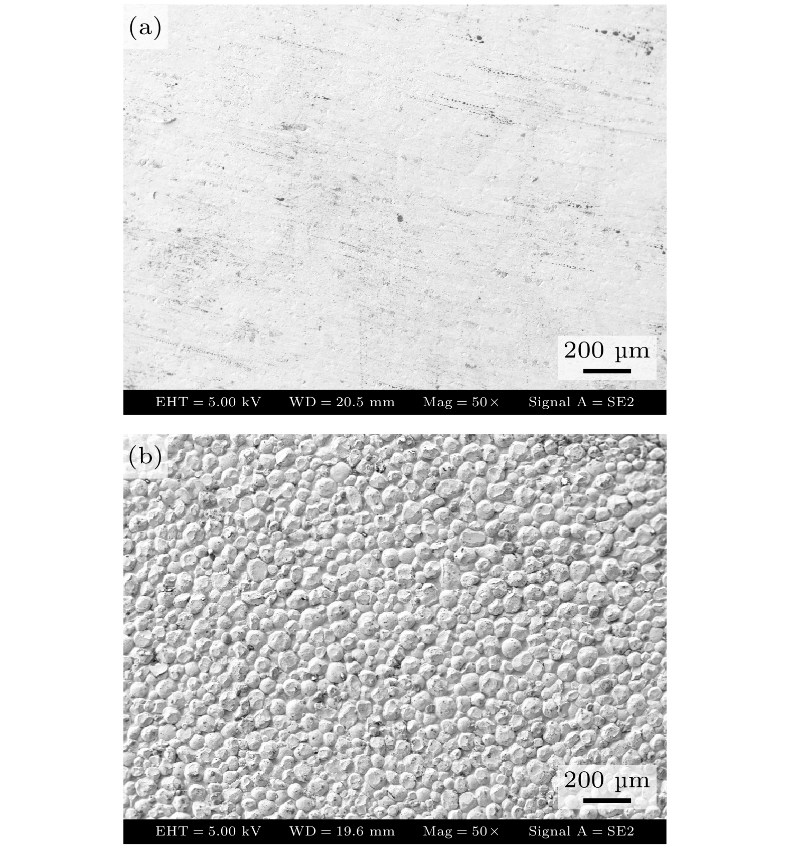
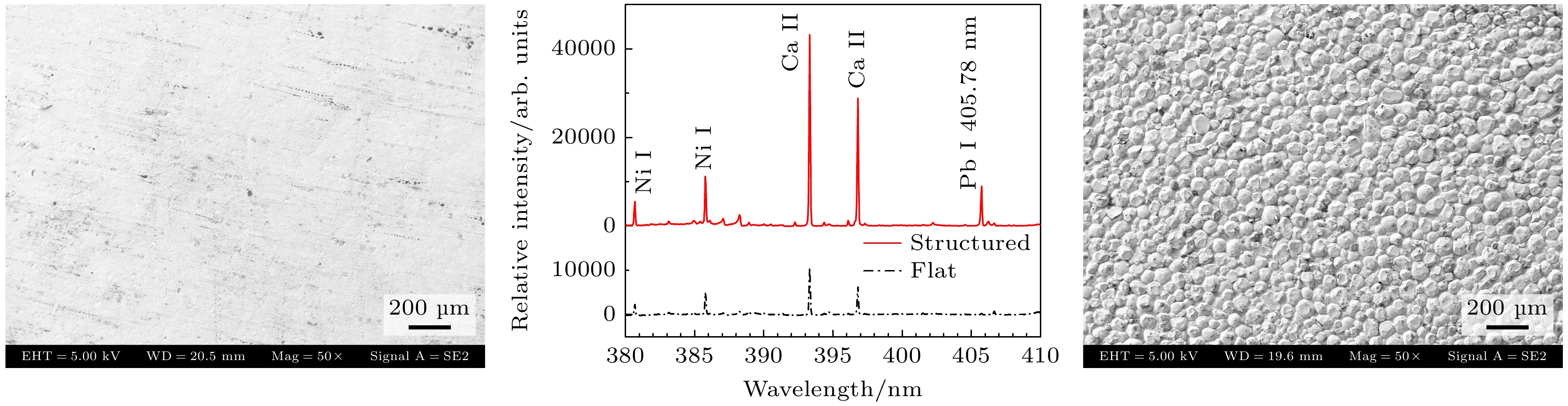
图 1 电镀出的镍板表面SEM图 (a) 光滑表面镍板; (b) 微结构表面的镍板
Fig. 1. SEM images of the nickel plates prepared by electroplating method: (a) Flat surface nickel plate; (b) surface micro-structured nickel plate.
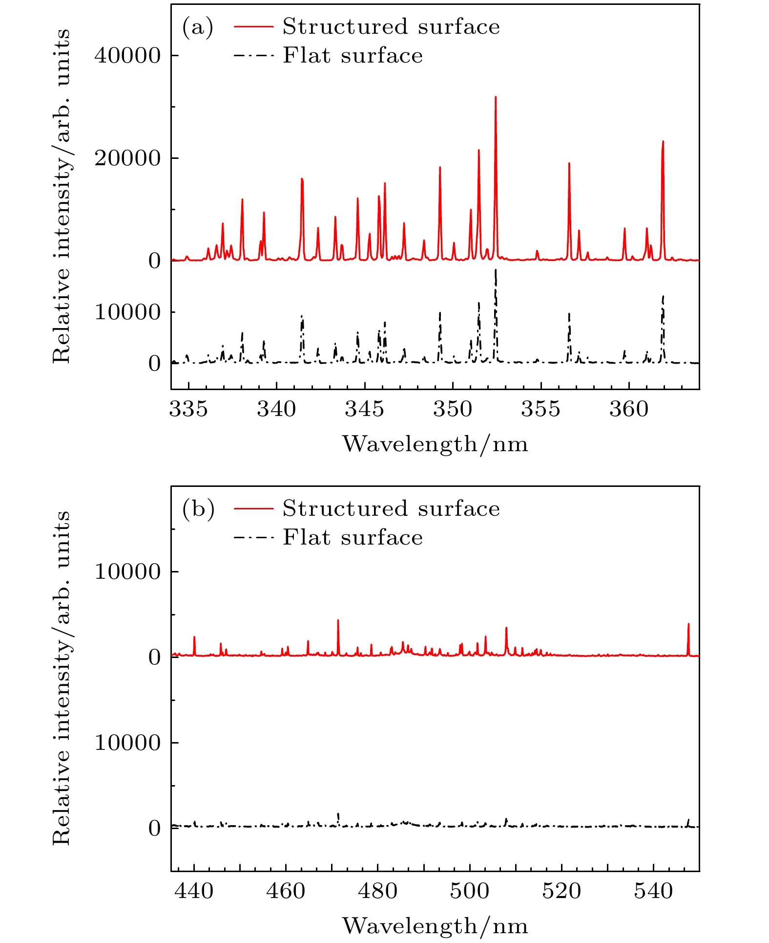
图 3 两种不同表面的Ni靶的局部LIBS光谱, 图中的谱线都是镍的谱线 (a) 334—364 nm; (b) 430—550 nm
Fig. 3. Comparison of the recorded LIBS spectra of two Ni targets with different surface morphology, all lines are atomic lines of nickel: (a) 334–364 nm region; (b) 430–550 nm region.
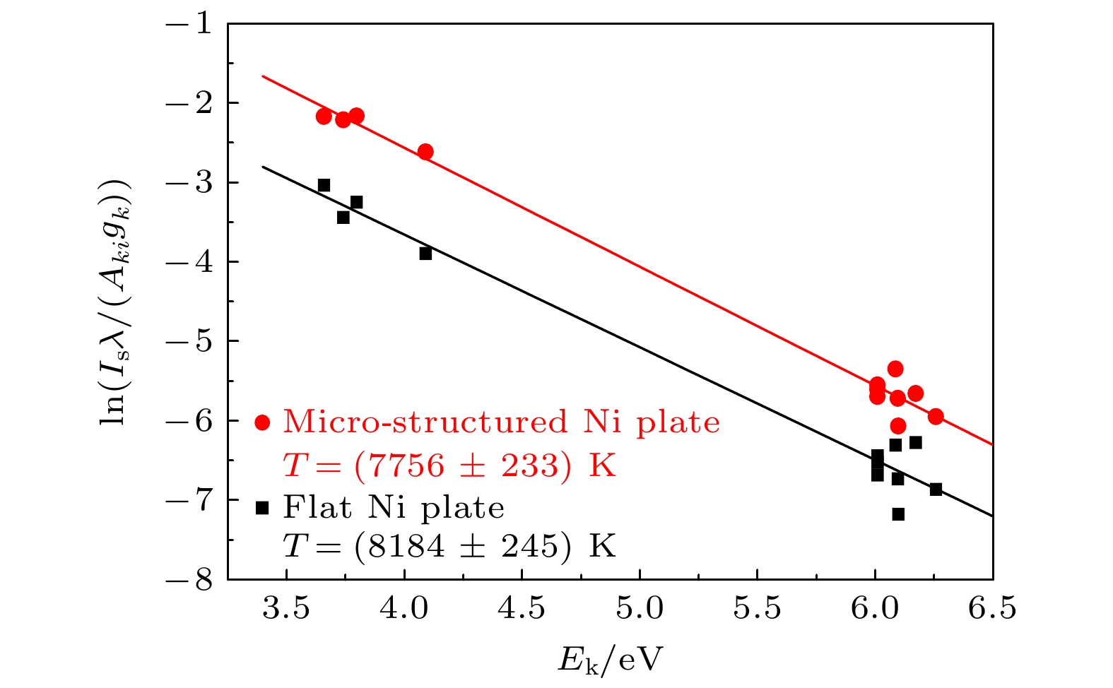
图 4 两种表面的Ni靶的等离子体的玻尔兹曼图
Fig. 4. Boltzmann plots of Ni plasma generated on targets with different surface morphology.
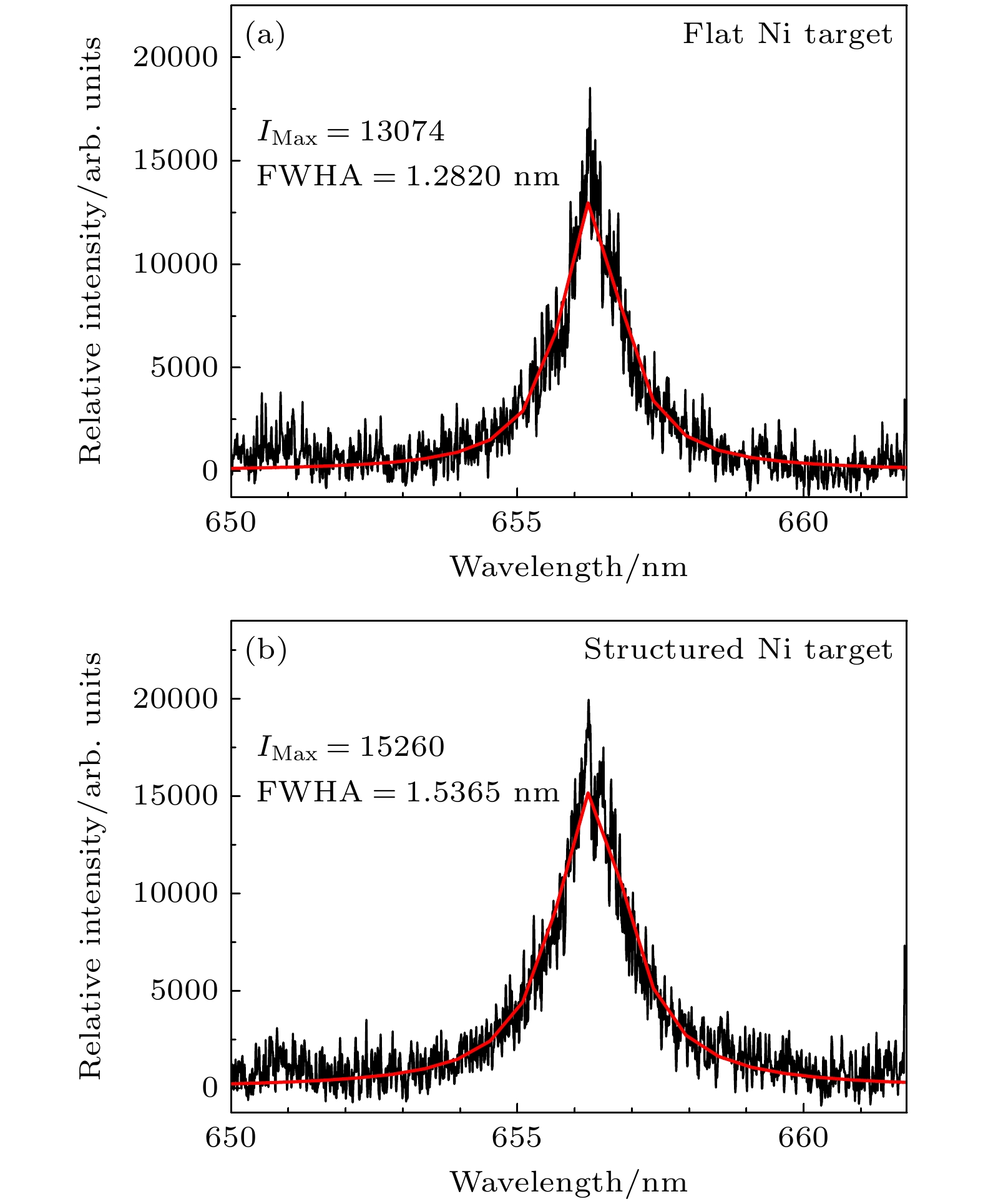
图 5 等离子体中观测到的氢Hα谱线的洛伦兹拟合结果 (a)光滑表面Ni 靶; (b)半球形结构的Ni靶
Fig. 5. Lorentzian fitting results of the observed Hα line of hydrogen from plasma: (a) Flat surface Ni target; (b) Ni target with hemispherical micro-structure on its surface.
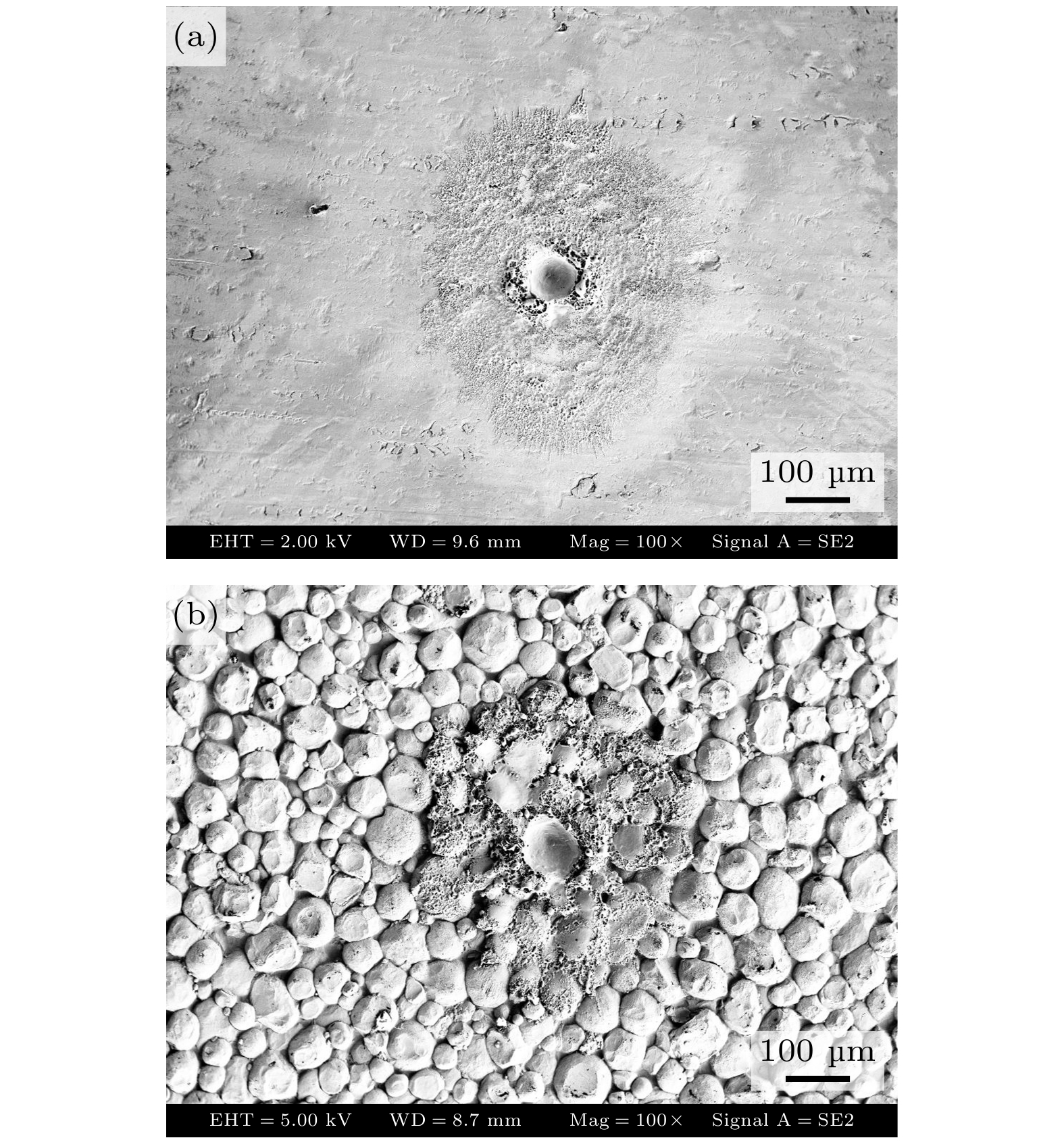
图 6 连续20个激光脉冲在镍板表面烧蚀出的坑洞的扫描电镜图 (a) 光滑表面镍板; (b) 具有半球形表面微结构的镍板
Fig. 6. SEM images of the craters generated by consecutive 20 laser shots on the surfaces of different nickel plates: (a) Flat surface Ni plate; (b) Ni plate with hemispherical micro-structure on its surface.
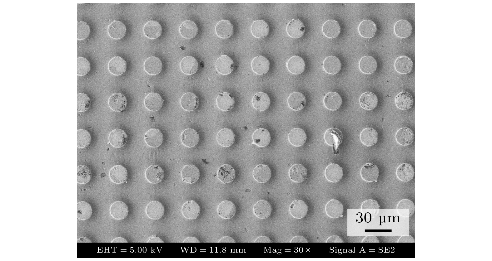
图 7 制造有直径20 μm、深15 μm、周期40 μm的圆柱形表面周期微结构镍板的扫描电镜图
Fig. 7. SEM image of a nickel plate fabricated with a periodic cylindrical surface micro-structure of 20 μm diameter, 15 μm depth and 40 μm period.
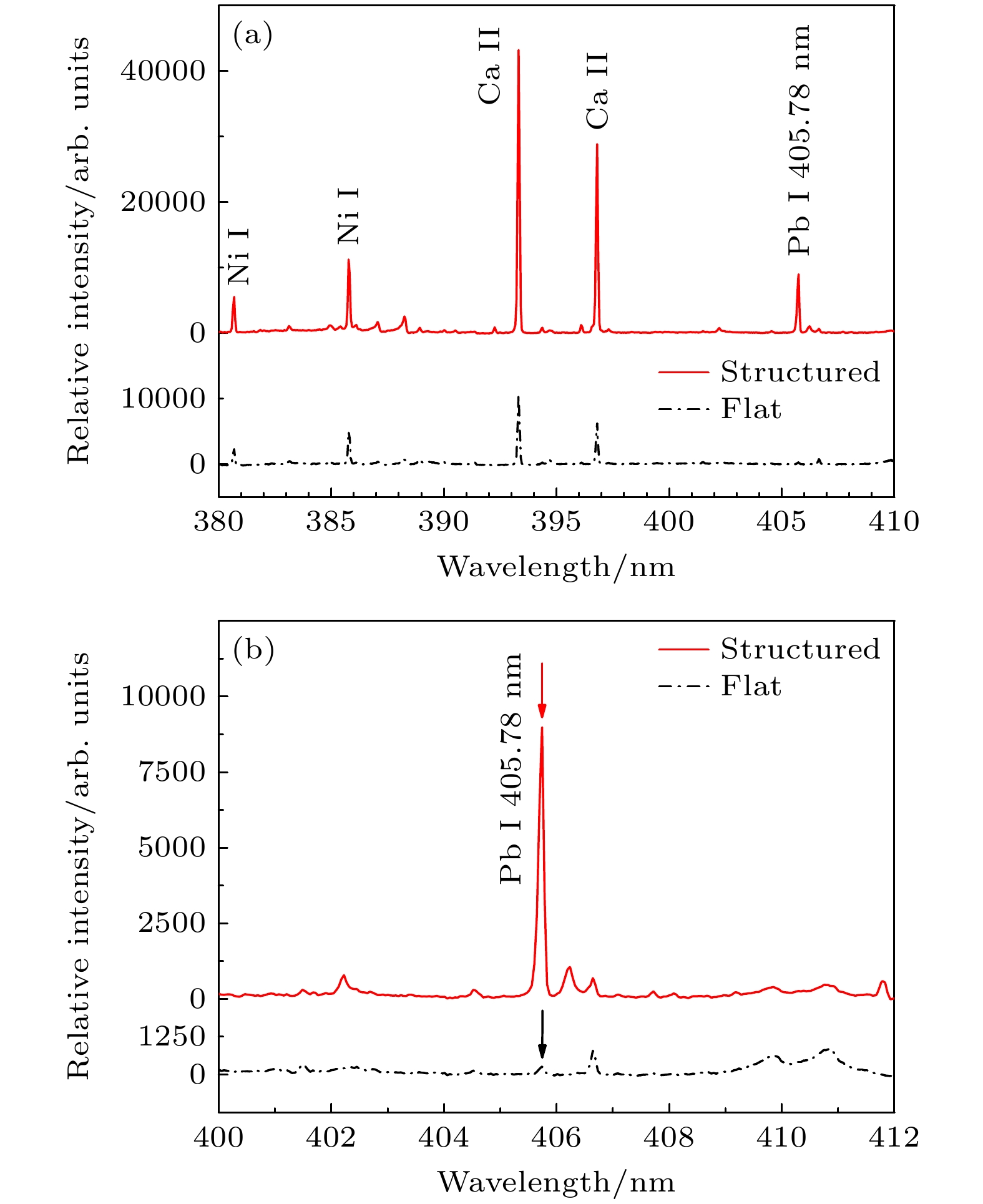
图 8 基于两种不同镍板的SENLIBS技术记录的局部光谱对照图, 样品为水溶液, 其中铅元素的浓度为20 ppm (a) 380—410 nm的光谱; (b)局部放大后400—412 nm波段的光谱
Fig. 8. Comparison of spectra recorded in SENLIBS experiments, the sample is aqueous solution containing 20 ppm lead: (a) Spectra of 380–410 nm; (b) locally amplified spectra of 400–412 nm in order to show lines more clearly.
表 1 图4中Ni原子谱线及对应的跃迁参数
Table 1. List of the selected Ni atomic lines in Fig.4 and the corresponding transition. parameters.
λki/nm Aki/(107 s–1) gk Ek/eV 367.415 0.2 5 3.80 373.681 0.14 5 3.74 383.169 0.15 3 3.66 440.155 3.8 11 6.01 447.048 1.9 7 6.17 464.866 2.4 9 6.09 478.654 1.8 11 6.01 498.016 1.9 11 6.09 501.759 2 11 6.01 503.537 5.7 9 6.10 511.540 2.2 9 6.26 547.691 0.95 3 4.09  下载: 导出CSV
-
[1] Babushok V I, DeLucia Jr. F C, Gottfried J L, Munson C A, Miziolek A W 2006 Spectrochim. Acta B 61 999
 Google Scholar
[2] 吴宜青, 刘津, 莫欣欣, 孙通, 刘木华 2017 66 054206
Google Scholar
Wu Y Q, Liu J, Mo X X, Sun T, Liu M H 2017 Acta Phys. Sin. 66 054206
Google Scholar
[3] 郑培超, 李晓娟, 王金梅, 郑爽, 赵怀冬 2019 68 125202
Google Scholar
Zheng P C, Li X J, Wang J M, Zheng S, Zhao H D 2019 Acta Phys. Sin. 68 125202
Google Scholar
[4] Nassef O A, Elsayed-Ali H E 2005 Spectrochim. Acta B 60 1564
Google Scholar
[5] Shen X K, Sun J, Ling H, Lu Y F 2007 Appl. Phys. Lett. 91 081501.
Google Scholar
[6] Popov A M, Colao F, Fantoni R 2009 J. Anal. At. Spectrom. 24 602
Google Scholar
[7] 戴宇佳, 李明亮, 宋超, 高勋, 郝作强, 林景全 2021 70 205204
Google Scholar
Dai Y J, Li M L, Song C, Gao X, Hao Z Q, Lin J Q 2021 Acta Phys. Sin. 70 205204
Google Scholar
[8] Rai V N, Zhang H S, Yueh F Y, Singh J P, Kumar A 2003 Appl. Opt. 42 3662
Google Scholar
[9] Liu Y, Baudelet M, Richardson M 2010 J. Anal. At. Spectrom. 25 1316
Google Scholar
[10] 胡慧琴, 徐雪红, 黄林, 姚明印, 陈添兵, 刘木华, 王彩虹 2016 光谱学与光谱分析 36 1180
Hu Q H, Xu X H, Huang L, Yao M Y, Chen T B, Liu M H, Wang C H 2016 Spectrosc. Spectral Anal. 36 1180
[11] De Giacomo A, Gaudiuso R, Koral C, Dell'Aglio M, Pascale O D 2013 Anal. Chem. 85 10180
Google Scholar
[12] Loudyi H, Rifai K, Laville S, Vidal F, Chaker M, Sabsabi M 2009 J. Anal. At. Spectrom. 24 1421
Google Scholar
[13] Li C M, Hao Z Q, Zou Z M, Zhou R, Li J M, Guo L B, Li X Y, Lu Y F, Zeng X Y 2016 Opt. Express 24 7850
Google Scholar
[14] Aguirre M A, Legnaioli S, Almodóvar F, Hidalgo M, Palleschi V, Canals A 2013 Spectrochim. Acta B 79-80 88
[15] Yang X Y, Hao Z Q, Li C M, Li J M, Yi R X, Shen M, Li K H, Guo L B, Li X Y, Lu Y F, Zeng X Y 2016 Opt. Express 24 13410
Google Scholar
[16] Ma S X, Tang Y, Ma Y Y, Chu Y W, Chen F, Hu Z L, Zhu Z H, Guo L B, Zeng X Y, Lu Y F 2019 Opt. Express 27 15091
Google Scholar
[17] 郑培超, 谭癸宁, 王金梅, 赵怀冬, 刘冉宁 2019 中国激光 46 0711002
Google Scholar
Zheng P C, Tan G N, Wang J M, Zhao H D, Liu R N 2019 Chin. J. Lasers 46 0711002
Google Scholar
[18] Bae D, Nam S H, Han S H, Yoo J, Lee Y H 2015 Spectrochim. Acta B 113 70
Google Scholar
[19] Matsumoto A, Shimazu Y, Yoshizumi S, Nakano H, Yae S 2020 J. Anal. At. Spectrom. 35 2239
Google Scholar
[20] Matsumoto A, Shimazu Y, Nakano H, Murakami K, Yae S 2021 Spectrochim. Acta B 178 106143
Google Scholar
[21] 陈凭, 王希林, 洪骁, 王晗, 赵晨龙, 贾志东, 邹林, 李彦民, 范建华 2019 光谱学与光谱分析 196 109568
Chen P, Wang X L, Hong X, Wang H, Zhao C L, J Z D, Zou L, Li Y M, Fan J H 2019 Spectrosc. Spectral Anal. 196 109568
[22] Kaplan D, Aras N, Yalcin S 2024 Microchem. J. 196 109568
Google Scholar
[23] Wang Q Y, Liu Y T, Jiang L Y, Chen A M, Han J H, Jin M X 2023 Anal. Chim. Acta 1241 340802
Google Scholar
[24] Wang Z H, Li Y F, Chen Y Q, Li R H 2025 Spectrochim. Acta B 226 107139
Google Scholar
[25] Bousquet B, Gardette V, Ros V M, Gaudiuso R, Dell’Aglio M, Giacomo A D 2023 Spectrochim. Acta B 204 106686
Google Scholar


 下载:
下载:


计量
- 文章访问数: 390
- PDF下载量: 6
- 被引次数: 0
