


-
高能量密度物理实验诊断中, 采用晶体谱仪等X射线分光元件可实现靶物质中心区温度和密度等参数的高谱线分辨要求, 通过优化靶点光源到探测器间的光输运效率, 可极大地提升在低光源发射度下实验诊断精度. 本文介绍了一种大口径锥型玻璃管的X光传输部件, 构建了对应的数学模型, 基于光线追迹法, 用MATLAB软件对其X射线的传输图像进行了数值模拟. 结果显示: 新设计的大口径锥型玻璃管传输部件对面型误差要求较低, 其焦斑范围可控; 可以有效提升光源的利用率, 其平均增益约为3.1. 另外, 本文还介绍了该大口径锥型玻璃管传输部件系统性的性能模拟和分析结果, 为实验室高能量密度物理研究中的低发射度X射线诊断技术升级提供了新的思路和参考.In high-energy density physics (HEDP) experiments, accurate diagnostics of physical parameters such as electron temperature, plasma density, and ionization state are essential for understanding matter behavior under extreme conditions. In these cases, X-ray spectroscopic technique, especially those using crystal spectrometers, is widely used to achieve high spectral resolution. However, a common challenge in such experiments lies in the inherent low brightness and poor spatial coherence of laboratory-based X-ray sources, which limit photon throughput, thus the diagnostic accuracy. Therefore, improving the X-ray optical transmission efficiency between the source and the detector is a key step to improve the performance of the whole system. Capillary X-ray optics, which function based on the principle of total internal reflection within hollow glass structures, provides a promising avenue for beam shaping, collimation, and focusing in the soft-to-hard X-ray range. These optical devices are usually divided into polycapillary type and monocapillary type. While polycapillary optics are composed of numerous micro-channels and used primarily for collimating or focusing divergent X-rays, monocapillary lenses—consisting of single curved channels—provide more precise beam control and are particularly suitable for customized X-ray pathways. Depending on the curvature of the inner reflective surface, monocapillaries are classified into conical, parabolic, and ellipsoidal geometries. In this study, we propose and analyze a novel design of a large-caliber conical glass tube, specifically tailored to address the issue of low light utilization in multi-channel focusing spectrographs with spatial resolution (FSSR). The proposed conical glass tube is made of a single large-diameter capillary structure, simplifying alignment requirements and reducing the surface manufacturing precision typically required by complex aspheric lenses. Its geometric configuration enables X-rays from extended or weak sources to be redirected and controlled to convergef, thereby improving photon collection without significantly altering beam divergence. To quantify the performance of this optical system, we develop a detailed mathematical ray-tracing model and implement it in MATLAB. The model combines physical parameters such as capillary inner diameter, taper angle, reflection loss, and source-detector geometry. Numerical simulations show that compared with traditional flat or slit based systems, the new conical design improves source utilization efficiency by 3.1 times. Furthermore, the lens exhibits a ring-shaped enhancement region in the output intensity profile, which can be regulated by adjusting the capillary geometry and source positioning. This feature enables the spatial customization of the beam profile, thereby facilitating optimized coupling with downstream spectroscopic components or imaging systems. In conclusion, the proposed large-aperture conical monocapillary X-ray lens provides a practical and efficient solution for enhancing X-ray optical transport in low-brightness source environments. Its simple construction, tunable focusing characteristics, and compatibility with diverse X-ray source types make it a compelling candidate for integration into a high-resolution X-ray diagnostic system, particularly in HEDP and laboratory-scale X-ray spectroscopy. This work not only introduces a novel optical approach but also provides a robust theoretical and simulation framework for guiding future experimental design and application of capillary-based X-ray optics.
-
Keywords:
- high energy density physics research /
- X-ray transmission efficiency /
- conical single capillary /
- ray tracing method /
- X-ray lens
[1] Reverdin C, Thais F, Loisel G, Bougeard M 2010 Rev. Sci. Instrum. 81 10E327
 Google Scholar
[2] Varentsov D, Ternovoi V Y, Kulish M, Fernengel D, Fertman A, Hug A, Menzel J, Ni P, Nikolaev D N, Shilkin N, Turtikov V, Udrea S, Fortov V E, Golubev A A, Gryaznov V K, Hoffmann D H H, Kim V, Lomonosov I V, Mintsev V, Sharkov By, Shutov A, Spiller P, Tahir N A, Wahl H 2007 Nucl. Instrum. Methods Phys. Res. , Sect. A 577 262
Google Scholar
[3] Ryazantsev S N, Skobelev I Y, Filippov E D, Martynenko A S, Mishchenko M D, Krůs M, Renner O, Pikuz S A 2021 Matter Radiat. Extremes 6 014402
Google Scholar
[4] Glenzer S H, Landen O L, Neumayer P, Lee R W, Widmann K, Pollaine S W, Wallace R J, Gregori G, Höll A, Bornath T, Thiele R, Schwarz V, Kraeft W D, Redmer R 2007 Phys. Rev. Lett. 98 065002
Google Scholar
[5] Regan S P, Falk K, Gregori G, Radha P B, Hu S X, Boehly T R, Crowley B J B, Glenzer S H, Landen O L, Gericke D O, Döppner T, Meyerhofer D D, Murphy C D, Sangster T C, Vorberger J 2012 Phys. Rev. Lett. 109 265003
Google Scholar
[6] Vinko S M, Ciricosta O, Cho B I, Engelhorn K, Chung H K, Brown C R D, Burian T, Chalupský J, Falcone R W, Graves C, Hájková V, Higginbotham A, Juha L, Krzywinski J, Lee H J, Messerschmidt M, Murphy C D, Ping Y, Scherz A, Schlotter W, Toleikis S, Turner J J, Vysin L, Wang T, Wu B, Zastrau U, Zhu D, Lee R W, Heimann P A, Nagler B, Wark J S 2012 Nature 482 59
Google Scholar
[7] Yi S Z, Du H Y, Si H X, Zhou Z X, Jiang L, Wang Z S, Cheng R 2023 Nucl. Instrum. Methods Phys. Res. , Sect. A 1057 168722
Google Scholar
[8] Yi Q, Meng S J, Ye F, Lu J, Yan X S, Yang R H, Jiang S Q, Ning J M, Zhou L, Chen F X, Yang J L, Xu Z P, Li Z H 2023 AIP Adv. 13 035216
Google Scholar
[9] Renner O, Šmíd M, Batani D, Antonelli L 2016 Plasma Phys. Controlled Fusion 58 75007
Google Scholar
[10] Eftekhari-Zadeh E, Loetzsch R, Manganelli L, Blümcke M S, Tauschwitz A, Uschmann I, Pukhov A, Rosmej O, Spielmann C, Kartashov D 2023 Phys. Scr. 98 115615
Google Scholar
[11] Hurricane O A, Herrmann M C 2017 Annu. Rev. Nucl. Part. Sci. 67 213
Google Scholar
[12] Zhao Y, Yang J M, Zhang J Y, Liu J S, Yuan X, Jin F T 2009 Rev. Sci. Instrum. 80 043505
Google Scholar
[13] Kumakhov M A, Komarov F F 1990 Phys. Rep. 191 289
Google Scholar
[14] Balaic D X, Nugent K A, Barnea Z, Garrett R, Wilkins W 1995 J. Synchrotron Radiat. 2 296
Google Scholar
[15] Yokomae S, Motoyama H, Mimura H 2018 Precis. Eng. 53 248
Google Scholar
[16] MacDonald C A 2010 X-Ray Opt. Instrum. 2010 867049
[17] Gibson W M, Kumakhov M 1993 Proc. SPIE. 172
[18] Bilderback D H, Hoffman S A, Thiel D J 1994 Science 263 201
Google Scholar
[19] Sowa K M, Jany B R, Korecki P 2018 Optica 5 577
Google Scholar
[20] Korecki P, Sowa K M, Jany B R, Krok F 2016 Phys. Rev. Lett. 116 233902
Google Scholar
[21] Szwedowski-Rammert V, Baumann J, Schlesiger C, Waldschläger U, Gross A, Kanngießer B, Mantouvalou I 2019 J. Anal. At. Spectrom. 34 922
Google Scholar
[22] Matsuyama T, Tanaka Y, Taniguchi N, Oh J S, Tsuji K 2024 J. Anal. At. Spectrom. 39 76
Google Scholar
[23] Matsuyama T, Tanaka Y, Mori Y, Tsuji K 2023 Talanta 265 124808
Google Scholar
[24] Peng S, Liu Z G, Sun T X, Ma Y Z, Ding X L 2014 Anal. Chem. 86 362
Google Scholar
[25] Fittschen U E A, Falkenberg G 2011 Spectrochim. Acta, Part B 66 567
Google Scholar
[26] Wallen S L, Pfund D M, Fulton J L, Yonker C R, Newville M, Ma Y 1996 Rev. Sci. Instrum. 67 2843
Google Scholar
[27] Alexandre B J, Gomes M G, Real S 2015 Mater. Struct. 48 2869
Google Scholar
[28] Lin X Y, Li Y D, Sun T X, Pan Q L 2010 Chin. Phys. B 19 070205
[29] Liu A D, Lin Y Z 2004 Math. Comput. Simul. 66 577
Google Scholar
[30] Peng S Q, Liu Z G, Sun T X, Wang K, Yi L T, Yang K, Chen M, Wang J B 2015 Nucl. Instrum. Methods Phys. Res., Sect. A 795 186
Google Scholar
[31] Sun T X, Ding X L 2015 Rev. Anal. Chem. 34 45
[32] Stern E A, Kalman Z, Lewis A, Lieberman K 1988 Appl. Opt. 27 5135
Google Scholar
[33] Wen H, Zhou M, Wu Y M, Yuan T Y, Liu Z G 2022 Appl. Opt. 61 3656
Google Scholar
[34] Motoyama H, Sato T, Iwasaki A, Takei Y, Kume T, Egawa S, Hiraguri K, Hashizume H, Yamanouchi K, Mimura H 2016 Rev. Sci. Instrum. 87 051803
Google Scholar
[35] Koch R J, Jozwiak C, Bostwick A, Stripe B, Cordier M, Hussain Z, Yun W, Rotenberg E 2018 J. Synchrotron Radiat. 31 50
Google Scholar
[36] 邵尚坤, 袁天语, 李惠泉, 孙学鹏, 华陆, 刘志国, 孙天希 2022 北京师范大学学报(自然科学版) 58 681
Shao S K, Yuan T Y, Li H Q, Sun X P, Hua L, Liu Z G, Sun T X 2022 J. Beijing Norm. Univ. (Nat. Sci.) 58 681
[37] Jiang B W, Liu Z G, Sun X P, Sun T X, Deng B, Li F Z, Yi L T, Yuan M N, Zhu Y, Zhang F S, Xiao T Q, Wang J, Tai R Z 2017 Opt. Commun. 398 91
Google Scholar
[38] Balaic D X, Barnea Z, Nugent K A, Garrett R F, Varghese J N, Wilkins S W 1996 J. Synchrotron Radiat. 3 289
Google Scholar
[39] Wang Y B, Li Y L, Shao S K, Zhang X Y, Li Y F, Sun X P, Tao F, Deng B, Sun T X 2020 Opt. Commun. 464 125544
Google Scholar
[40] Wang X Y, Li Y D, Luo H, Ye L, Zhou M, Duan J Y, Lin X Y 2019 Nucl. Instrum. Methods Phys. Res., Sect. A 947 162762
Google Scholar
[41] Sun X P, Zhang X Y, Zhu Y, Wang Y B, Shang H Z, Zhang F S, Liu Z G, Sun T X 2018 Nucl. Instrum. Methods Phys. Res. , Sect. A 888 13
Google Scholar
[42] Zhou Z X, Cheng R, Du H Y, Yi S Z, Fu F, Wang G D, Shi L L, Wang Z, Jin X J, Chen Y H, Zhang Y S, Chen L W, Yang J, Su M G 2024 J. Anal. At. Spectrom. 39 31
[43] 孙天希 2022 光学学报 42 1134002
Google Scholar
Sun T X 2022 Acta Opt. Sin. 42 1134002
Google Scholar
[44] Shao S K, Li H Q, Yuan T Y, Zhang X Y, Hua L, Sun X P, Liu Z G, Sun T X 2022 Front. Phys. 10 816981
Google Scholar
[45] Zymierska D 1996 Acta. Phys. Pol. A 89 347
Google Scholar
-
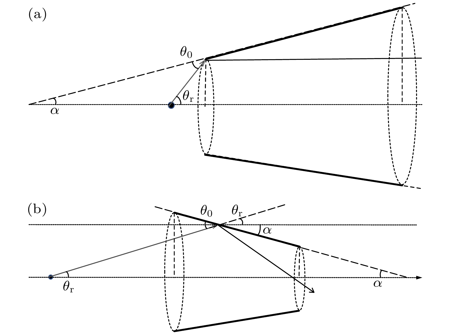
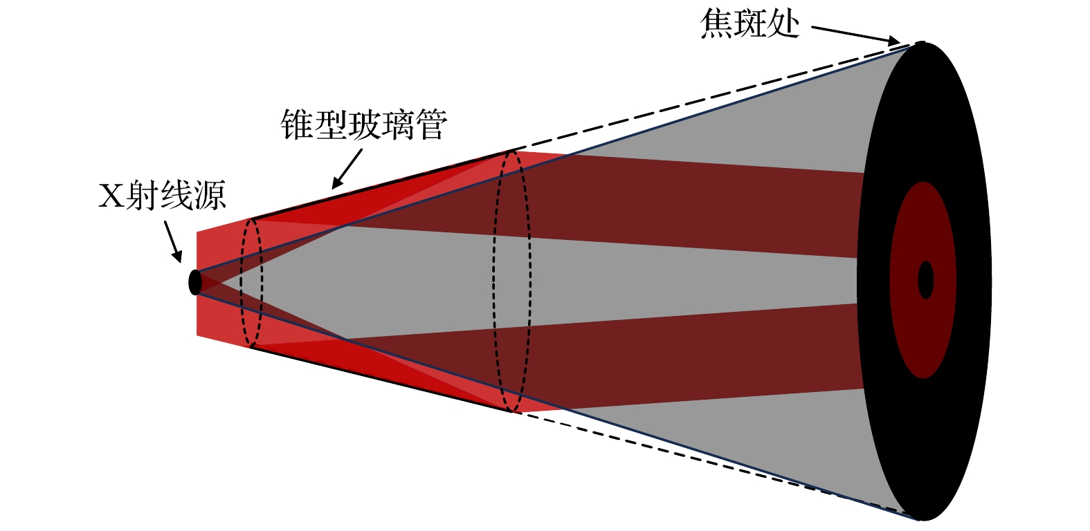
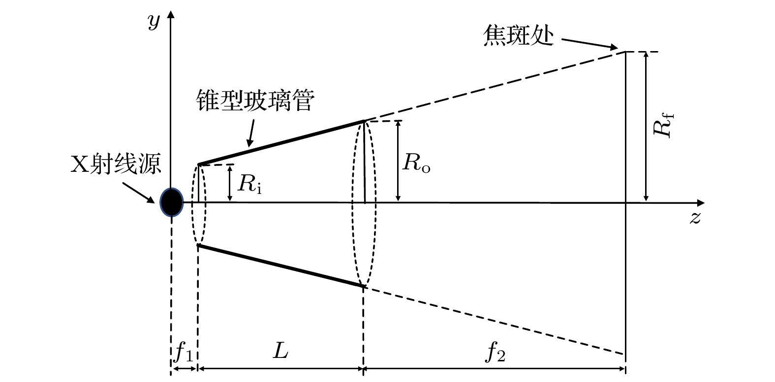
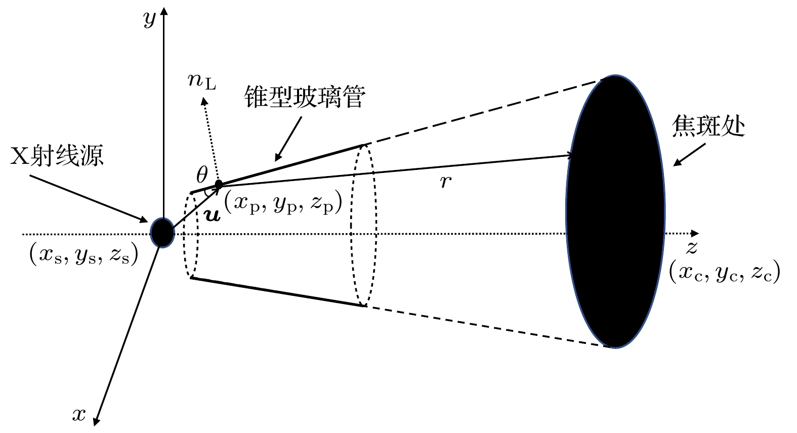
图 1 锥型玻璃管使用对比图 (a)锥管逆向使用的光路图; (b)锥管正向使用的光路图
Fig. 1. Comparison of conical glass tube usage: (a) Light path with reverse use of the tube; (b) light path with forward use of the tube.
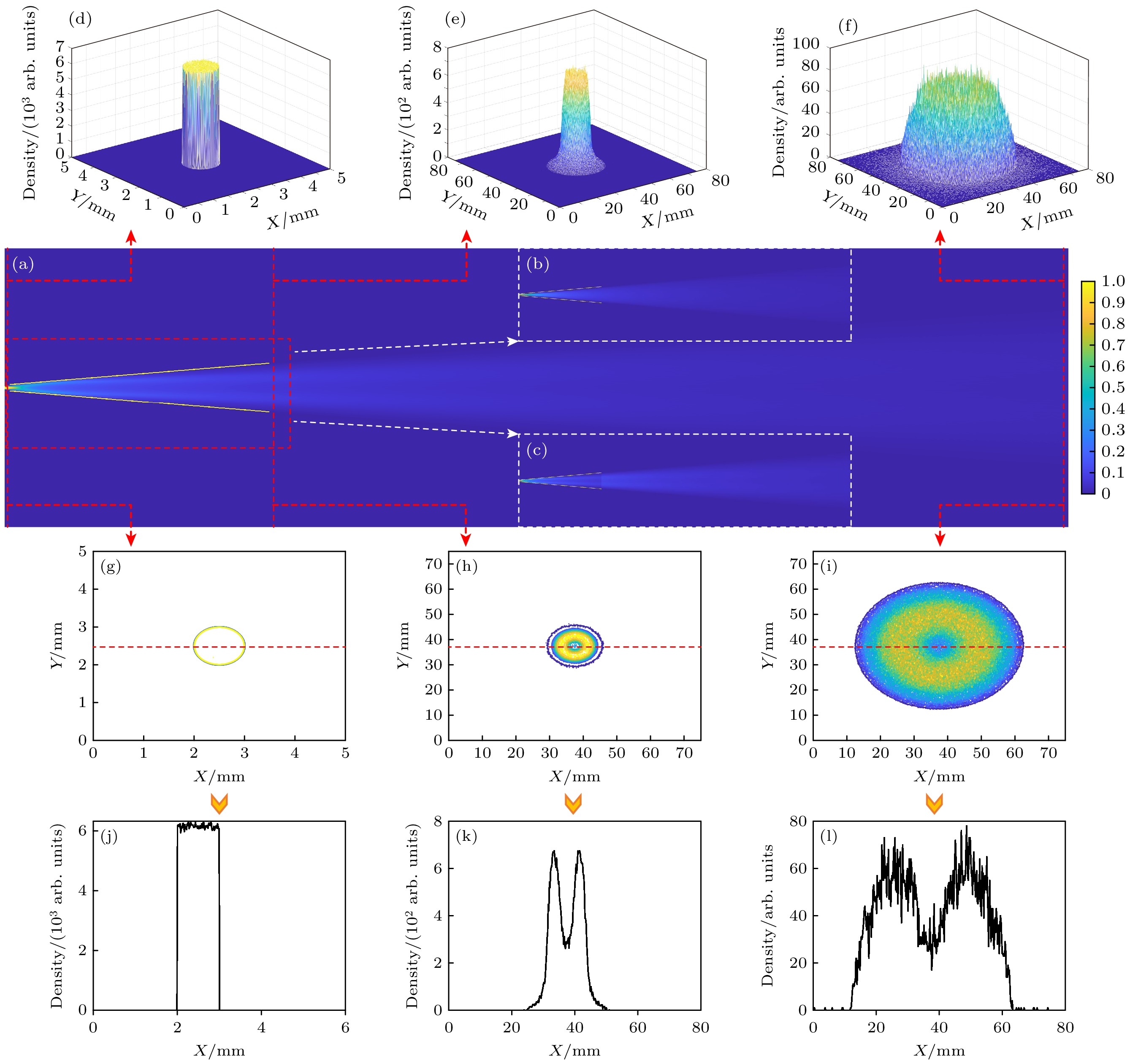
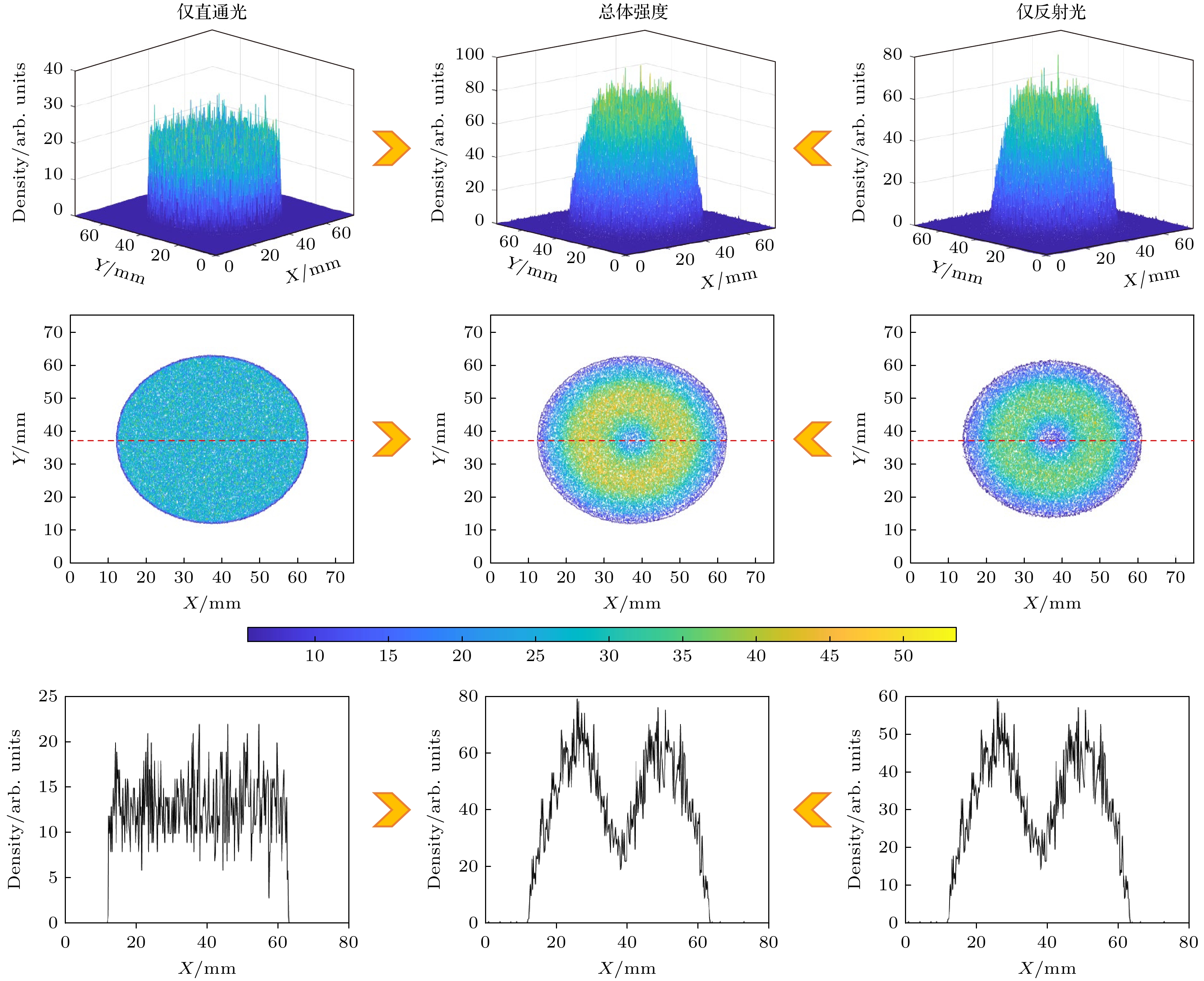
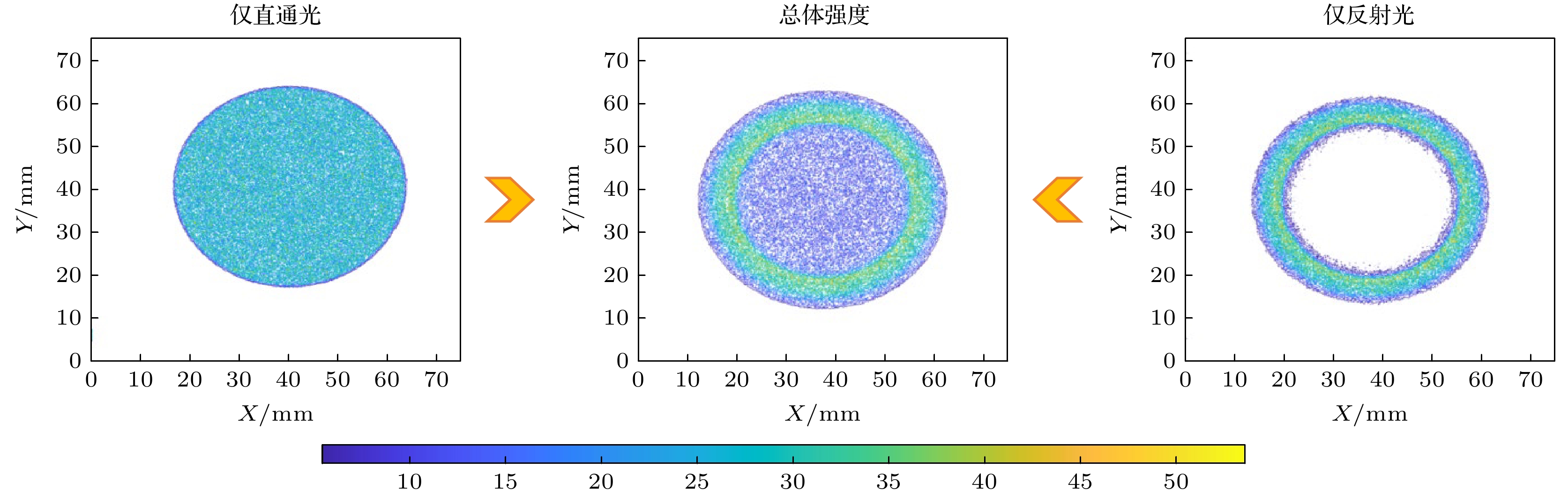
图 5 LCGTXL X射线传输的可视化 (a) X射线传输的整体剖面图; (b)直通光的部分传输图; (c)反射光的部分传输图; (d)—(f)在图(a)中红色虚线的三维强度分布图; (g)—(i)在图(a)中红色虚线的二维分布; (j)—(l)分别沿着图(g)—(i)中的红色虚线的强度分布
Fig. 5. Visualization of X-ray transmission in LCGTXL: (a) Overall profile of X-ray transmission; (b) partial transmission diagram of straight light; (c) partial transmission diagram of reflected light; (d)–(f) three-dimensional intensity distribution of the red dotted line in Figure (a); (g)–(i) two-dimensional distribution of the red dotted line in Figure (a); (j)–(l) intensity distribution along red dotted lines in Figure(g)–(i), respectively.
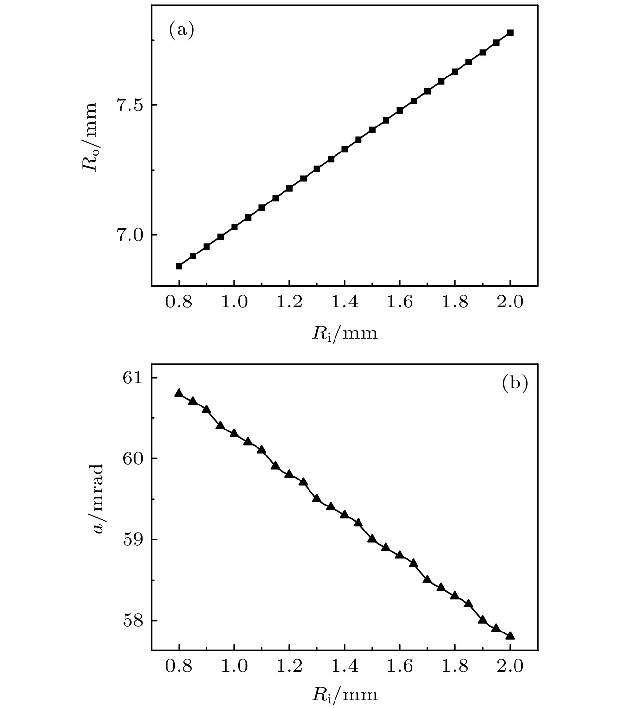
图 9 斜率$ a $ 和出口半径$ {R}_{{\mathrm{o}}} $随入口半径$ {R}_{{\mathrm{i}}} $的变化关系
Fig. 9. Relationship between slope $ a $ and outlet radius $ {R}_{{\mathrm{o}}} $ with inlet radius $ {R}_{{\mathrm{i}}} $.
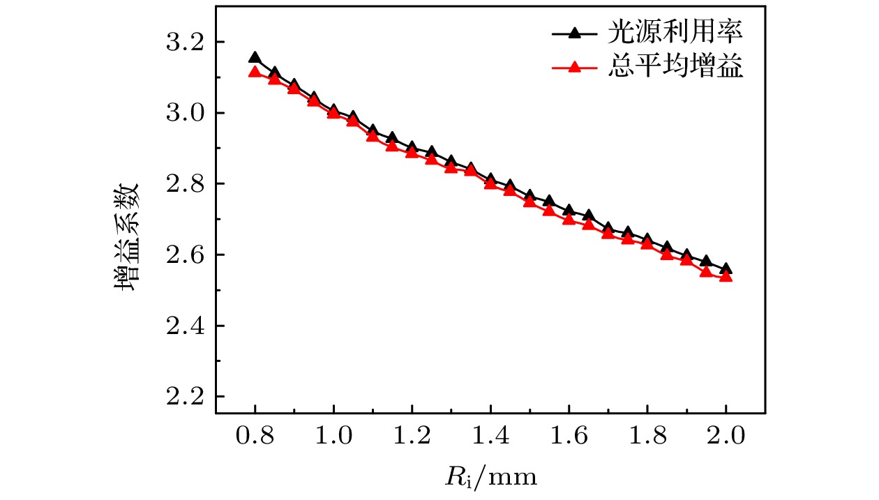
图 10 LCGTXL传输性能与入口半径的关系
Fig. 10. Relationship between LCGTXL transmission performance and inlet radius.
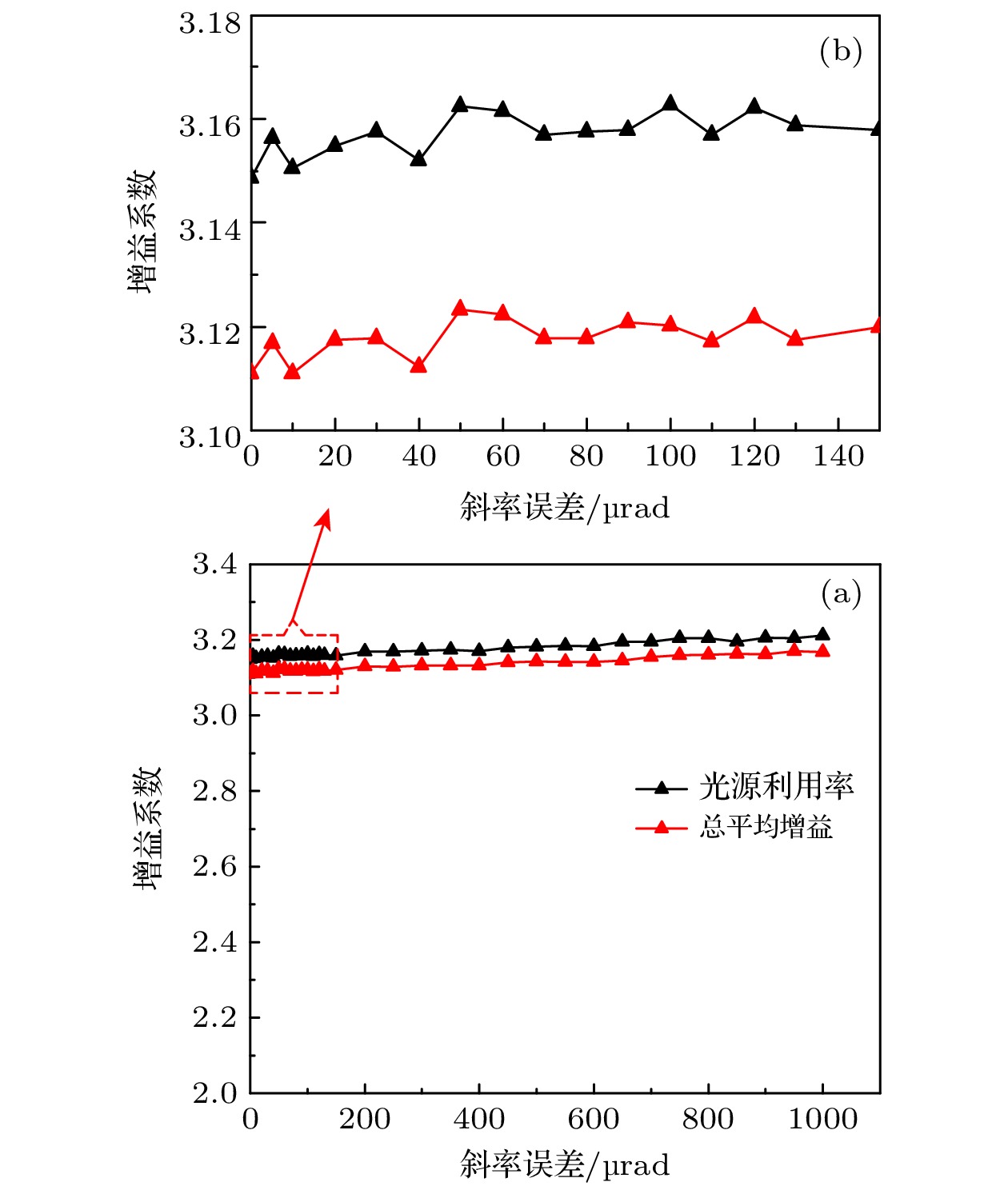
图 11 LCGTXL传输性能与斜率误差的关系
Fig. 11. Relationship between LCGTXL transmission performance and slope error.
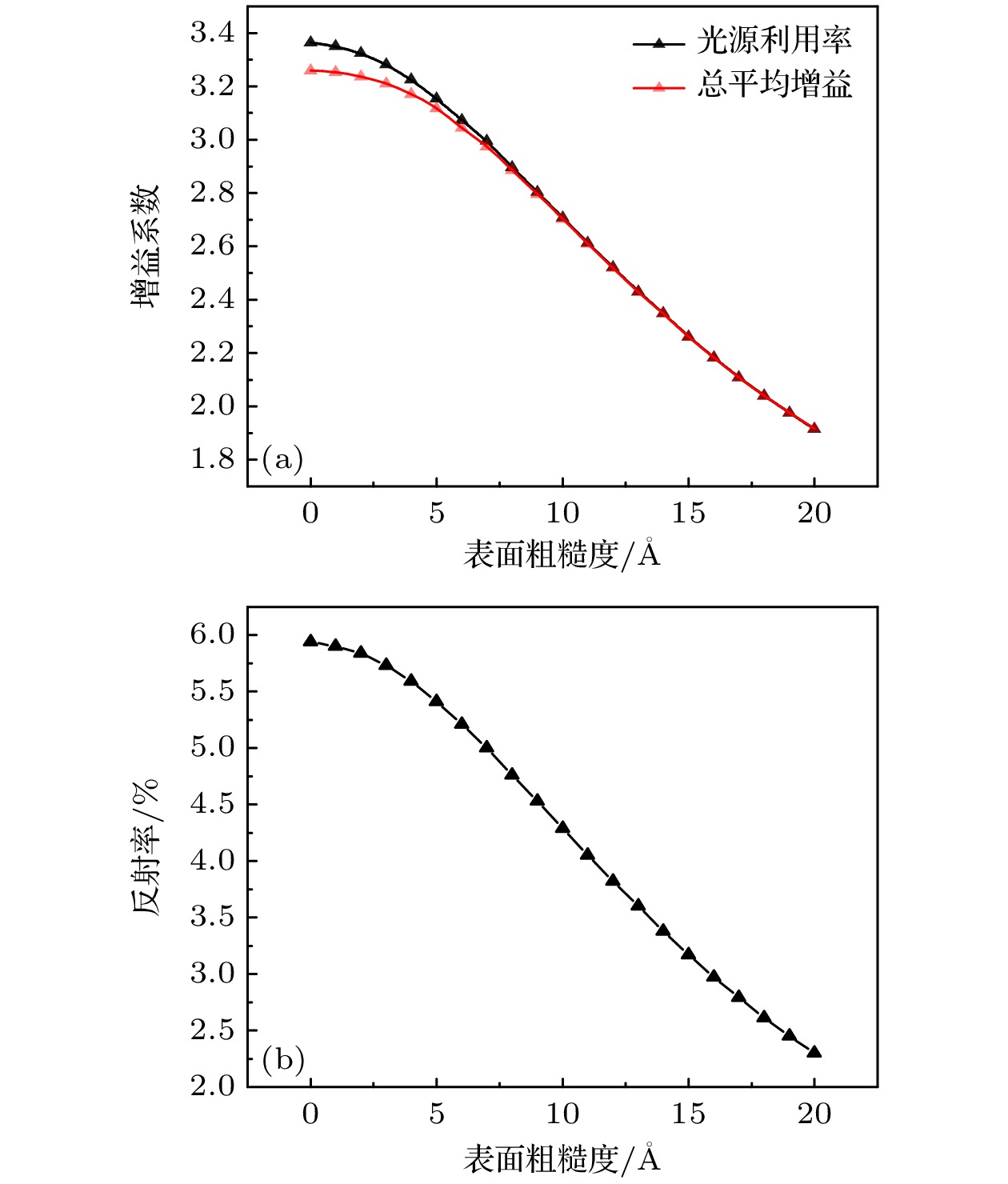
图 12 LCGTXL传输性能与表面粗糙度的关系 (a)光源利用率和总平均增益随表面粗糙度的变化关系; (b)表面粗糙度对反射率的影响
Fig. 12. Relationship between LCGTXL transmission performance and surface roughness: (a) Variation of source utilization and total average gain with surface roughness; (b) effect of surface roughness on reflectivity.
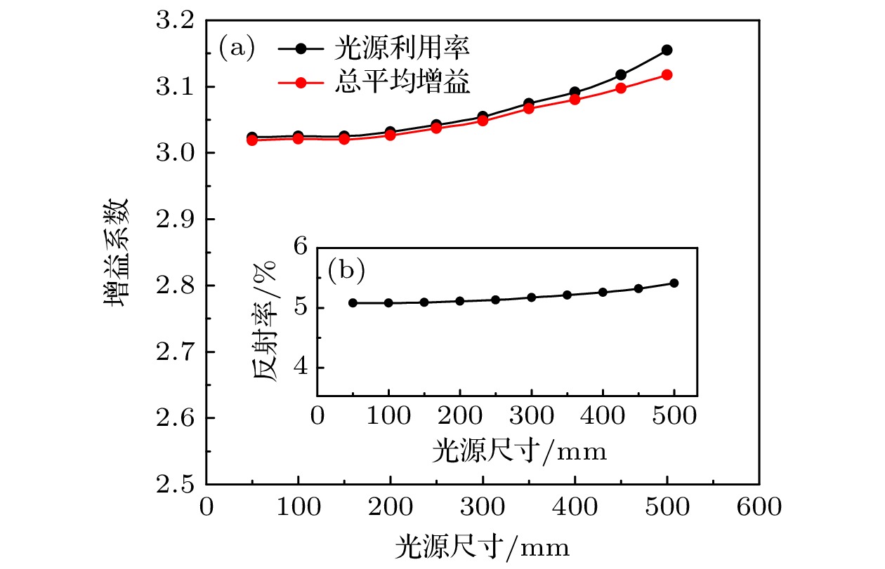
图 13 LCGTXL传输性能与光源尺寸的关系 (a)光源尺寸对光源利用率和总平均增益的影响; (b) 光源尺寸对反射率的影响
Fig. 13. Relationship between LCGTXL transmission performance and source size: (a) Effect of source size on source utilization and total average gain; (b) effect of source size on reflectivity.
表 1 LCGTXL中的几何仿真参数
Table 1. Geometric simulation parameters in LCGTXL.
参数 $ {f}_{1} $ $ L $ $ {f}_{2} $ $ {R}_{{\mathrm{i}}} $ $ {R}_{{\mathrm{o}}} $ $ {R}_{{\mathrm{f}}} $ 单位/mm 2 100 298 0.8 6.88 25  下载: 导出CSV
表 2 不同条件下的传输性能
Table 2. Transmission performance under different conditions.
种类 X射线能量/keV 光源利用率 增益 不加透镜 1.50 1.000 1.000 玻璃管 1.50 1.588 1.599 镀铱玻璃管 1.50 3.155 3.118
下载: 导出CSV
-
[1] Reverdin C, Thais F, Loisel G, Bougeard M 2010 Rev. Sci. Instrum. 81 10E327
Google Scholar
[2] Varentsov D, Ternovoi V Y, Kulish M, Fernengel D, Fertman A, Hug A, Menzel J, Ni P, Nikolaev D N, Shilkin N, Turtikov V, Udrea S, Fortov V E, Golubev A A, Gryaznov V K, Hoffmann D H H, Kim V, Lomonosov I V, Mintsev V, Sharkov By, Shutov A, Spiller P, Tahir N A, Wahl H 2007 Nucl. Instrum. Methods Phys. Res. , Sect. A 577 262
Google Scholar
[3] Ryazantsev S N, Skobelev I Y, Filippov E D, Martynenko A S, Mishchenko M D, Krůs M, Renner O, Pikuz S A 2021 Matter Radiat. Extremes 6 014402
Google Scholar
[4] Glenzer S H, Landen O L, Neumayer P, Lee R W, Widmann K, Pollaine S W, Wallace R J, Gregori G, Höll A, Bornath T, Thiele R, Schwarz V, Kraeft W D, Redmer R 2007 Phys. Rev. Lett. 98 065002
Google Scholar
[5] Regan S P, Falk K, Gregori G, Radha P B, Hu S X, Boehly T R, Crowley B J B, Glenzer S H, Landen O L, Gericke D O, Döppner T, Meyerhofer D D, Murphy C D, Sangster T C, Vorberger J 2012 Phys. Rev. Lett. 109 265003
Google Scholar
[6] Vinko S M, Ciricosta O, Cho B I, Engelhorn K, Chung H K, Brown C R D, Burian T, Chalupský J, Falcone R W, Graves C, Hájková V, Higginbotham A, Juha L, Krzywinski J, Lee H J, Messerschmidt M, Murphy C D, Ping Y, Scherz A, Schlotter W, Toleikis S, Turner J J, Vysin L, Wang T, Wu B, Zastrau U, Zhu D, Lee R W, Heimann P A, Nagler B, Wark J S 2012 Nature 482 59
Google Scholar
[7] Yi S Z, Du H Y, Si H X, Zhou Z X, Jiang L, Wang Z S, Cheng R 2023 Nucl. Instrum. Methods Phys. Res. , Sect. A 1057 168722
Google Scholar
[8] Yi Q, Meng S J, Ye F, Lu J, Yan X S, Yang R H, Jiang S Q, Ning J M, Zhou L, Chen F X, Yang J L, Xu Z P, Li Z H 2023 AIP Adv. 13 035216
Google Scholar
[9] Renner O, Šmíd M, Batani D, Antonelli L 2016 Plasma Phys. Controlled Fusion 58 75007
Google Scholar
[10] Eftekhari-Zadeh E, Loetzsch R, Manganelli L, Blümcke M S, Tauschwitz A, Uschmann I, Pukhov A, Rosmej O, Spielmann C, Kartashov D 2023 Phys. Scr. 98 115615
Google Scholar
[11] Hurricane O A, Herrmann M C 2017 Annu. Rev. Nucl. Part. Sci. 67 213
Google Scholar
[12] Zhao Y, Yang J M, Zhang J Y, Liu J S, Yuan X, Jin F T 2009 Rev. Sci. Instrum. 80 043505
Google Scholar
[13] Kumakhov M A, Komarov F F 1990 Phys. Rep. 191 289
Google Scholar
[14] Balaic D X, Nugent K A, Barnea Z, Garrett R, Wilkins W 1995 J. Synchrotron Radiat. 2 296
Google Scholar
[15] Yokomae S, Motoyama H, Mimura H 2018 Precis. Eng. 53 248
Google Scholar
[16] MacDonald C A 2010 X-Ray Opt. Instrum. 2010 867049
[17] Gibson W M, Kumakhov M 1993 Proc. SPIE. 172
[18] Bilderback D H, Hoffman S A, Thiel D J 1994 Science 263 201
Google Scholar
[19] Sowa K M, Jany B R, Korecki P 2018 Optica 5 577
Google Scholar
[20] Korecki P, Sowa K M, Jany B R, Krok F 2016 Phys. Rev. Lett. 116 233902
Google Scholar
[21] Szwedowski-Rammert V, Baumann J, Schlesiger C, Waldschläger U, Gross A, Kanngießer B, Mantouvalou I 2019 J. Anal. At. Spectrom. 34 922
Google Scholar
[22] Matsuyama T, Tanaka Y, Taniguchi N, Oh J S, Tsuji K 2024 J. Anal. At. Spectrom. 39 76
Google Scholar
[23] Matsuyama T, Tanaka Y, Mori Y, Tsuji K 2023 Talanta 265 124808
Google Scholar
[24] Peng S, Liu Z G, Sun T X, Ma Y Z, Ding X L 2014 Anal. Chem. 86 362
Google Scholar
[25] Fittschen U E A, Falkenberg G 2011 Spectrochim. Acta, Part B 66 567
Google Scholar
[26] Wallen S L, Pfund D M, Fulton J L, Yonker C R, Newville M, Ma Y 1996 Rev. Sci. Instrum. 67 2843
Google Scholar
[27] Alexandre B J, Gomes M G, Real S 2015 Mater. Struct. 48 2869
Google Scholar
[28] Lin X Y, Li Y D, Sun T X, Pan Q L 2010 Chin. Phys. B 19 070205
[29] Liu A D, Lin Y Z 2004 Math. Comput. Simul. 66 577
Google Scholar
[30] Peng S Q, Liu Z G, Sun T X, Wang K, Yi L T, Yang K, Chen M, Wang J B 2015 Nucl. Instrum. Methods Phys. Res., Sect. A 795 186
Google Scholar
[31] Sun T X, Ding X L 2015 Rev. Anal. Chem. 34 45
[32] Stern E A, Kalman Z, Lewis A, Lieberman K 1988 Appl. Opt. 27 5135
Google Scholar
[33] Wen H, Zhou M, Wu Y M, Yuan T Y, Liu Z G 2022 Appl. Opt. 61 3656
Google Scholar
[34] Motoyama H, Sato T, Iwasaki A, Takei Y, Kume T, Egawa S, Hiraguri K, Hashizume H, Yamanouchi K, Mimura H 2016 Rev. Sci. Instrum. 87 051803
Google Scholar
[35] Koch R J, Jozwiak C, Bostwick A, Stripe B, Cordier M, Hussain Z, Yun W, Rotenberg E 2018 J. Synchrotron Radiat. 31 50
Google Scholar
[36] 邵尚坤, 袁天语, 李惠泉, 孙学鹏, 华陆, 刘志国, 孙天希 2022 北京师范大学学报(自然科学版) 58 681
Shao S K, Yuan T Y, Li H Q, Sun X P, Hua L, Liu Z G, Sun T X 2022 J. Beijing Norm. Univ. (Nat. Sci.) 58 681
[37] Jiang B W, Liu Z G, Sun X P, Sun T X, Deng B, Li F Z, Yi L T, Yuan M N, Zhu Y, Zhang F S, Xiao T Q, Wang J, Tai R Z 2017 Opt. Commun. 398 91
Google Scholar
[38] Balaic D X, Barnea Z, Nugent K A, Garrett R F, Varghese J N, Wilkins S W 1996 J. Synchrotron Radiat. 3 289
Google Scholar
[39] Wang Y B, Li Y L, Shao S K, Zhang X Y, Li Y F, Sun X P, Tao F, Deng B, Sun T X 2020 Opt. Commun. 464 125544
Google Scholar
[40] Wang X Y, Li Y D, Luo H, Ye L, Zhou M, Duan J Y, Lin X Y 2019 Nucl. Instrum. Methods Phys. Res., Sect. A 947 162762
Google Scholar
[41] Sun X P, Zhang X Y, Zhu Y, Wang Y B, Shang H Z, Zhang F S, Liu Z G, Sun T X 2018 Nucl. Instrum. Methods Phys. Res. , Sect. A 888 13
Google Scholar
[42] Zhou Z X, Cheng R, Du H Y, Yi S Z, Fu F, Wang G D, Shi L L, Wang Z, Jin X J, Chen Y H, Zhang Y S, Chen L W, Yang J, Su M G 2024 J. Anal. At. Spectrom. 39 31
[43] 孙天希 2022 光学学报 42 1134002
Google Scholar
Sun T X 2022 Acta Opt. Sin. 42 1134002
Google Scholar
[44] Shao S K, Li H Q, Yuan T Y, Zhang X Y, Hua L, Sun X P, Liu Z G, Sun T X 2022 Front. Phys. 10 816981
Google Scholar
[45] Zymierska D 1996 Acta. Phys. Pol. A 89 347
Google Scholar


 下载:
下载:


计量
- 文章访问数: 350
- PDF下载量: 12
- 被引次数: 0
