-
在激光扫描共聚焦显微镜的基础上, 线扫描荧光显微术利用线扫描代替点扫描, 提升图像获取速度, 具有系统结构简单、成像速度快、光毒性弱、更适合于活体厚样品的高分辨快速成像, 对于生命科学和生物医学等领域的研究具有重要的意义. 然而, 目前的线扫描显微技术在系统灵活性、成像速度、分辨率和光学层析能力等方面仍面临着许多亟需解决的问题. 因此, 本文提出一种基于数字微镜器件(digital micromirror device, DMD)的数字线扫描荧光显微(digital line-scanning fluorescence microscopy, DLSFM)成像方法和系统, 在照明光路中引入高速空间光调制器DMD实现多线并行扫描激发, 简化光学系统, 提升系统灵活性和扫描速度; 提出基于荧光信号标准差的DLSFM图像重构算法, 结合三维Landweber解卷积算法实现了三维高分辨光切片图像重构. 在此基础上, 利用搭建DLSFM开展了荧光珠和老鼠肾切片标准样品的成像实验, 实验结果表明, DLSFM具有快速三维高分辨层析成像能力.Laser scanning confocal microscope (LSCM) is one of the most important tools for biological imaging due to its strong optical sectioning capability, high signal-to-noise ratio, and high resolution. On the basis of LSCM, line-scanning fluorescence microscopy (LSFM) uses linear scanning instead of point scanning to improve the speed of image acquisition. It has the advantages of simple system structure, fast imaging speed, and weak phototoxicity, and in addition, it is more suitable for high-resolution and fast imaging of living thick samples. It is of great significance for studying the life science, biomedicine, and others. However, the current LSFM technology still faces many urgent problems in terms of system flexibility, imaging speed, resolution and optical sectioning capabilities. Therefore, based on the existing multifocal structured illumination microscopy (MSIM) in our laboratory, a digital line-scanning fluorescence microscopy (DLSFM) based on digital micromirror device(DMD) is presented in this paper. In the illumination path, a high-speed spatial light modulator DMD is adopted to realize multi-line parallel scanning excitation, which simplies the optical system and improves the flexibility and scanning speed of the system. A DLSFM image reconstruction algorithm based on the standard deviation of fluorescence signal is proposed, which is combined withthree-dimensional (3D) Landweber deconvolution algorithm to achieve 3D high-resolution optical slice image reconstruction. On this basis, the imaging experiments on fluorescent beads and standard samples of mouse kidney section are carried out by using DLSFM. The experimental results show that the resolution of DLSFM in the x, y and z directions is 1.33 times, 1.42 times and 1.19 times that of wide field microscope, respectively, and the fast 3D high-resolution optical sectioning imaging of biological samples is realized, which lays a technical foundation for further developing the rapid high-resolution imaging of the whole cells and tissues in vivo.
[1] Minsky M 1988 Scanning 10 128
 Google Scholar
[2] Mitsuhiro I, Hiromi S 1998 SPIE 34 68
[3] Gustafsson M G 2010 J. Microsc. 198 82
[4] 赵启韬, 苗俊英 2003 北京生物医学工程 22 52
 Google Scholar
Zhao Q T, Miao J Y 2003 Beijing Biomed. Eng. 22 52
 Google Scholar
[5] Rainer H, Christoph G C 1999 SPIE 35 185
[6] Sauermann K, Gambichler T, Wilmert M 2002 Skin Res.Technol. 8 141
 Google Scholar
[7] Taejoong K, DaeGab G, Jun-Hee L 2009 Meas. Sci. Technol. 20 055501
 Google Scholar
[8] SchulzO, Pieper C, Clever M, et al. 2013 Proc. Natl. Acad. Sci. U.S.A. 110 21000
 Google Scholar
[9] Fan G Y, Fujisaki H, Miywwaki A, Tsay R K, Tsien R Y, Ellisman M H 1999 Biophys. J. 76 2412
 Google Scholar
[10] Rahadhyaksha M, Anderson R R, Webb R H 1999 Appl. Opt. 38 2105
 Google Scholar
[11] Li Y X, Gautam V, Brüstle A, Cockburn I A, Daria VR, Gillespie C, Gaus K, Alt C, Lee W M 2017 J. Bophotonics 10 1526
 Google Scholar
[12] Sheppard C JR, Mao X Q 1988 J. Mod. Optic. 35 1169
 Google Scholar
[13] Puash W C, Wasser M 2016 Methods 96 103
 Google Scholar
[14] Yazawa M, Hsueh B, Jia X, Pasca A M, Bernstein J A, Hallmayer J, Dolmetsch R E 2011 Nature 471 230
 Google Scholar
[15] Reto F, Andreas S, Yury B 2007 Histochem. Cell Biol. 128 499
 Google Scholar
[16] Kang B I, Sumin H, Hwajoon P, Dongsun K, Beop M K 2005 Opt. Express 13 5151
 Google Scholar
[17] Ondrej M, Martin K, Kai Wicker, Gerhard K, Ingo K, Rainer H 2012 Opt. Express 20 24167
 Google Scholar
[18] Lu R W, Wang B Q, Zhang QX, Yao XC 2013 Biomed.Opt. Express 4 1673
 Google Scholar
[19] Wang B, Lu R, Zhang Q, Yao X 2013 Quant. Imag. Med. Surg. 3 243
[20] Yanan Z, Lu R W, Wang B Q, Zhang Q X, Yao X C 2015 Opt. Lett. 40 1683
[21] Poher V, Zhang HX, Kennedy GT 2007 Opt. Express 15 11196
 Google Scholar
[22] Chamma I, Levet F, Sibarita J B, Sainlos M, Thoumine O 2016 Neurophotonics 3 041810
 Google Scholar
[23] Endesfelder U, Heilemann M 2014 Nat. Methods 11 235
 Google Scholar
[24] Flors C 2011 Biopolymers 95 290
 Google Scholar
[25] Patterson G, Davidson M, Manley S 2010 Annu. Rev. Phys. Chem. 61 345
 Google Scholar
[26] Hossain S, Hashimoto M, Katakura M, Mamun A A, Shido O 2015 Bmc. Complem. Altern. M 15 1
[27] Rainer H, Pier A, Benedetti 2006 Appl. Opt. 45 5037
 Google Scholar
[28] Ren Y X, Lu R D, Gong L 2015 Ann. Phys.(Berlin) 527 447
 Google Scholar
[29] 冯维, 张福民, 王惟婧, 曲兴华 2017 66 234201
 Google Scholar
Feng W, Zhang F M, Wang W J, Qu X H 2017 Acta Phys. Sin. 66 234201
 Google Scholar
[30] Vienola K V, Damodaran M, Braaf B, Vermeer K A, Johannes F 2015 Opt. Lett. 40 5335
 Google Scholar
[31] Wu J J, Li S W, Cao H Q, Lin D Y, Yu B, Qu J L 2018 Opt. Express 26 31430
 Google Scholar
[32] Vonesch C, Unser M 2008 IEEE Trans. Image Process. 17 539
[33] Zhang W, Yu B, Lin D Y, Yu H H, Li S W, Qu J L 2020 Opt. Express 28 10919
 Google Scholar
-
图 3 DMD不同填充因子的信噪比分析 (a) 焦面信号和背景信号强度分布图; (b) 焦面信号和背景信号强度与DMD填充因子的关系曲线; (c) DMD不同填充因子的信噪比曲线
Fig. 3. SNR analysis of DMD with different fill factor: (a) The intensity distributions of focal plane signal and background signal intensity; (b) the intensity curves of focal planne signal (black) and background signal (red); (c) the curve of the SNR versus fill factor of DMD.
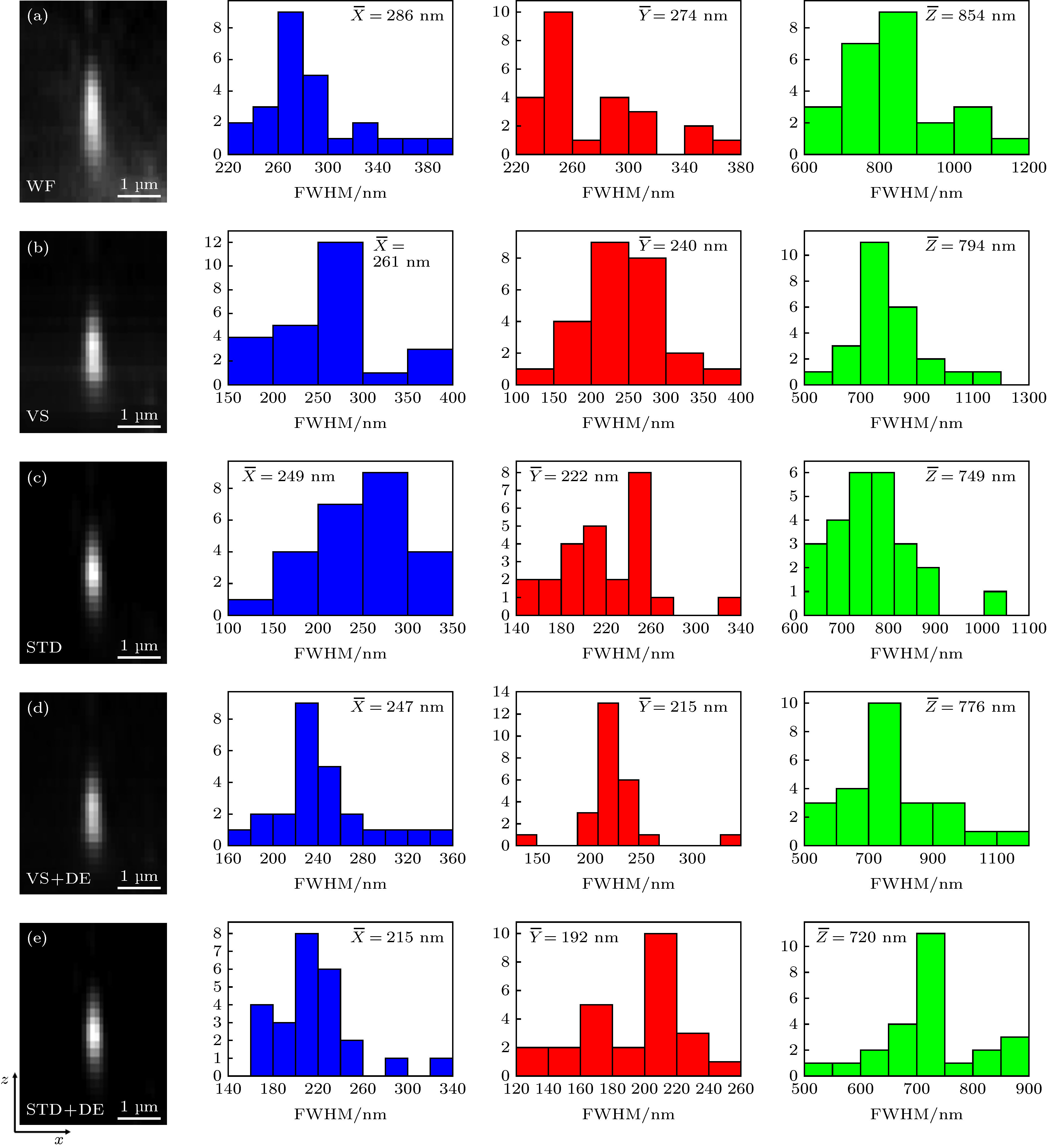
图 4 100 nm荧光珠标定的系统三维空间分辨率 (a)宽场图像; (b)虚拟狭缝重构图像; (c)标准差重构图像; (d)虚拟狭缝与LW解卷积组合重构图像; (e)标准差与LW解卷积组合重构图像. 其右边对应的直方图为25颗荧光珠在沿
$x, y, z$ 方向的FWHM分布,$\overline X, \overline Y, \overline Z $ 分别为其平均值Fig. 4. 3D spatial resolution of the system calibrated by the 100 nm fluorescent bead: (a) WF image; (b) VS image; (c) STD image; (d) VS + DE image; (e) STD + DE image. The corresponding histograms on the right show the measured distribution of
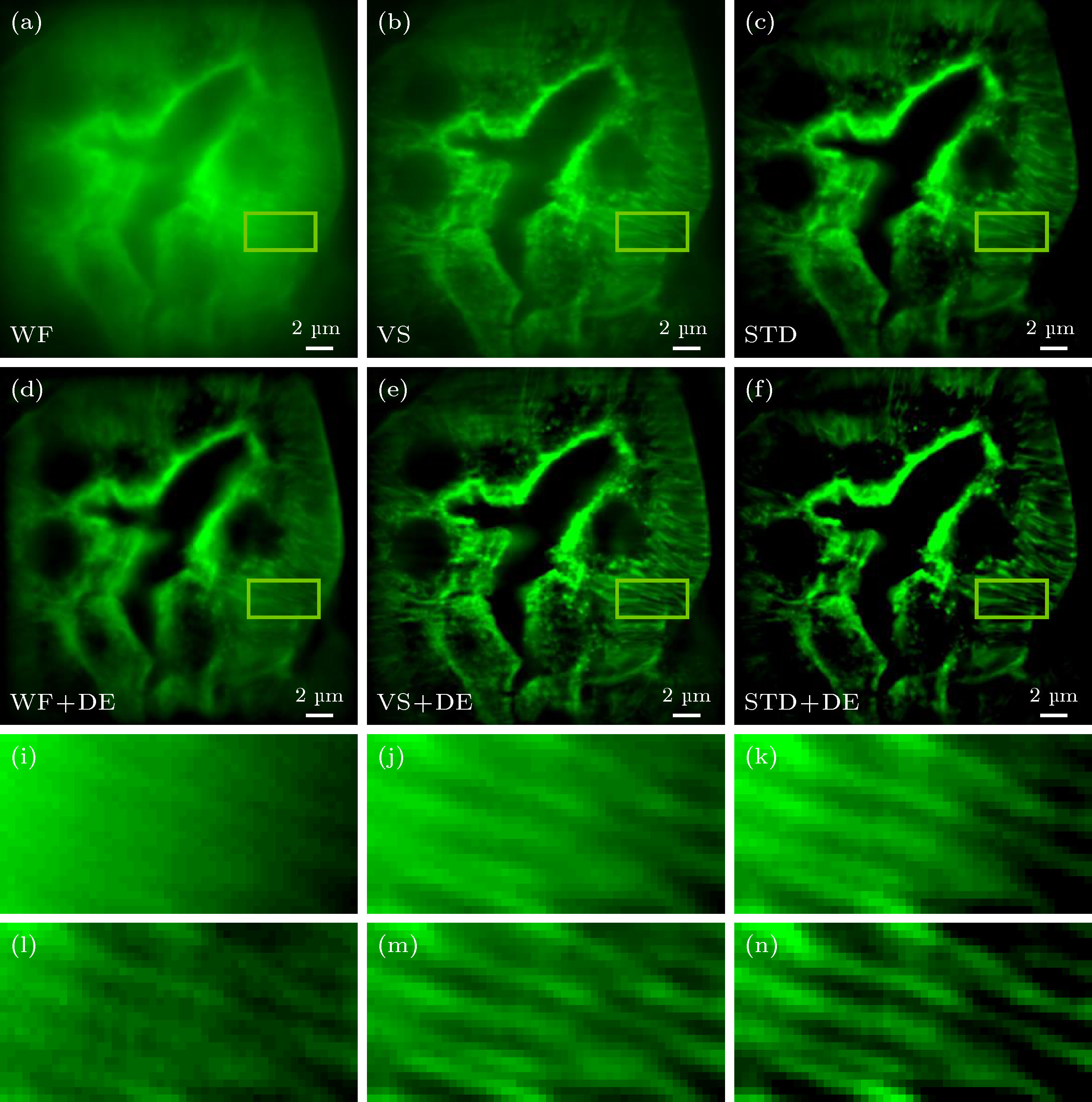
$x, y, z$ FWHM values from 25 beads.图 6 不同算法的图像重构细节比较 (a)−(c) 分别为WF图像、VS图像和STD图像; (d)−(f) 分别由(a)−(c)经Landweber DE算法处理成像; (i)−(n) 分别为图(a)−(f)黄色线框区域的放大图
Fig. 6. Comparison of image reconstruction details of different algorithms: (a)−(f) WF image, VS image, and STD image; (d)−(f) the Landweber DE images for (a)−(c); (i)−(n) magnified view of the yellow rectangular areas in (a)−(f).
图 7 不同算法重构图像的对比度比较 (a)−(c) 分别为WF图像、VS图像和STD图像; (d)−(f) 分别为图(a)−(c)经Landwebe解卷积算法处理成像; (g)−(h) 分别为图(a)−(f)白色虚线位置的归一化强度轮廓图
Fig. 7. Contrast comparison of reconstructed images with different algorithms: (a)−(c) WF image, VS image, and STD image; (d)−(f) images obtained from (a)−(c) processed by Landwebe deconvolution algorithm; (g)−(h) the normalized intensity profiles through the white dotted line position in (a)−(f).
-
[1] Minsky M 1988 Scanning 10 128
 Google Scholar
[2] Mitsuhiro I, Hiromi S 1998 SPIE 34 68
[3] Gustafsson M G 2010 J. Microsc. 198 82
[4] 赵启韬, 苗俊英 2003 北京生物医学工程 22 52
 Google Scholar
Zhao Q T, Miao J Y 2003 Beijing Biomed. Eng. 22 52
 Google Scholar
[5] Rainer H, Christoph G C 1999 SPIE 35 185
[6] Sauermann K, Gambichler T, Wilmert M 2002 Skin Res.Technol. 8 141
 Google Scholar
[7] Taejoong K, DaeGab G, Jun-Hee L 2009 Meas. Sci. Technol. 20 055501
 Google Scholar
[8] SchulzO, Pieper C, Clever M, et al. 2013 Proc. Natl. Acad. Sci. U.S.A. 110 21000
 Google Scholar
[9] Fan G Y, Fujisaki H, Miywwaki A, Tsay R K, Tsien R Y, Ellisman M H 1999 Biophys. J. 76 2412
 Google Scholar
[10] Rahadhyaksha M, Anderson R R, Webb R H 1999 Appl. Opt. 38 2105
 Google Scholar
[11] Li Y X, Gautam V, Brüstle A, Cockburn I A, Daria VR, Gillespie C, Gaus K, Alt C, Lee W M 2017 J. Bophotonics 10 1526
 Google Scholar
[12] Sheppard C JR, Mao X Q 1988 J. Mod. Optic. 35 1169
 Google Scholar
[13] Puash W C, Wasser M 2016 Methods 96 103
 Google Scholar
[14] Yazawa M, Hsueh B, Jia X, Pasca A M, Bernstein J A, Hallmayer J, Dolmetsch R E 2011 Nature 471 230
 Google Scholar
[15] Reto F, Andreas S, Yury B 2007 Histochem. Cell Biol. 128 499
 Google Scholar
[16] Kang B I, Sumin H, Hwajoon P, Dongsun K, Beop M K 2005 Opt. Express 13 5151
 Google Scholar
[17] Ondrej M, Martin K, Kai Wicker, Gerhard K, Ingo K, Rainer H 2012 Opt. Express 20 24167
 Google Scholar
[18] Lu R W, Wang B Q, Zhang QX, Yao XC 2013 Biomed.Opt. Express 4 1673
 Google Scholar
[19] Wang B, Lu R, Zhang Q, Yao X 2013 Quant. Imag. Med. Surg. 3 243
[20] Yanan Z, Lu R W, Wang B Q, Zhang Q X, Yao X C 2015 Opt. Lett. 40 1683
[21] Poher V, Zhang HX, Kennedy GT 2007 Opt. Express 15 11196
 Google Scholar
[22] Chamma I, Levet F, Sibarita J B, Sainlos M, Thoumine O 2016 Neurophotonics 3 041810
 Google Scholar
[23] Endesfelder U, Heilemann M 2014 Nat. Methods 11 235
 Google Scholar
[24] Flors C 2011 Biopolymers 95 290
 Google Scholar
[25] Patterson G, Davidson M, Manley S 2010 Annu. Rev. Phys. Chem. 61 345
 Google Scholar
[26] Hossain S, Hashimoto M, Katakura M, Mamun A A, Shido O 2015 Bmc. Complem. Altern. M 15 1
[27] Rainer H, Pier A, Benedetti 2006 Appl. Opt. 45 5037
 Google Scholar
[28] Ren Y X, Lu R D, Gong L 2015 Ann. Phys.(Berlin) 527 447
 Google Scholar
[29] 冯维, 张福民, 王惟婧, 曲兴华 2017 66 234201
 Google Scholar
Feng W, Zhang F M, Wang W J, Qu X H 2017 Acta Phys. Sin. 66 234201
 Google Scholar
[30] Vienola K V, Damodaran M, Braaf B, Vermeer K A, Johannes F 2015 Opt. Lett. 40 5335
 Google Scholar
[31] Wu J J, Li S W, Cao H Q, Lin D Y, Yu B, Qu J L 2018 Opt. Express 26 31430
 Google Scholar
[32] Vonesch C, Unser M 2008 IEEE Trans. Image Process. 17 539
[33] Zhang W, Yu B, Lin D Y, Yu H H, Li S W, Qu J L 2020 Opt. Express 28 10919
 Google Scholar
计量
- 文章访问数: 11355
- PDF下载量: 218
- 被引次数: 0


 下载:
下载:


