-
Plasma nanostructures are of particular significance for serving as a substrate for spectroscopic detection and identification of individual molecules. By combining the excitation wavelength of the molecule with the resonance wavelength of the nanostructure, the sensitive single-molecule Raman detection can be achieved. A high and stable plasma substrate for coherent anti-Stokes Raman scattering(CARS) is very useful for developing the surface-enhanced coherent anti-Stokes Raman scattering (SECARS). In the plasma nanostructures, the strong coupling of plasmonic nanoparticles with an inter-particle gap smaller than the diameter of the individual nanoparticles results in the hybridization of the optical properties of these individual nanoparticles. There are also the charge transfer plasmons(CTP) appearing in conductive bridging nanoparticles. Their unique properties make linked nanosystems a suitable candidate for building artificial molecules, nanomotors, sensors, and other optoelectronic devices. In this work, we, starting from reality, theoretically design a new linked nanosystem SECARS substrate where Fano resonance can be generated by the plasmon hybridization (PH) model resonance and the charge transfer plasmon resonance. The introduction of charge transfer plasma improves the tunability of structural resonance. By adjusting the conductivity of the conductive junction, the wavelength of the charge transfer plasma resonance can be easily adjusted to change the wavelength position of the Fano resonance. The data obtained by numerical simulation of the Raman mode at 1557 cm–1 of L-tryptophan when a 1064 nm light source is used as the pump light show that this spatially symmetrical structure can generate multiple high-enhancement hot spots that do not depend on the polarization direction of the incident light. Ordinary CARS signal can generally be enhanced by 1012, and its maximum can reach 1014. Due to the ultrastrong field enhancement and insensitive-to-polarization, this method of using charge transfer plasma to design a substrate can be used in the practical substrate of SECARS and provide new ideas for designing other nonlinear optical processes such as four wave mixing and stimulated Raman scattering.
-
Keywords:
- surface enhancement coherent anti-Stokes Raman scattering /
- Raman scattering /
- surface plasmon resonance
[1] Minck R W, Terhune R W, Rado W G 1963 Appl. Phys. Lett. 3 181
[2] Begley R F, Harvey A B, Byer R L 1974 Appl. Phys. Lett. 25 387
 Google Scholar
[3] Duncan M D, Reintjes J, Manuccia T J 1982 Opt. Lett. 7 350
 Google Scholar
[4] Shi K, Li H, Xu Q, Psaltis D, Liu Z 2010 Phys. Rev. Lett. 104 093902
 Google Scholar
[5] 刘双龙, 刘伟, 陈丹妮, 屈军乐, 牛憨笨 2016 65 064204
 Google Scholar
Liu S L, Liu W, Chen D N, Qu J L, Niu H B, 2016 Acta Phys. Sin. 65 064204
 Google Scholar
[6] Steuwe C, Kaminski C F, Baumberg J J, Mahajan S 2011 Nano Lett. 11 5339
 Google Scholar
[7] Krafft C, Dietzek B, Schmitt M, Popp J 2012 J Biomed. Opt. 17 040801
 Google Scholar
[8] Koo T W, Chan S, Berlin A A 2005 Opt. Lett. 30 1024
 Google Scholar
[9] Chew H, Wang D, Kerker M 1984 J. Opt. Soc. Am. B: Opt. Phys. 1 56
 Google Scholar
[10] Addison C J, Konorov S O, Brolo A G, Blades M W, Turner R F B 2009 J. Phys.Chem. C 113 3586
 Google Scholar
[11] Dmitri V V, Alexander M S, Xia H, Kai W, Pankaj K J, Elango M, Steven E W, George W, Alexei V S, Marlan O S 2012 Sci. Rep. 2 891
 Google Scholar
[12] Shutov A D, Yi Z, Wang J, Sinyukov A M, He Z, Tang C, Chen J, Ocola E J, Laane J, Sokolov A V, Voronine D V, Scully M O 2018 ACS Photonics 5 4960
 Google Scholar
[13] Prodan E, Nordlander P 2004 J. Chem. Phys. 120 5444
 Google Scholar
[14] Halas N J, Lal S, Wei-Shun C, Link S, Nordlander P 2011 Chem. Rev. 111 3913
 Google Scholar
[15] Fontana J, Charipar N, Flom S R, Naciri J, Piqué A, Ratna B R 2016 ACS Photonics 3 904
 Google Scholar
[16] Fontana J, Ratna B R 2014 Appl. Phys. Lett. 105 011107
 Google Scholar
[17] Huang Y, Ma L, Hou M, Xie Z, Zhang Z 2016 Phys. Chem. Chem. Phys. 18 2319
 Google Scholar
[18] Liu L, Wang Y, Fang Z, Zhao K 2013 J. Chem. Phys. 139 064310
 Google Scholar
[19] Pérez-González O, Zabala N, Borisov A G, Halas N J, Nordlander P, Aizpurua J 2010 Nano Lett. 10 3090
 Google Scholar
[20] Zhang Y, Wen F, Zhen Y R, Nordlander P, Halas N J 2013 Proc. Natl. Acad. Sci. U. S. A. 110 9215
 Google Scholar
[21] Zhang Y, Zhen Y R, Neumann O, Day J K, Nordlander P, Halas N J 2014 Nat. Commun. 5 4424
 Google Scholar
[22] He J N, Fan C Z, Ding P, Zhu S M, Liang E J 2016 Sci. Rep. 6 20777
 Google Scholar
[23] Kim K H, Rim W S 2019 Appl. Phys. A 125 1
[24] Arpan D, Erik M V 2020 JEOS:RP 16 1
 Google Scholar
[25] Tian M, Zhao Y, Wan M, Ji P, Li Y, Song Y, Yuan S, Zhou F, He J, Ding P 2018 Phys. Lett. A. 382 3187
 Google Scholar
[26] Maiti N, Thomas S, Jacob J A, Chadha R, Mukherjee T, Kapoor S 2012 J. Colloid Interface Sci. 380 141
 Google Scholar
[27] 李亚琴, 简国树, 吴世法 2006 中国光学快报(英文版) 4 671
li Y Q, Jian G S, Wu S F, 2006 Chin. Opt. Lett. 4 671
[28] Hentschel M, Saliba M, Vogelgesang R, Giessen H, Alivisatos A P, Liu N 2010 Nano Lett. 10 2721
 Google Scholar
[29] Hentschel M, Dregely D, Vogelgesang R, Giessen H, Liu N 2011 ACS Nano 5 2042
 Google Scholar
[30] Encina E R, Coronado E A 2011 J. Phys. Chem. C 115 15908
 Google Scholar
[31] Lovera A, Gallinet B, Nordlander P, Martin O J F 2013 ACS Nano 7 4527
 Google Scholar
-
图 2 圆盘的参数不变(r1 = 63 nm, r2 = 97 nm, d = 10 nm, h1 = 50 nm)改变导电结参数时散射系数的变化 (a)
$ \theta $ = 450, h2 = 50 nm, O(x, y) = (100, 100), 改变结的宽度l从30到50 nm; (b) l = 40 nm, h2 = 50 nm, O(x, y) = (100, 100), 改变倾斜角度$ \theta $ 从250到450; (c) l = 40 nm,$ \theta $ = 450, h2 = 50 nm, 改变中心坐标O(x, y)从(70, 70)到(110, 110); (d) l = 40 nm,$ \theta $ = 450, O(x, y) = (100, 100), 改变结厚度h2从30到50 nmFigure 2. When the parameters of the disc are unchanged (r1 = 63 nm, r2 = 97 nm, d = 10 nm, h1 = 50 nm) that the scattering spectrum depond on geometrical parameters: (a) Vary l with
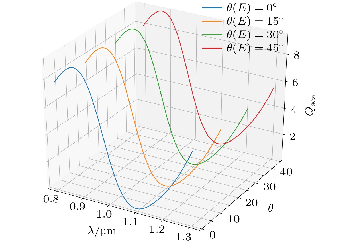
$ \theta $ = 450, h2 = 50 nm, O(x, y) = (100, 100); (b) vary$ \theta $ with l = 40 nm, h2 = 50 nm, O(x, y) = (100, 100); (c) vary O(x, y) with l = 40 nm,$ \theta $ = 450, h2 = 50 nm; (d) vary$ {h}_{2} $ with l = 40 nm,$ \theta $ = 450, O(x, y) = (100, 100).图 3 入射光不同偏振角度时的相同参数(l = 35 nm, h2 = 50 nm, O(x, y) = (100, 100), r1 = 63 nm, r2 = 97 nm, d = 10 nm)结构的散射系数, 偏振角度定义为入射光偏振方向与结构Y轴夹角
Figure 3. . Scattering spectra for various excitation polarizations with the same parameters (l = 35 nm, h2 = 50 nm, O(x, y) = (100, 100), r1 = 63 nm, r2 = 97 nm, d = 10 nm), and the polarization angle is defined as the angle between the polarization direction and the Y-axis.
图 4 (a) 入射光偏振方向沿基底的Y轴方向时与入射光偏振方向与基底的Y轴的夹角为450时基底表面912 nm, 1064 nm, 1275 nm三个波长处与1064 nm时基底中心YZ横截面处的电场强度空间分布; (b) 当入射光偏振方向沿基底的Y轴方向夹角θ为0°, 15°, 30°, 45°时基底表面对应的增强GSECARS因子的对数空间分布图
Figure 4. (a)The spatial distributions of enhanced electric-filed amplitude (|E/E0|) in the top surface plane of the structure at three characteristic wavelengths for two polarizations; (b) the corresponding SECARS map for various polarizations. From the top to bottom, the polarization angle θ equals to 0°, 15°, 30°, 45°, respectively.
-
[1] Minck R W, Terhune R W, Rado W G 1963 Appl. Phys. Lett. 3 181
[2] Begley R F, Harvey A B, Byer R L 1974 Appl. Phys. Lett. 25 387
 Google Scholar
[3] Duncan M D, Reintjes J, Manuccia T J 1982 Opt. Lett. 7 350
 Google Scholar
[4] Shi K, Li H, Xu Q, Psaltis D, Liu Z 2010 Phys. Rev. Lett. 104 093902
 Google Scholar
[5] 刘双龙, 刘伟, 陈丹妮, 屈军乐, 牛憨笨 2016 65 064204
 Google Scholar
Liu S L, Liu W, Chen D N, Qu J L, Niu H B, 2016 Acta Phys. Sin. 65 064204
 Google Scholar
[6] Steuwe C, Kaminski C F, Baumberg J J, Mahajan S 2011 Nano Lett. 11 5339
 Google Scholar
[7] Krafft C, Dietzek B, Schmitt M, Popp J 2012 J Biomed. Opt. 17 040801
 Google Scholar
[8] Koo T W, Chan S, Berlin A A 2005 Opt. Lett. 30 1024
 Google Scholar
[9] Chew H, Wang D, Kerker M 1984 J. Opt. Soc. Am. B: Opt. Phys. 1 56
 Google Scholar
[10] Addison C J, Konorov S O, Brolo A G, Blades M W, Turner R F B 2009 J. Phys.Chem. C 113 3586
 Google Scholar
[11] Dmitri V V, Alexander M S, Xia H, Kai W, Pankaj K J, Elango M, Steven E W, George W, Alexei V S, Marlan O S 2012 Sci. Rep. 2 891
 Google Scholar
[12] Shutov A D, Yi Z, Wang J, Sinyukov A M, He Z, Tang C, Chen J, Ocola E J, Laane J, Sokolov A V, Voronine D V, Scully M O 2018 ACS Photonics 5 4960
 Google Scholar
[13] Prodan E, Nordlander P 2004 J. Chem. Phys. 120 5444
 Google Scholar
[14] Halas N J, Lal S, Wei-Shun C, Link S, Nordlander P 2011 Chem. Rev. 111 3913
 Google Scholar
[15] Fontana J, Charipar N, Flom S R, Naciri J, Piqué A, Ratna B R 2016 ACS Photonics 3 904
 Google Scholar
[16] Fontana J, Ratna B R 2014 Appl. Phys. Lett. 105 011107
 Google Scholar
[17] Huang Y, Ma L, Hou M, Xie Z, Zhang Z 2016 Phys. Chem. Chem. Phys. 18 2319
 Google Scholar
[18] Liu L, Wang Y, Fang Z, Zhao K 2013 J. Chem. Phys. 139 064310
 Google Scholar
[19] Pérez-González O, Zabala N, Borisov A G, Halas N J, Nordlander P, Aizpurua J 2010 Nano Lett. 10 3090
 Google Scholar
[20] Zhang Y, Wen F, Zhen Y R, Nordlander P, Halas N J 2013 Proc. Natl. Acad. Sci. U. S. A. 110 9215
 Google Scholar
[21] Zhang Y, Zhen Y R, Neumann O, Day J K, Nordlander P, Halas N J 2014 Nat. Commun. 5 4424
 Google Scholar
[22] He J N, Fan C Z, Ding P, Zhu S M, Liang E J 2016 Sci. Rep. 6 20777
 Google Scholar
[23] Kim K H, Rim W S 2019 Appl. Phys. A 125 1
[24] Arpan D, Erik M V 2020 JEOS:RP 16 1
 Google Scholar
[25] Tian M, Zhao Y, Wan M, Ji P, Li Y, Song Y, Yuan S, Zhou F, He J, Ding P 2018 Phys. Lett. A. 382 3187
 Google Scholar
[26] Maiti N, Thomas S, Jacob J A, Chadha R, Mukherjee T, Kapoor S 2012 J. Colloid Interface Sci. 380 141
 Google Scholar
[27] 李亚琴, 简国树, 吴世法 2006 中国光学快报(英文版) 4 671
li Y Q, Jian G S, Wu S F, 2006 Chin. Opt. Lett. 4 671
[28] Hentschel M, Saliba M, Vogelgesang R, Giessen H, Alivisatos A P, Liu N 2010 Nano Lett. 10 2721
 Google Scholar
[29] Hentschel M, Dregely D, Vogelgesang R, Giessen H, Liu N 2011 ACS Nano 5 2042
 Google Scholar
[30] Encina E R, Coronado E A 2011 J. Phys. Chem. C 115 15908
 Google Scholar
[31] Lovera A, Gallinet B, Nordlander P, Martin O J F 2013 ACS Nano 7 4527
 Google Scholar
Catalog
Metrics
- Abstract views: 7041
- PDF Downloads: 80
- Cited By: 0


 DownLoad:


