-
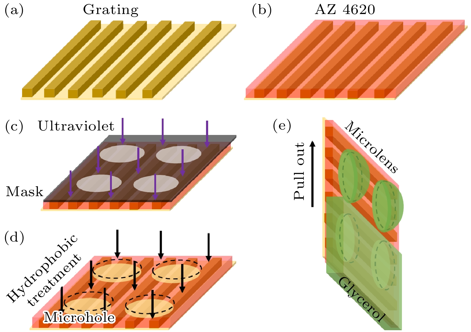
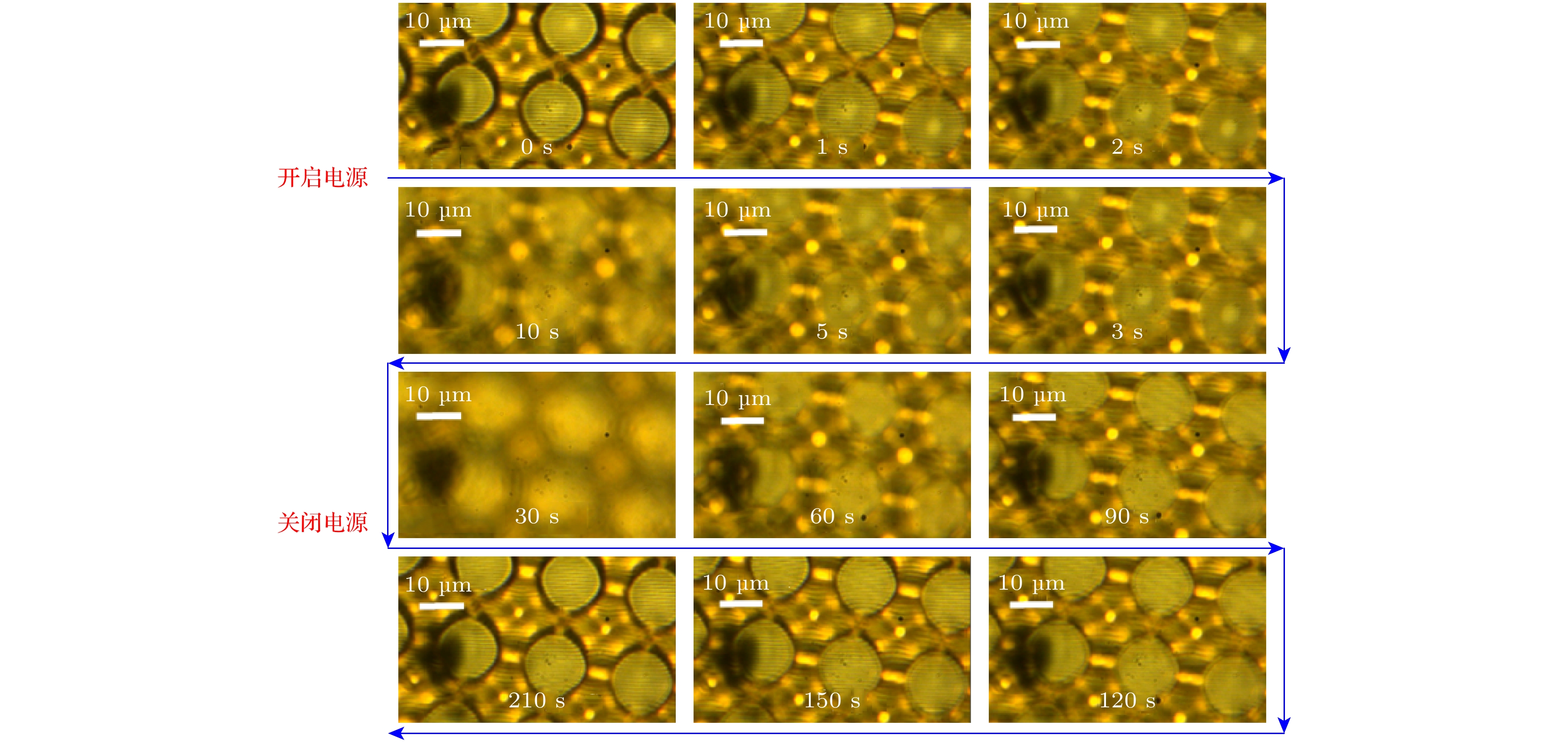
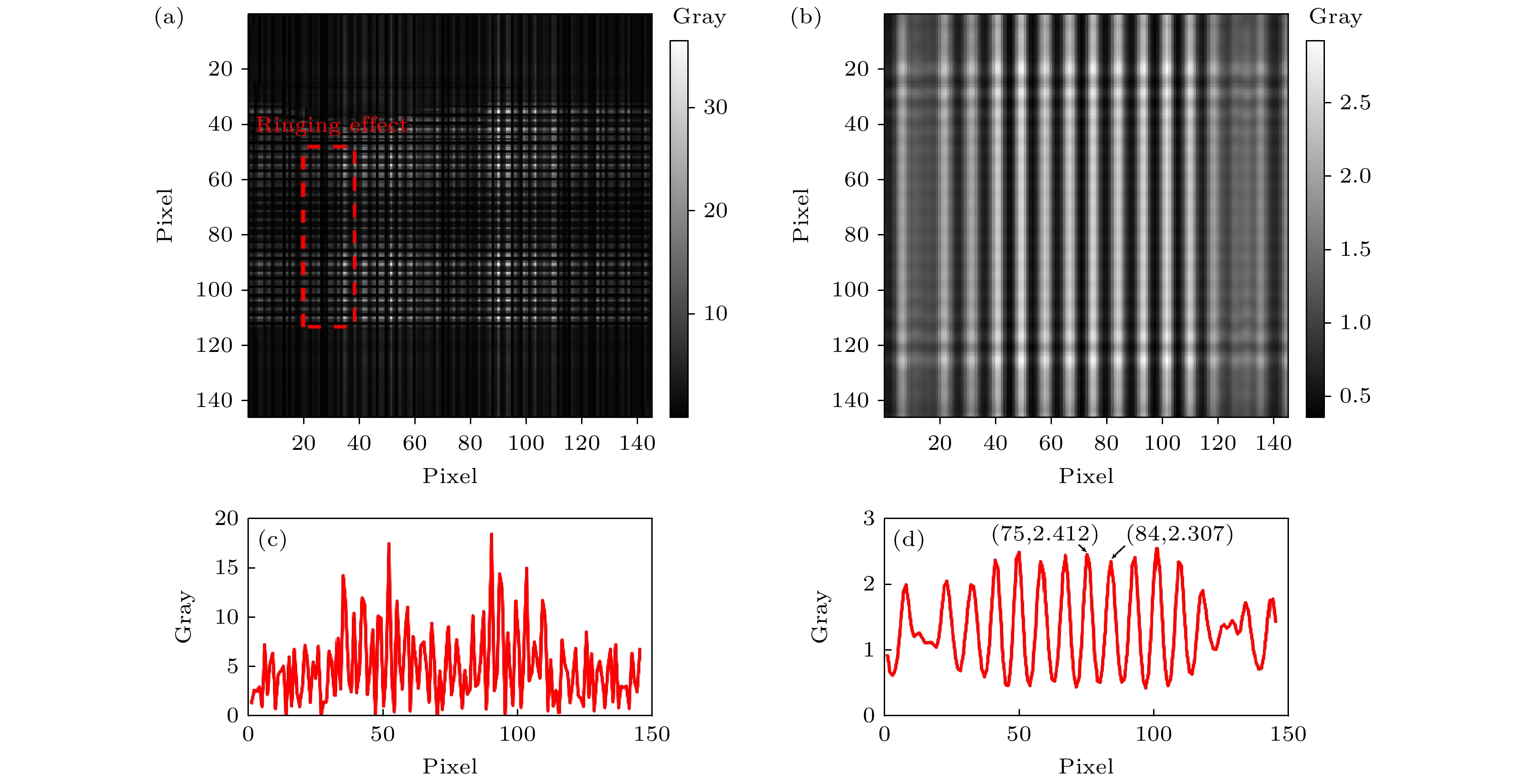
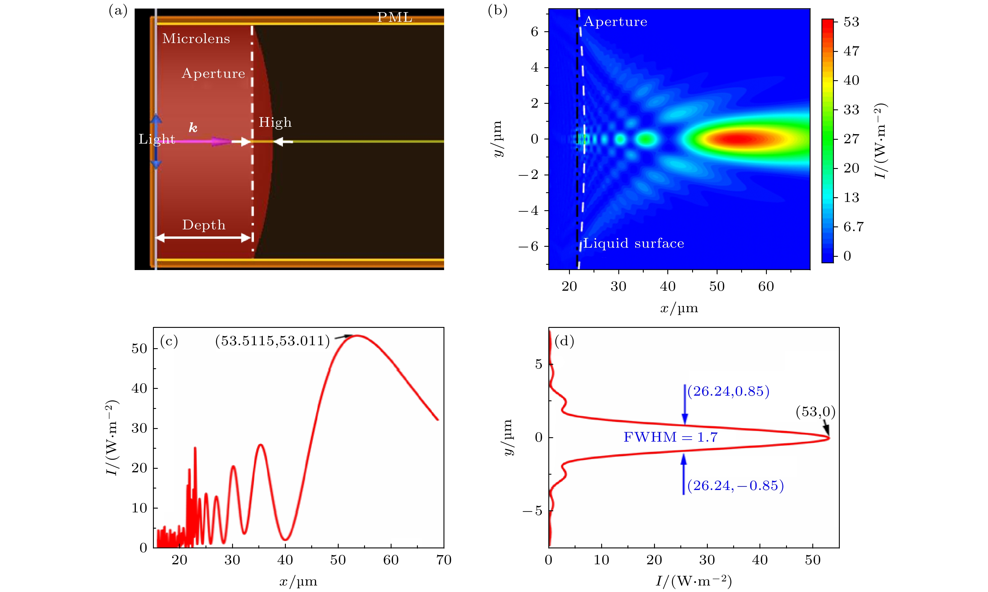
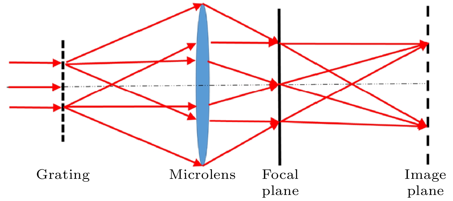
微透镜辅助显微镜实现超分辨成像观测, 具有免标记、无损伤、实时、定域和环境兼容性好等优势. 液体微透镜阵列具有均一、易操控的特性, 可实现无复杂机械扫描与驱动的超分辨成像. 然而, 简单高效地精确控制成像距离是微透镜实现超分辨成像的关键技术挑战. 本文利用紫外曝光技术, 实现了光盘上光刻胶微孔深度的均一性. 结合液体自组装技术, 在微孔中填充甘油液滴, 保证微透镜辅助超分辨的成像距离. 在光学显微镜下实现了对226 nm光栅栅线的可重构超分辨观测与1.59倍成像放大. 本文从液体微透镜的阿贝显微成像原理出发, 通过理论与模拟解释了液体微透镜的成像放大与超分辨特性. 由此可见, 光盘上集成的液体微透镜阵列在光学纳米测量与传感等器件中展现了巨大的应用潜力.The microlens-assisted microscope realizes super-resolution imaging and observation, and has the advantages of no marking, no damage, real-time, localization, and good environmental compatibility. Liquid microlens arrays with uniformity and easy manipulation can realize super-resolution imaging without complicated mechanical scanning and driving. However, simply and efficiently controlling the imaging distance is a key technical challenge to the realization of super-resolution imaging of microlens. In this paper, the uniform depths of photoresist microholes on light disk are fabricated by ultraviolet exposure technology. Using liquid self-assembly technology, the microholes are filled with glycerol droplets, and thus ensuring the near-field imaging distance of the microlens. The reconfigurable super-resolution of 226-nm-wide grating line and the imaging magnification of 1.59 times are observed under the optical microscope. At present, the theory of super-resolution imaging based on microlens is not unified and perfect. In this paper, the Abbe imaging principle is used to explain the imaging magnification and super-resolution characteristics. Therefore, the liquid microlens arrays integrated on the light disk show great potential application in optical nanometer measurements and sensing devices.
-
Keywords:
- liquid microlens arrays /
- super resolution /
- imaging magnification /
- Abbe imaging.
[1] Ling J Z, Wang Y C, Liu X, Wang X R 2021 Opt. Lett. 46 1265
 Google Scholar
[2] Chen L W, Zhou Y, Li Y, Hong M H 2019 Appl. Phys. Rev. 6 021304
 Google Scholar
[3] Hüser L, Pahl T, Künne M, Lehmann P 2022 J. Opt. Microsyst. 2 044501
 Google Scholar
[4] Wang Z B, Guo W, Li L, Luk'yanchuk B S, Khan A, Liu Z, Chun Z C, Hong M H 2011 Nat. Commun. 2 218
 Google Scholar
[5] 周锐, 吴梦雪, 沈飞, 洪明辉 2017 66 140702
 Google Scholar
Zhou R, Wu M X, Shen F, Hong M H 2017 Acta Phys. Sin. 66 140702
 Google Scholar
[6] 宋扬, 杨西斌, 闫冰, 王驰, 孙建美, 熊大曦 2020 69 134201
 Google Scholar
Song Y, Yang X B, Yan B, Wang C, Sun J M, Xiong D X 2020 Acta Phys. Sin. 69 134201
 Google Scholar
[7] 王建国, 杨松林, 叶永红 2018 67 214209
 Google Scholar
Wang J G, Yang S L, Ye Y H 2018 Acta Phys. Sin. 67 214209
 Google Scholar
[8] Darafsheh A 2022 J. Appl. Phys. 131 031102
 Google Scholar
[9] Pei Y, Zang J J, Yang S L, Wang J G, Cao Y Y, Ye Y H 2021 ACS Appl. Nano Mater. 4 11281
 Google Scholar
[10] Yang S L, Ye Y H, Shi Q F, Zhang J Y 2020 J. Phys. Chem. C 124 25951
 Google Scholar
[11] Gu G Q, Zhang P C, Chen S H, Zhang Y, Yang H 2021 Photonics. Res. 9 1157
 Google Scholar
[12] Zhang P P, Yan B, Gu G Q, Yu Z T, Chen X, Wang Z B, Yang H 2022 Sensor. Actuat. B-Chem. 357 131401
 Google Scholar
[13] Kwon S, Park J, Kim K, Cho Y, Lee M 2022 Light Sci. Appl. 11 32
 Google Scholar
[14] Gu G Q, Song J, Ming C, Xiao P, Liang H D, Qu J L 2018 Nanoscale 10 14182
 Google Scholar
[15] Xie Y, Cai D, Pan J, Zhou N, Guo X, Wang P, Tong L 2022 Adv. Opt. Mater. 10 2102269
 Google Scholar
[16] Su S J, Liang J S, Li X J, Xin W W, Chen L, Yin P H, Wang Z Z, Ye X S, Xiao J P, Wang D 2021 Adv. Mater. Technol-US. 6 2100449
 Google Scholar
[17] Darafsheh A 2021 J. Phys. Photonics. 3 022001
 Google Scholar
[18] Wang F F, Liu L Q, Yu H B, Wen Y D, Yu P, Liu Z, Wang Y C, Li W J 2016 Nat. Commun. 7 13748
 Google Scholar
[19] Wang S Y, Zhang D X, Zhang H J, Han X, Xu R 2015 Microsc. Res. Techniq. 78 1128
 Google Scholar
[20] Zhang T Y, Yu H B, Li P, Wang X D, Wang F F, Shi J L, Liu Z, Yu P, Yang W G, Wang Y C, Liu L Q 2020 ACS Appl. Mater. Inter. 12 48093
 Google Scholar
[21] Chen X X, Wu T L, Gong Z Y, Guo J H, Liu X S, Zhang Y, Li Y C, Ferraro P, Li B J 2021 Light Sci. Appl. 12 242
 Google Scholar
[22] 李姮, 张熙熙, 张垚, 李宇超, 李宝军 2022 光学学报 42 0411003
 Google Scholar
Li H, Chen X X, Zhang Y, Li Y C, Li B J 2022 Acta. Opt. Sin. 42 0411003
 Google Scholar
[23] Jia B L, Wang F F, Chan H Y, Zhang G L, Li W J 2019 Microsyst. Nanoeng. 5 13
 Google Scholar
[24] Gu T K, Wang L L, Li R, Dong Y Z, Zhang Y J, Jia M C, Jiang W T, Liu H Z 2018 Opt. Commun. 428 89
 Google Scholar
[25] Zhang H C, Qi T Y, Zhu X Y, Zhou L J, Li Z H, Zhang Y F, Yang W C, Yang J J, Peng Z L, Zhang G M, Wang F, Guo P F, Lan H B 2021 ACS Appl. Mater. Inter. 13 36295
 Google Scholar
[26] Wang L, Luo Y, Liu Z Z, Feng X M, Lu B H 2018 Appl. Surf. Sci. 442 417
 Google Scholar
[27] Wang L L, Liu H Z, Jiang W T, Li R, Li F, Yang Z B, Yin L, Shi Y S, Chen B D 2015 J. Mater. Chem. C 3 5896
 Google Scholar
[28] Wang L L, Li F, Liu H Z, Jiang W T, Niu D, Li R, Yin L, Shi Y S, Chen B D 2015 ACS Appl. Mater. Inter. 7 21416
 Google Scholar
[29] Xu M, Zhou Z W, Wang Z, Lu H B 2020 ACS Appl. Mater. Inter. 12 7826
 Google Scholar
[30] Chen X X, Wu T L, Gong Z Y, Li Y C, Zhang Y, Li B J 2020 Photonics. Res. 8 225
 Google Scholar
[31] Yang H, Trouillon R, Huszka G, Gijs M A 2016 Nano. Lett. 16 4862
 Google Scholar
[32] 叶燃, 许楚, 汤芬, 尚晴晴, 范瑶, 李加基, 叶永红, 左超 2022 红外与激光工程 51 20220086
 Google Scholar
Ye R, Xu C, Tang F, Shang Q Q, Fan Y, Li J J, Ye Y H, Zuo C 2022 Infrared Laser Eng. 51 20220086
 Google Scholar
[33] Duan Y, Barbastathis G, Zhang B 2013 Opt. Lett. 38 2988
 Google Scholar
[34] Zhou S, Deng Y B, Zhou W C, Yu M X, Urbach H P, Wu Y H 2017 Appl. Phys. B 123 236
 Google Scholar
-
图 4 液体自组装前后模板的共聚焦扫描结果 (a) 微孔阵列; (b)微孔轮廓和结构参数; (c)微透镜阵列; (d) 微透镜轮廓和结构参数
Fig. 4. Observation of template before and after liquid self-assembly process through CLSM: (a) Microhole arrays; (b) profile and parameters of microholes; (c) liquid microlens arrays (LMLAs); (d) profile and parameters of microlenses.
-
[1] Ling J Z, Wang Y C, Liu X, Wang X R 2021 Opt. Lett. 46 1265
 Google Scholar
[2] Chen L W, Zhou Y, Li Y, Hong M H 2019 Appl. Phys. Rev. 6 021304
 Google Scholar
[3] Hüser L, Pahl T, Künne M, Lehmann P 2022 J. Opt. Microsyst. 2 044501
 Google Scholar
[4] Wang Z B, Guo W, Li L, Luk'yanchuk B S, Khan A, Liu Z, Chun Z C, Hong M H 2011 Nat. Commun. 2 218
 Google Scholar
[5] 周锐, 吴梦雪, 沈飞, 洪明辉 2017 66 140702
 Google Scholar
Zhou R, Wu M X, Shen F, Hong M H 2017 Acta Phys. Sin. 66 140702
 Google Scholar
[6] 宋扬, 杨西斌, 闫冰, 王驰, 孙建美, 熊大曦 2020 69 134201
 Google Scholar
Song Y, Yang X B, Yan B, Wang C, Sun J M, Xiong D X 2020 Acta Phys. Sin. 69 134201
 Google Scholar
[7] 王建国, 杨松林, 叶永红 2018 67 214209
 Google Scholar
Wang J G, Yang S L, Ye Y H 2018 Acta Phys. Sin. 67 214209
 Google Scholar
[8] Darafsheh A 2022 J. Appl. Phys. 131 031102
 Google Scholar
[9] Pei Y, Zang J J, Yang S L, Wang J G, Cao Y Y, Ye Y H 2021 ACS Appl. Nano Mater. 4 11281
 Google Scholar
[10] Yang S L, Ye Y H, Shi Q F, Zhang J Y 2020 J. Phys. Chem. C 124 25951
 Google Scholar
[11] Gu G Q, Zhang P C, Chen S H, Zhang Y, Yang H 2021 Photonics. Res. 9 1157
 Google Scholar
[12] Zhang P P, Yan B, Gu G Q, Yu Z T, Chen X, Wang Z B, Yang H 2022 Sensor. Actuat. B-Chem. 357 131401
 Google Scholar
[13] Kwon S, Park J, Kim K, Cho Y, Lee M 2022 Light Sci. Appl. 11 32
 Google Scholar
[14] Gu G Q, Song J, Ming C, Xiao P, Liang H D, Qu J L 2018 Nanoscale 10 14182
 Google Scholar
[15] Xie Y, Cai D, Pan J, Zhou N, Guo X, Wang P, Tong L 2022 Adv. Opt. Mater. 10 2102269
 Google Scholar
[16] Su S J, Liang J S, Li X J, Xin W W, Chen L, Yin P H, Wang Z Z, Ye X S, Xiao J P, Wang D 2021 Adv. Mater. Technol-US. 6 2100449
 Google Scholar
[17] Darafsheh A 2021 J. Phys. Photonics. 3 022001
 Google Scholar
[18] Wang F F, Liu L Q, Yu H B, Wen Y D, Yu P, Liu Z, Wang Y C, Li W J 2016 Nat. Commun. 7 13748
 Google Scholar
[19] Wang S Y, Zhang D X, Zhang H J, Han X, Xu R 2015 Microsc. Res. Techniq. 78 1128
 Google Scholar
[20] Zhang T Y, Yu H B, Li P, Wang X D, Wang F F, Shi J L, Liu Z, Yu P, Yang W G, Wang Y C, Liu L Q 2020 ACS Appl. Mater. Inter. 12 48093
 Google Scholar
[21] Chen X X, Wu T L, Gong Z Y, Guo J H, Liu X S, Zhang Y, Li Y C, Ferraro P, Li B J 2021 Light Sci. Appl. 12 242
 Google Scholar
[22] 李姮, 张熙熙, 张垚, 李宇超, 李宝军 2022 光学学报 42 0411003
 Google Scholar
Li H, Chen X X, Zhang Y, Li Y C, Li B J 2022 Acta. Opt. Sin. 42 0411003
 Google Scholar
[23] Jia B L, Wang F F, Chan H Y, Zhang G L, Li W J 2019 Microsyst. Nanoeng. 5 13
 Google Scholar
[24] Gu T K, Wang L L, Li R, Dong Y Z, Zhang Y J, Jia M C, Jiang W T, Liu H Z 2018 Opt. Commun. 428 89
 Google Scholar
[25] Zhang H C, Qi T Y, Zhu X Y, Zhou L J, Li Z H, Zhang Y F, Yang W C, Yang J J, Peng Z L, Zhang G M, Wang F, Guo P F, Lan H B 2021 ACS Appl. Mater. Inter. 13 36295
 Google Scholar
[26] Wang L, Luo Y, Liu Z Z, Feng X M, Lu B H 2018 Appl. Surf. Sci. 442 417
 Google Scholar
[27] Wang L L, Liu H Z, Jiang W T, Li R, Li F, Yang Z B, Yin L, Shi Y S, Chen B D 2015 J. Mater. Chem. C 3 5896
 Google Scholar
[28] Wang L L, Li F, Liu H Z, Jiang W T, Niu D, Li R, Yin L, Shi Y S, Chen B D 2015 ACS Appl. Mater. Inter. 7 21416
 Google Scholar
[29] Xu M, Zhou Z W, Wang Z, Lu H B 2020 ACS Appl. Mater. Inter. 12 7826
 Google Scholar
[30] Chen X X, Wu T L, Gong Z Y, Li Y C, Zhang Y, Li B J 2020 Photonics. Res. 8 225
 Google Scholar
[31] Yang H, Trouillon R, Huszka G, Gijs M A 2016 Nano. Lett. 16 4862
 Google Scholar
[32] 叶燃, 许楚, 汤芬, 尚晴晴, 范瑶, 李加基, 叶永红, 左超 2022 红外与激光工程 51 20220086
 Google Scholar
Ye R, Xu C, Tang F, Shang Q Q, Fan Y, Li J J, Ye Y H, Zuo C 2022 Infrared Laser Eng. 51 20220086
 Google Scholar
[33] Duan Y, Barbastathis G, Zhang B 2013 Opt. Lett. 38 2988
 Google Scholar
[34] Zhou S, Deng Y B, Zhou W C, Yu M X, Urbach H P, Wu Y H 2017 Appl. Phys. B 123 236
 Google Scholar
计量
- 文章访问数: 7744
- PDF下载量: 235
- 被引次数: 0


 下载:
下载:
