


-
Over the last two decades, the grating-based phase-contrast imaging has aroused the interest of a number of researchers. It could provide an access to three complementary signals simultaneously: the conventional absorption contrast, the differential phase contrast related to refraction of incident wave, and the dark-field contrast that relates to ultra small angle scattering in a sample. The grating-based phase-contrast signals have higher contrast sensitivity for some types of soft samples than the absorption signals. Dark-field signals have better diagnostic effects in the detection of lung tumors, pneumothorax and the identification of microcalcifications in breast. There are two main phase retrieval methods in grating-based X-ray phase-contrast imaging, i.e. phase stepping method and Fourier transform method. The phase signals retrieved by phase stepping is high precise and has low noise. But the sample suffers high dose due to at least three exposures. The phase signals retrieved by Fourier transform is low-dose due to the fact that only one image with sample is needed, but it is easily affected by artifacts when the size of the filtering window is too large. However, when the size of the filtering window is too small, the high-frequency information of the phase-contrast image will be lost, and the image will become blurred. A trade-off between definitions of the image and artifacts should be made. Since the phase-contrast signal and the dark-field signal of the sample are modulated by carrier fringes, the frequency spectrum of the detected image consists of many different harmonics. The artifacts in the retrieved signals originate from the spectrum aliasing between primary peak around zero spatial frequency and first-order harmonic peaks. Therefore, the subtraction between two images with phase difference can remove the primary peak, and the artifacts in the phase-contrast signals and dark-field signals will be suppressed. In order to further suppress the artifacts, we increase the frequency of carrier fringes, which results in a larger distance between first-order harmonic peaks in frequency domain. We finally attain artifact-free phase-contrast images and dark-field images while maintaining high definition of the images. The method proposed here is not only applicable to incoherent imaging system, but also to Talbot-Lau interferometer, and it would be useful in fast and low-dose X-ray phase-contrast and dark-field imaging.
-
Keywords:
- X-ray phase-contrast imaging /
- Fourier transform /
- dark-field imaging
[1] David C, Nohammer E, Solak H H, Ziegler E 2002 Appl. Phys. Lett. 81 3287
 Google Scholar
[2] Momose A, Kawamoto S, Koyama I, Hamaishi Y, Takai K, Suzuki Y 2003 Jpn. J. Appl. Phys. 42 L866
Google Scholar
[3] Pfeiffer F, Weitkamp T, Bunk O, David C 2006 Nat. Phys. 2 258
Google Scholar
[4] Pfeiffer F, Bech M, Bunk O, Kraft P, Eikenberry E F, Bronnimann C, Grunzweig C, David C 2008 Nat. Mater. 7 134
Google Scholar
[5] Bech M, Tapfer A, Pauwels B, Bruyndonckx P, Sasov A, Pfeiffer F 2013 Sci. Rep. 3 3209
Google Scholar
[6] Anton G, Michel T, Pelzer G, Radicke M, Rieger J, Weber T 2013 Z. Med. Phys. 23 228
Google Scholar
[7] Yang J, Guo J C, Lei Y H, Yi M H, Chen L 2017 Chin. Phys. B 26 028701
Google Scholar
[8] Weitkamp T, Diaz A, David C, Pfeiffer F, Stampanoni M, Cloetens P, Ziegler E 2005 Opt. Express 13 6296
Google Scholar
[9] Takeda M, Ina H, Kobayashi S 1982 J. Opt. Soc. Am. 72 156
Google Scholar
[10] Wen H, Bennett E E, Hegedus M M, Carroll S C 2008 IEEE Trans. Med. Imaging 27 997
Google Scholar
[11] Wen H, Bennett E E, Hegedus M M, Rapacchi S 2009 Radiology 251 910
Google Scholar
[12] Lim H, Park Y, Cho H, Je U, Hong D, Park C, Woo T, Lee M, Kim J, Chung N, Kim J, Kim J 2015 Opt. Commun. 348 85
Google Scholar
[13] Lim H W, Lee H W, Cho H S, Je U K, Park C K, Kim K S, Kim G A, Park S Y, Lee D Y, Park Y O, Woo T H, Lee S H, Chung W H, Kim J W, Kim J G 2017 Nucl. Instrum. Methods Phys. Res., Sect. A 850 89
Google Scholar
[14] Lim H, Lee H, Cho H, Seo C, Je U, Park C, Kim K, Kim G, Park S, Lee D, Kang S, Lee M 2017 J. Korean Phys. Soc. 71 722
Google Scholar
[15] Seifert M, Gallersdörfer M, Ludwig V, Schuster M, Horn F, Pelzer G, Rieger J, Michel T, Anton G 2018 J. Imaging 4 62
Google Scholar
[16] Seifert M, Ludwig V, Gallersdorfer M, Hauke C, Hellbach K, Horn F, Pelzer G, Radicke M, Rieger J, Sutter S M, Michel T, Anton G 2018 Phys. Med. Biol. 63 185010
Google Scholar
[17] Li J, Su X Y, Guo L R 1990 Opt. Eng. 29 1439
Google Scholar
[18] 陈文静, 苏显渝, 曹益平, 向立群 2004 中国激光 31 740
Google Scholar
Chen W J, Su X Y, Cao Y P, Xiang L Q 2004 Chin. J. Las. 31 740
Google Scholar
[19] Zhu P, Zhang K, Wang Z, Liu Y, Liu X, Wu Z, McDonald S A, Marone F, Stampanoni M 2010 Proc. Natl. Acad. Sci. U. S. A. 107 13576
Google Scholar
[20] Wang Z, Gao K, Ge X, Wu Z, Chen H, Wang S, Zhu P, Yuan Q, Huang W, Zhang K, Wu Z 2013 J. Phys. D: Appl. Phys. 46 494003
Google Scholar
[21] 杜杨, 雷耀虎, 刘鑫, 郭金川, 牛憨笨 2013 62 06872
Google Scholar
Yang D, Lei Y H, Liu X, Guo J C, Niu H B 2013 Acta Phys. Sin. 62 06872
Google Scholar
[22] Momose A, Yashiro W, Takeda Y, Suzuki Y, Hattori T 2006 Jpn. J. Appl. Phys. 45 5254
Google Scholar
-

图 2 一般情形下载波条纹的频谱图
Fig. 2. The Fourier spectrum of carrier fringe patterns in general case.

图 3 实际情况下发生的频谱混叠
Fig. 3. Spectrum aliasing between different harmonic peaks in practice.
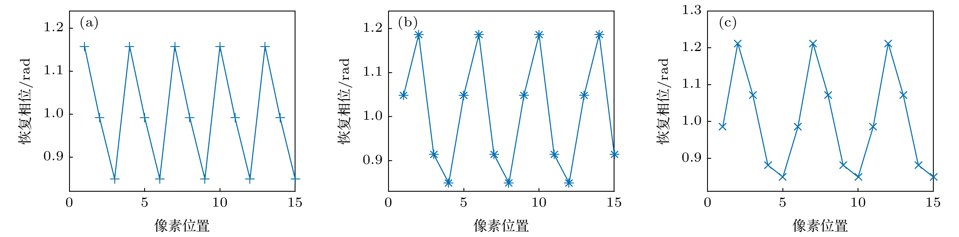
图 4 频谱混叠对恢复出相位的影响 (a), (b)和(c)分别代表载波条纹周期与探测器像素尺寸比值r为3, 4和5时的情形
Fig. 4. The impact of spectrum aliasing on phase retrieval. (a), (b) and (c) denote the cases, in which the ratios of the carrier fringe period to size of detector pixel are 3, 4 and 5, respectively.
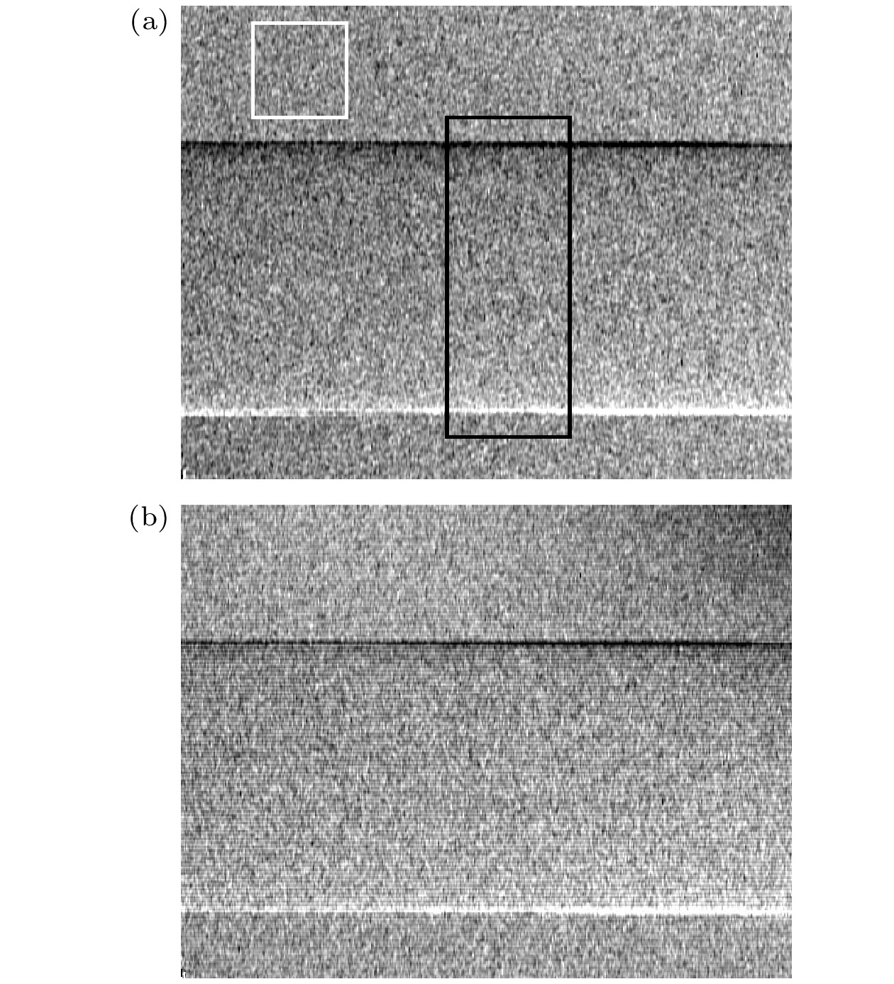
图 6 (a) 两幅图像傅里叶变换法所恢复出的PMMA相衬像; (b)单幅图像傅里叶变换法所恢复出的PMMA相衬像
Fig. 6. (a) The phase-contrast image of PMMA retrieved by Fourier transform with two images; (b) the phase-contrast image of PMMA retrieved by Fourier transform with one image.

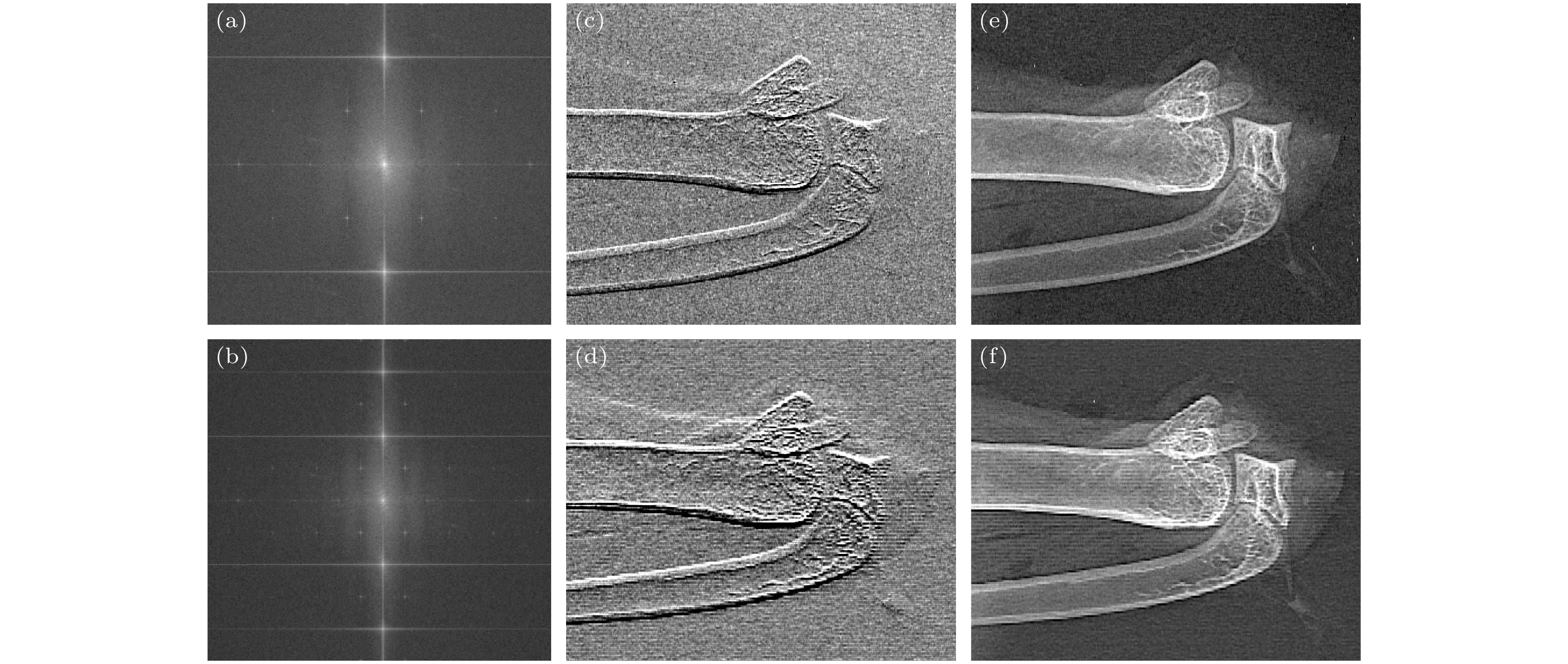
图 8 载波条纹周期与探测器像素尺寸比值r = 3时, 鸡翅的频谱 (a)、相衬像(c)和暗场像(e). r = 5时, 鸡翅的频谱(b)、相衬像(d)和暗场像(f)
Fig. 8. Fourier spectrum (a), phase-contrast image (c) and dark-field image (e) of a chicken wing when r = 3. Fourier spectrum (b), phase-contrast image (d) and dark-field image (f) of a chicken wing when r = 5.
表 1 两种不同傅里叶变换方法的定量比较
Table 1. Quantitative comparison between two kinds of Fourier transform algorithms.
背景相位
均值/rad背景相位标
准差/rad横截面峰
峰值/rad单幅图像
傅里叶变换0.3502 0.0059 0.2412 两幅图像
傅里叶变换0.2526 0.0017 0.1112  下载: 导出CSV
-
[1] David C, Nohammer E, Solak H H, Ziegler E 2002 Appl. Phys. Lett. 81 3287
Google Scholar
[2] Momose A, Kawamoto S, Koyama I, Hamaishi Y, Takai K, Suzuki Y 2003 Jpn. J. Appl. Phys. 42 L866
Google Scholar
[3] Pfeiffer F, Weitkamp T, Bunk O, David C 2006 Nat. Phys. 2 258
Google Scholar
[4] Pfeiffer F, Bech M, Bunk O, Kraft P, Eikenberry E F, Bronnimann C, Grunzweig C, David C 2008 Nat. Mater. 7 134
Google Scholar
[5] Bech M, Tapfer A, Pauwels B, Bruyndonckx P, Sasov A, Pfeiffer F 2013 Sci. Rep. 3 3209
Google Scholar
[6] Anton G, Michel T, Pelzer G, Radicke M, Rieger J, Weber T 2013 Z. Med. Phys. 23 228
Google Scholar
[7] Yang J, Guo J C, Lei Y H, Yi M H, Chen L 2017 Chin. Phys. B 26 028701
Google Scholar
[8] Weitkamp T, Diaz A, David C, Pfeiffer F, Stampanoni M, Cloetens P, Ziegler E 2005 Opt. Express 13 6296
Google Scholar
[9] Takeda M, Ina H, Kobayashi S 1982 J. Opt. Soc. Am. 72 156
Google Scholar
[10] Wen H, Bennett E E, Hegedus M M, Carroll S C 2008 IEEE Trans. Med. Imaging 27 997
Google Scholar
[11] Wen H, Bennett E E, Hegedus M M, Rapacchi S 2009 Radiology 251 910
Google Scholar
[12] Lim H, Park Y, Cho H, Je U, Hong D, Park C, Woo T, Lee M, Kim J, Chung N, Kim J, Kim J 2015 Opt. Commun. 348 85
Google Scholar
[13] Lim H W, Lee H W, Cho H S, Je U K, Park C K, Kim K S, Kim G A, Park S Y, Lee D Y, Park Y O, Woo T H, Lee S H, Chung W H, Kim J W, Kim J G 2017 Nucl. Instrum. Methods Phys. Res., Sect. A 850 89
Google Scholar
[14] Lim H, Lee H, Cho H, Seo C, Je U, Park C, Kim K, Kim G, Park S, Lee D, Kang S, Lee M 2017 J. Korean Phys. Soc. 71 722
Google Scholar
[15] Seifert M, Gallersdörfer M, Ludwig V, Schuster M, Horn F, Pelzer G, Rieger J, Michel T, Anton G 2018 J. Imaging 4 62
Google Scholar
[16] Seifert M, Ludwig V, Gallersdorfer M, Hauke C, Hellbach K, Horn F, Pelzer G, Radicke M, Rieger J, Sutter S M, Michel T, Anton G 2018 Phys. Med. Biol. 63 185010
Google Scholar
[17] Li J, Su X Y, Guo L R 1990 Opt. Eng. 29 1439
Google Scholar
[18] 陈文静, 苏显渝, 曹益平, 向立群 2004 中国激光 31 740
Google Scholar
Chen W J, Su X Y, Cao Y P, Xiang L Q 2004 Chin. J. Las. 31 740
Google Scholar
[19] Zhu P, Zhang K, Wang Z, Liu Y, Liu X, Wu Z, McDonald S A, Marone F, Stampanoni M 2010 Proc. Natl. Acad. Sci. U. S. A. 107 13576
Google Scholar
[20] Wang Z, Gao K, Ge X, Wu Z, Chen H, Wang S, Zhu P, Yuan Q, Huang W, Zhang K, Wu Z 2013 J. Phys. D: Appl. Phys. 46 494003
Google Scholar
[21] 杜杨, 雷耀虎, 刘鑫, 郭金川, 牛憨笨 2013 62 06872
Google Scholar
Yang D, Lei Y H, Liu X, Guo J C, Niu H B 2013 Acta Phys. Sin. 62 06872
Google Scholar
[22] Momose A, Yashiro W, Takeda Y, Suzuki Y, Hattori T 2006 Jpn. J. Appl. Phys. 45 5254
Google Scholar


 下载:
下载:


计量
- 文章访问数: 7835
- PDF下载量: 91
- 被引次数: 0
